
Main menu

  • Home
  • Content
    • Current Issue
    • Ahead of Print
    • Past Issues
    • Supplements
    • Article Type
  • Specialty
    • Articles by Specialty
  • CME/MOC
    • Articles
    • Calendar
  • Info For
    • Manuscript Submission
    • Authors & Reviewers
    • Subscriptions
    • About CCJM
    • Contact Us
    • Media Kit
  • Conversations with Leaders
  • Conference Coverage
    • Kidney Week 2024
    • CHEST 2024
    • ACR Convergence 2023
    • Kidney Week 2023
    • ObesityWeek 2023
    • IDWeek 2023
    • CHEST 2023
    • MDS 2023
    • IAS 2023
    • ACP 2023
    • AAN 2023
    • ACC / WCC 2023
    • AAAAI Meeting 2023
    • ACR Convergence 2022
    • Kidney Week 2022
    • AIDS 2022
  • Other Publications
    • www.clevelandclinic.org

User menu

  • Register
  • Log in

Search

  • Advanced search
Cleveland Clinic Journal of Medicine
  • Other Publications
    • www.clevelandclinic.org
  • Register
  • Log in
Cleveland Clinic Journal of Medicine

Advanced Search

  • Home
  • Content
    • Current Issue
    • Ahead of Print
    • Past Issues
    • Supplements
    • Article Type
  • Specialty
    • Articles by Specialty
  • CME/MOC
    • Articles
    • Calendar
  • Info For
    • Manuscript Submission
    • Authors & Reviewers
    • Subscriptions
    • About CCJM
    • Contact Us
    • Media Kit
  • Conversations with Leaders
  • Conference Coverage
    • Kidney Week 2024
    • CHEST 2024
    • ACR Convergence 2023
    • Kidney Week 2023
    • ObesityWeek 2023
    • IDWeek 2023
    • CHEST 2023
    • MDS 2023
    • IAS 2023
    • ACP 2023
    • AAN 2023
    • ACC / WCC 2023
    • AAAAI Meeting 2023
    • ACR Convergence 2022
    • Kidney Week 2022
    • AIDS 2022
Review

Hypertrophic cardiomyopathy: A complex disease

Laura Young, MD, Nicholas G. Smedira, MD, Albree Tower-Rader, MD, Harry Lever, MD and Milind Y. Desai, MD
Cleveland Clinic Journal of Medicine May 2018, 85 (5) 399-411; DOI: https://doi.org/10.3949/ccjm.85a.17076
Laura Young
Robert and Suzanne Tomsich Department of Cardiovascular Medicine, Heart and Vascular Institute, Cleveland Clinic
  • Find this author on Google Scholar
  • Find this author on PubMed
  • Search for this author on this site
Nicholas G. Smedira
  • Find this author on Google Scholar
  • Find this author on PubMed
  • Search for this author on this site
Albree Tower-Rader
  • Find this author on Google Scholar
  • Find this author on PubMed
  • Search for this author on this site
Harry Lever
  • Find this author on Google Scholar
  • Find this author on PubMed
  • Search for this author on this site
Milind Y. Desai
  • Find this author on Google Scholar
  • Find this author on PubMed
  • Search for this author on this site
  • For correspondence: [email protected]
  • Article
  • Figures & Data
  • Info & Metrics
  • PDF
Loading

Article Figures & Data

Figures

  • Tables
  • Figure 1
    • Download figure
    • Open in new tab
    • Download powerpoint
    Figure 1

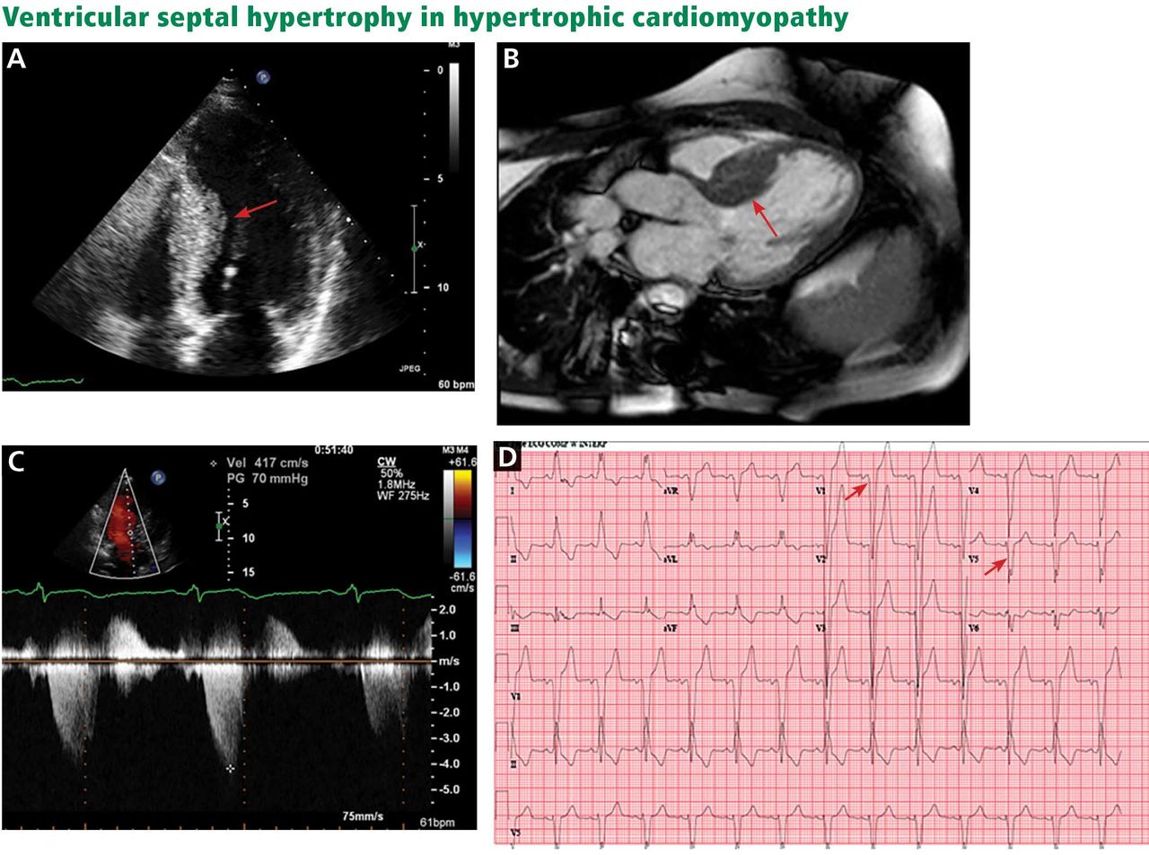
    A, echocardiography, apical 4-chamber view, demonstrates septal hypertrophy (arrow). B, cardiac magnetic resonance imaging of the left ventricular outflow tract also demonstrates septal hypertrophy (arrow). C, echocardiography with continuous-wave Doppler across the left ventricular outflow tract demonstrates a gradient of 70 mm Hg, consistent with obstruction. D, electrocardiography reveals signs of left ventricular hypertrophy by Sokolov-Lynon criteria with S wave depth in V1 plus R wave height in V5 > 35 mm (arrows).

  • Figure 2
    • Download figure
    • Open in new tab
    • Download powerpoint
    Figure 2

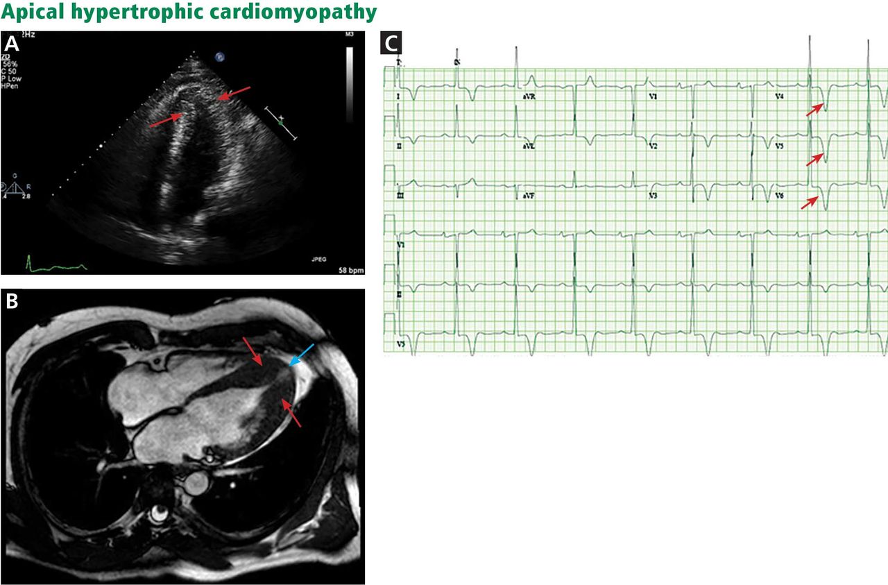
    A, echocardiography, apical 4-chamber view, shows apical hypertrophy (arrows). B, cardiac magnetic resonance imaging (4-chamber view) shows apical hypertrophy (red arrows), as well as an apical aneurysm (blue arrow). C, electrocardiography demonstrates giant T-wave inversions in the left precordial leads, characteristic of apical hypertrophic cardiomyopathy (arrows).

  • Figure 3
    • Download figure
    • Open in new tab
    • Download powerpoint
    Figure 3

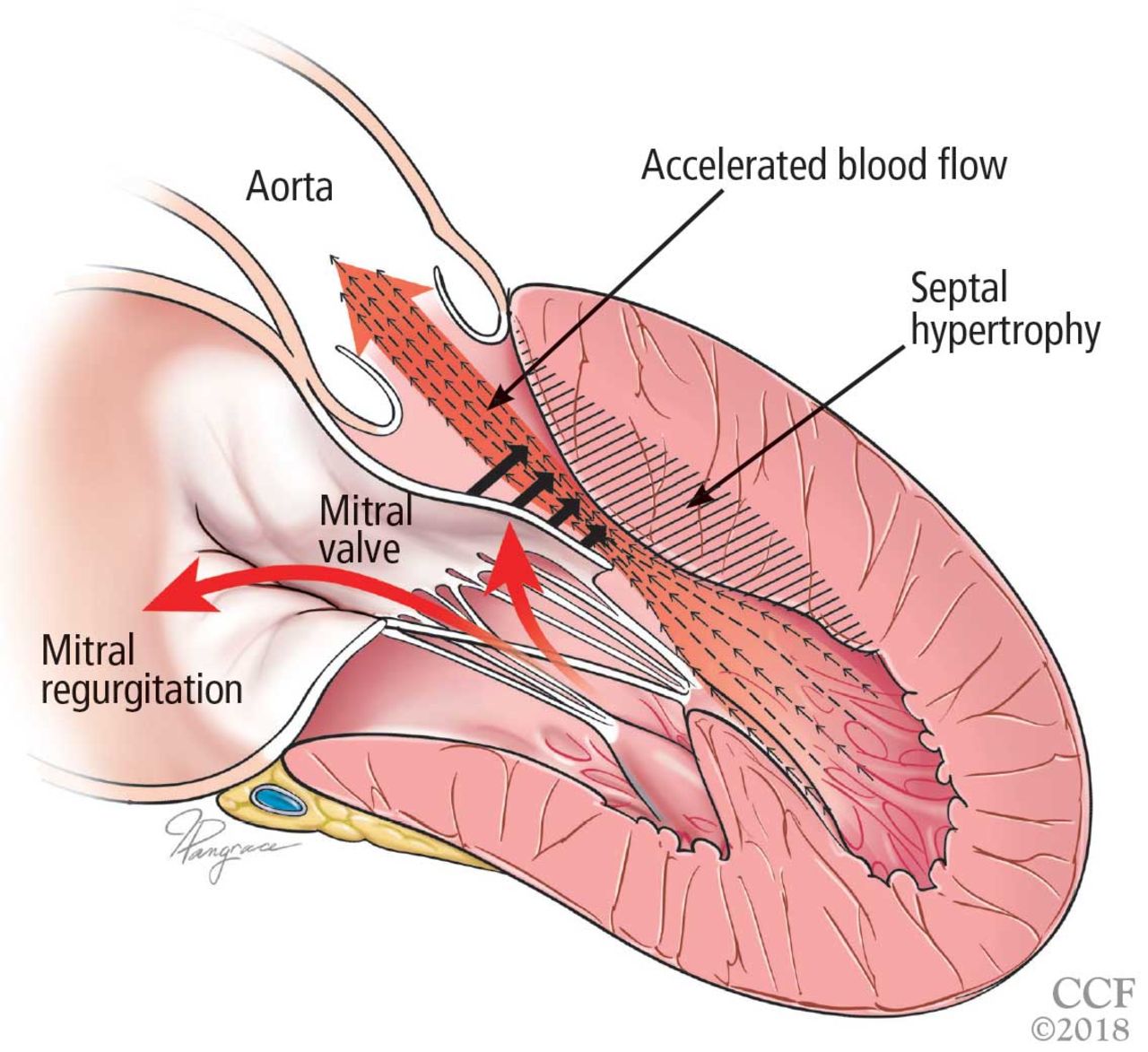
    Left ventricular outflow tract obstruction due to ventricular septal hypertrophy. The obstruction is dynamic, as the blood flow sweeps the mitral valve toward the septum.

  • Figure 4
    • Download figure
    • Open in new tab
    • Download powerpoint
    Figure 4

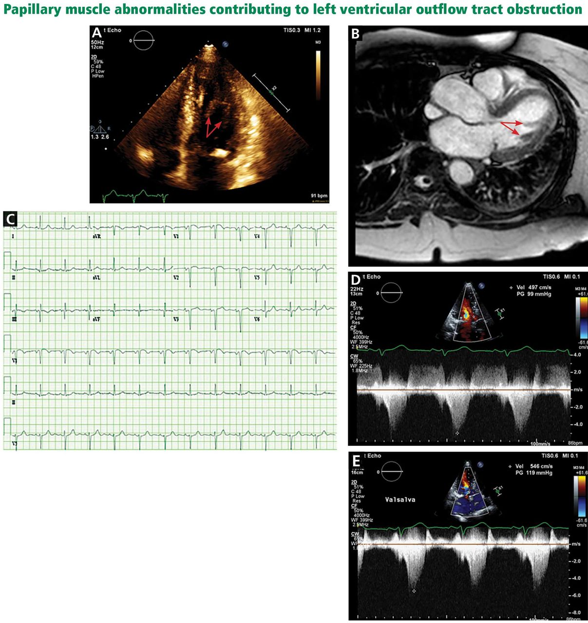
    A, echocardiography, apical 4-chamber view, demonstrates a bifid papillary muscle resulting in left ventricular outflow tract obstruction (arrows). B, cardiac magnetic resonance imaging (left ventricular outflow tract view) demonstrates a bifid papillary muscle (arrows). C, an electrocardiogram of a patient with obstruction related to abnormal papillary muscle morphology demonstrates a lack of significant left ventricular hypertrophy. D, continuous-wave Doppler through the left ventricular outflow tract demonstrates a peak gradient of 99 mm Hg, consistent with obstruction, which increases with the Valsalva maneuver to 119 mm Hg (E).

  • Figure 5
    • Download figure
    • Open in new tab
    • Download powerpoint
    Figure 5

    Left ventricular outflow tract (LVOT) obstruction without significant left ventricular hypertrophy. The prominent bifid papillary muscles lead to systolic anterior motion of the mitral valve, causing LVOT obstruction and simultaneous mitral regurgitation.
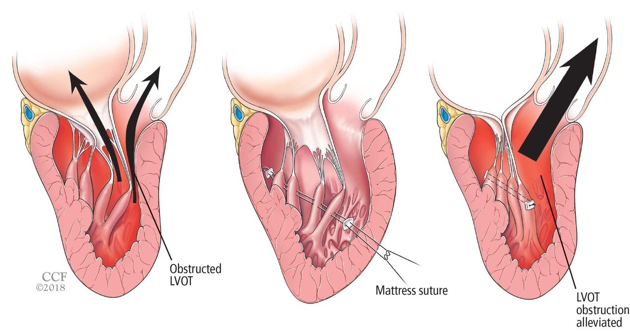
  • Figure 6
    • Download figure
    • Open in new tab
    • Download powerpoint
    Figure 6

    Reorientation surgery reduces mobility of bifid hypermobile papillary muscles, reducing left ventricular outflow tract (LVOT) obstruction.

Tables

  • Figures
    • View popup
    TABLE 1

    Differential diagnosis of hypertrophic cardiomyopathy

    Hypertensive cardiomyopathy
    Aortic valvulopathy
    Aortic stenosis
    Supra-aortic or subaortic membranes
    Infiltrative cardiomyopathy
    Amyloidosis
    Fabry disease
    Lysosomal diseases (eg, Danon disease)
    Glycogen storage disorders (eg, Pompe disease)
    Hemochromatosis
    ‘Athlete’s heart’
    Noncompaction cardiomyopathy
    • View popup
    TABLE 2

    Main causative genes of hypertrophic cardiomyopathy (HCM)

    Sarcomeric proteinsGeneGene prevalence in HCM probands
    Myosin-binding protein CMYPBC315%
    Beta myosin heavy chainMYH715%
    Cardiac troponin TTNNT2  7%
    Alpha-tropomyosinTPM1  7%
    Regulatory myosin light chainMYL2< 5%
    Essential myosin light chainMYL3< 5%
    Cardiac troponin ITNNI3< 5%
    Nonsarcomeric proteinsGeneInheritanceAssociated phenotype
    TransthyretinTTRDominant; 1%–10%Amyloidosis
    Lysosome-associated membrane glycoprotein 2LAMP2X-linked; rareDanon disease
    Alpha-galactosidase AGLAX-linked; 1%–2% of malesFabry disease
    Lysosomal alpha-glucosidaseGAARecessive; rarePompe disease
    FrataxinFXNRecessive; rareFriedrich ataxia
    • Based on information in reference 3.

    • View popup
    TABLE 3

    Risk-stratification models for primary prevention of sudden cardiac death in hypertrophic cardiomyopathy

    North American modelEuropean model
    An implantable cardioverter-defibrillator (ICD) is reasonable (class IIa recommendation, level of evidence C—limited evidence) if any of the following are present:
    • Family history of sudden death

    • Unexplained syncope

    • Maximum left ventricular wall thickness ≥ 30 mm


    Or if the patient has any other risk factor or modifier for sudden cardiac death and either of the following:
    • Nonsustained ventricular tachycardia

    • Abnormal blood pressure response during exercise (decrease or failure to increase systolic blood pressure ≥ 20 mm Hg during exercise stress test)

    The following factors are used to electronically calculate the 5-year risk of sudden cardiac death:
    • Family history of sudden death

    • Unexplained syncope

    • Maximum left ventricular wall thickness

    • Nonsustained ventricular tachycardia

    • Age

    • Left atrial diameter

    • Left ventricular outflow gradient


    5-year risk < 4%: an ICD is generally not indicated
    5-year risk ≥ 4% to < 6%: an ICD may be considered
    5-year risk ≥ 6%: an ICD should be considered
    • Based on information in references 9 and 30.

PreviousNext
Back to top

In this issue

Cleveland Clinic Journal of Medicine: 85 (5)
Cleveland Clinic Journal of Medicine
Vol. 85, Issue 5
1 May 2018
  • Table of Contents
  • Table of Contents (PDF)
  • Index by author
Print
Download PDF
Article Alerts
Sign In to Email Alerts with your Email Address
Email Article

Thank you for your interest in spreading the word on Cleveland Clinic Journal of Medicine.

NOTE: We only request your email address so that the person you are recommending the page to knows that you wanted them to see it, and that it is not junk mail. We do not capture any email address.

Enter multiple addresses on separate lines or separate them with commas.
Hypertrophic cardiomyopathy: A complex disease
(Your Name) has sent you a message from Cleveland Clinic Journal of Medicine
(Your Name) thought you would like to see the Cleveland Clinic Journal of Medicine web site.
CAPTCHA
This question is for testing whether or not you are a human visitor and to prevent automated spam submissions.
Citation Tools
Hypertrophic cardiomyopathy: A complex disease
Laura Young, Nicholas G. Smedira, Albree Tower-Rader, Harry Lever, Milind Y. Desai
Cleveland Clinic Journal of Medicine May 2018, 85 (5) 399-411; DOI: 10.3949/ccjm.85a.17076

Citation Manager Formats

  • BibTeX
  • Bookends
  • EasyBib
  • EndNote (tagged)
  • EndNote 8 (xml)
  • Medlars
  • Mendeley
  • Papers
  • RefWorks Tagged
  • Ref Manager
  • RIS
  • Zotero
Share
Hypertrophic cardiomyopathy: A complex disease
Laura Young, Nicholas G. Smedira, Albree Tower-Rader, Harry Lever, Milind Y. Desai
Cleveland Clinic Journal of Medicine May 2018, 85 (5) 399-411; DOI: 10.3949/ccjm.85a.17076
Twitter logo Facebook logo Mendeley logo
  • Tweet Widget
  • Facebook Like
  • Linkedin Share Button

Jump to section

  • Article
    • ABSTRACT
    • A PLETHORA OF MUTATIONS IN CARDIAC SARCOMERIC GENES
    • LEFT VENTRICULAR OUTFLOW TRACT OBSTRUCTION
    • DIAGNOSTIC EVALUATION
    • GENETIC TESTING, COUNSELING, AND FAMILY SCREENING
    • MANAGEMENT
    • REFERENCES
  • Figures & Data
  • Info & Metrics
  • PDF

Related Articles

  • No related articles found.
  • PubMed
  • Google Scholar

Cited By...

  • Hypertrophic cardiomyopathy (May 2018)
  • Google Scholar

More in this TOC Section

  • Alpha-gal syndrome: Recognizing and managing a tick-bite–related meat allergy
  • Managing right ventricular failure in the setting of pulmonary embolism
  • Psychedelics, spirituality, and existential distress in patients at the end of life
Show more Review

Similar Articles

Subjects

  • Adolescent Medicine
  • Cardiology
  • Genetics
  • Imaging

Navigate

  • Current Issue
  • Past Issues
  • Supplements
  • Article Type
  • Specialty
  • CME/MOC Articles
  • CME/MOC Calendar
  • Media Kit

Authors & Reviewers

  • Manuscript Submission
  • Authors & Reviewers
  • Subscriptions
  • About CCJM
  • Contact Us
  • Cleveland Clinic Center for Continuing Education
  • Consult QD

Share your suggestions!

Copyright © 2025 The Cleveland Clinic Foundation. All rights reserved. The information provided is for educational purposes only. Use of this website is subject to the website terms of use and privacy policy. 

Powered by HighWire