
Main menu

  • Home
  • Content
    • Current Issue
    • Ahead of Print
    • Past Issues
    • Supplements
    • Article Type
  • Specialty
    • Articles by Specialty
  • CME/MOC
    • Articles
    • Calendar
  • Info For
    • Manuscript Submission
    • Authors & Reviewers
    • Subscriptions
    • About CCJM
    • Contact Us
    • Media Kit
  • Conversations with Leaders
  • Conference Coverage
    • Kidney Week 2024
    • CHEST 2024
    • ACR Convergence 2023
    • Kidney Week 2023
    • ObesityWeek 2023
    • IDWeek 2023
    • CHEST 2023
    • MDS 2023
    • IAS 2023
    • ACP 2023
    • AAN 2023
    • ACC / WCC 2023
    • AAAAI Meeting 2023
    • ACR Convergence 2022
    • Kidney Week 2022
    • AIDS 2022
  • Other Publications
    • www.clevelandclinic.org

User menu

  • Register
  • Log in

Search

  • Advanced search
Cleveland Clinic Journal of Medicine
  • Other Publications
    • www.clevelandclinic.org
  • Register
  • Log in
Cleveland Clinic Journal of Medicine

Advanced Search

  • Home
  • Content
    • Current Issue
    • Ahead of Print
    • Past Issues
    • Supplements
    • Article Type
  • Specialty
    • Articles by Specialty
  • CME/MOC
    • Articles
    • Calendar
  • Info For
    • Manuscript Submission
    • Authors & Reviewers
    • Subscriptions
    • About CCJM
    • Contact Us
    • Media Kit
  • Conversations with Leaders
  • Conference Coverage
    • Kidney Week 2024
    • CHEST 2024
    • ACR Convergence 2023
    • Kidney Week 2023
    • ObesityWeek 2023
    • IDWeek 2023
    • CHEST 2023
    • MDS 2023
    • IAS 2023
    • ACP 2023
    • AAN 2023
    • ACC / WCC 2023
    • AAAAI Meeting 2023
    • ACR Convergence 2022
    • Kidney Week 2022
    • AIDS 2022
Review

Thoracic aortic aneurysm: How to counsel, when to refer

Frank Cikach, MD, Milind Y. Desai, MD, FACC, FAHA, FESC, Eric E. Roselli, MD, FACS and Vidyasagar Kalahasti, MD
Cleveland Clinic Journal of Medicine June 2018, 85 (6) 481-492; DOI: https://doi.org/10.3949/ccjm.85a.17039
Frank Cikach
Cleveland Clinic Lerner College of Medicine of Case Western Reserve University, Cleveland, OH
  • Find this author on Google Scholar
  • Find this author on PubMed
  • Search for this author on this site
Milind Y. Desai
  • Find this author on Google Scholar
  • Find this author on PubMed
  • Search for this author on this site
Eric E. Roselli
  • Find this author on Google Scholar
  • Find this author on PubMed
  • Search for this author on this site
Vidyasagar Kalahasti
  • Find this author on Google Scholar
  • Find this author on PubMed
  • Search for this author on this site
  • For correspondence: [email protected]
  • Article
  • Figures & Data
  • Info & Metrics
  • PDF
Loading

Article Figures & Data

Figures

  • Tables
  • Figure 1
    • Download figure
    • Open in new tab
    • Download powerpoint
    Figure 1
  • Figure 2
    • Download figure
    • Open in new tab
    • Download powerpoint
    Figure 2

    Echocardiographic image of an aneurysmal aortic root (white arrow) that tapers to normal dimensions at the sinotubular junction (yellow arrow) and ascending aorta. See videos (this image and a bicuspid aortic valve).

  • Figure 3
    • Download figure
    • Open in new tab
    • Download powerpoint
    Figure 3

    An aortic root aneurysm in a patient with Mar-fan syndrome using magnetic resonance angiography. See videos (this image, follow-up after surgical repair, and a computed tomographic study).
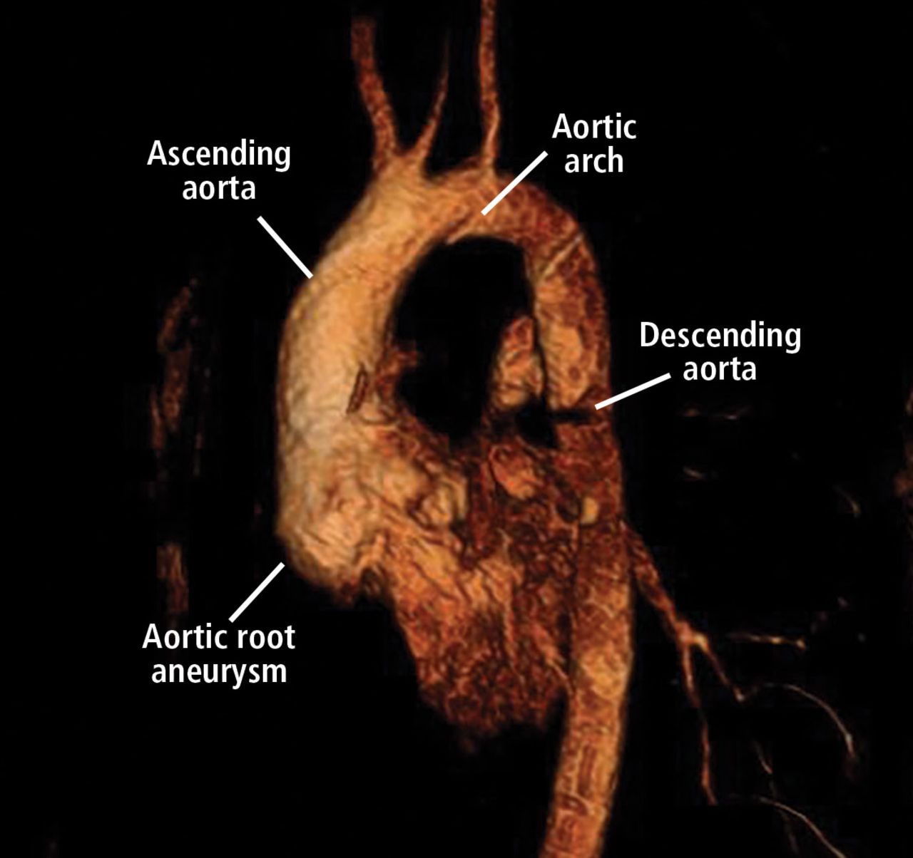
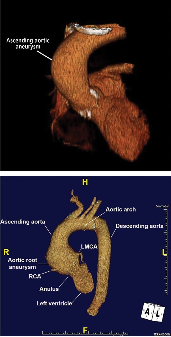
  • Figure 4
    • Download figure
    • Open in new tab
    • Download powerpoint
    Figure 4

    Top, 3D reconstruction of an ascending aortic aneurysm in a patient with a bicuspid aortic valve. See video. Bottom, 3D reconstruction of a young patient with an aortic root aneurysm. Note the tapering from the aneurysmal aortic root to the normalsized ascending aorta. LMCA = left main coronary artery; RCA = right coronary artery.
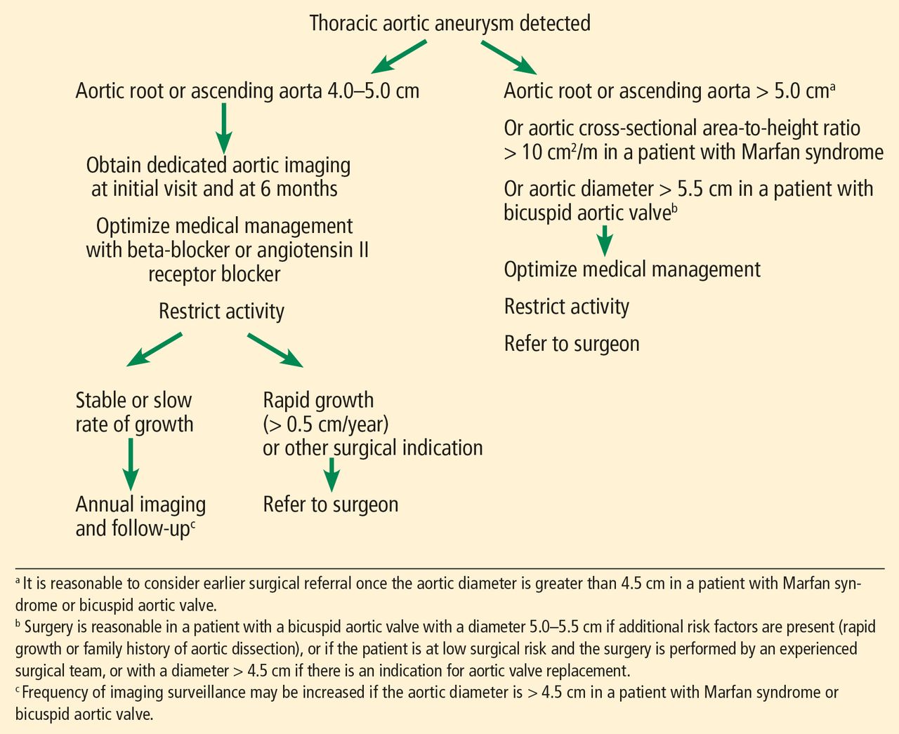
  • Figure 5
    • Download figure
    • Open in new tab
    • Download powerpoint
    Figure 5

    Initial screening and follow-up of thoracic aortic aneurysm.

Tables

  • Figures
    • View popup
    TABLE 1

    Aortic diameters: Upper limits of normala

    Age (years)BSA (m2)Ascending aorta (mm) Descending aorta (mm)
    Women (n = 1,147)Men (n = 1,805)Women (n = 736)Men (n = 1,195)
    < 45< 1.7033.833.023.0NA
    1.70–1.8934.436.324.626.6
    1.90–2.0935.036.322.726.7
    > 2.1NA38.3NA28.3
    45–54< 1.7035.238.624.324.2
    1.70–1.8937.238.125.427.5
    1.90–2.0938.939.727.229.2
    > 2.140.640.628.329.6
    55–64< 1.7036.936.325.926.1
    1.70–1.8937.039.727.128.6
    1.90–2.0939.041.227.829.9
    > 2.142.043.131.731.6
    ≥ 65< 1.7037.538.527.0NA
    1.70–1.8939.241.027.432.4
    1.90–2.0942.742.229.031.0
    > 2.1NA42.429.832.5
    • ↵a Upper limits of normal are 2 standard deviations above the mean. Not calculated if there were fewer than 6 patients in a group. BSA = body surface area; NA = not available

    • Information from Wolak A, Gransar H, Thomson LE, et al. Aortic size assessment by noncontrast cardiac computed tomography: normal limits by age, gender, and body surface area. J Am Coll Cardiol Cardiovasc Imaging 2008; 1(2):200–209. doi:10.1016/j.jcmg.2007.11.005

    • View popup
    TABLE 2

    Common causes of thoracic aortic aneurysm

    Idiopathic
    Bicuspid aortic valve
    Genetic syndromes
    Marfan syndrome
    Loeys-Dietz syndrome
    Ehlers-Danlos syndrome
    Familial thoracic aortic aneurysm and dissection
    Autosomal dominant polycystic kidney disease
    Turner syndrome
    Inflammatory vasculitis
    Takayasu arteritis
    Giant cell arteritis
    Behçet arteritis
    Ankylosing spondylitis
    Other
    Atherosclerosis
    Syphilis
    Previous dissection
    Infectious aortitis
    Trauma
    Risk factors
    Hypertension
    Smoking
    Advanced age
    • View popup
    TABLE 3

    Inherited connective tissue diseases and thoracic aortic aneurysm

    Inherited conditionImplicated geneClinical features
    Marfan syndromeFBN1Aneurysm of aortic root, dilation of pulmonary artery, and aortic dissection
    Ehlers-Danlos syndromeCOL5A1, COL5A2, COL3A1Arterial mid-sized rupture, especially involving thoracic vasculature
    Loeys-Dietz syndromeTGFBR1, TGFBR2, SMAD3, TGFB2, TGFB3Premature and aggressive aneurysm and dissection; aneurysm may involve aortic segments other than the root
    Familial thoracic aortic aneurysm and dissectionACTA2, MYH11, PRKG1, MYLK, TGFBR2Thoracic aortic aneurysm and dissection; associated vascular disease (eg, patent ductus arteriosus)
    Bicuspid aortic valveUnknown (may be associated with ACTA2, MYH11, syndromic connective tissue diseases)Aortic dilation typically involving the aortic root and ascending aorta
    Autosomal dominant polycystic kidney diseasePKD1, PKD2Dilation of the aorta and thoracic aortic dissection
    Turner syndrome45,XThoracic aortic aneurysms and dissections, bicuspid aortic valve, aortic coarctation
    • Reprinted from Cury M, Zeidan F, Lobato AC. Aortic disease in the young: genetic aneurysm syndromes, connective tissue disorders, and familial aortic aneurysms and dissections. Int J Vasc Med 2013(2013); 2013:267215. doi:10.1155/2013/267215

    • View popup
    TABLE 4

    Imaging studies for aortic aneurysm

    Imaging studyAdvantagesDisadvantages
    Cardiac-gated multidetector computed tomographic angiographyHighly specific and sensitive Can diagnose vascular anomalies, tortuosityLarge doses of ionizing radiation and contrast
    Magnetic resonance angiographyHighly specific and sensitive Can be accurate without using contrast Ability to assess valve diseaseProlonged scanning time Difficult for those with claustrophobia
    Transthoracic echocardiographyNoninvasive, readily available Can assess valve diseaseLimited visualization of ascending aorta Accuracy reduced with “oblique” measurements
    Transesophageal echocardiographyHighly specific and sensitive Ability to assess valve diseaseRequires skilled personnel Invasive, requires procedural sedation
    AngiographyHighly sensitive and specificInvasive Requires contrast
    • Based on Smith AD, Schoenhagen P. CT imaging for acute aortic syndrome. Cleve Clin J Med 2008; 75(1):7–17. pmid:18236724

PreviousNext
Back to top

In this issue

Cleveland Clinic Journal of Medicine: 85 (6)
Cleveland Clinic Journal of Medicine
Vol. 85, Issue 6
1 Jun 2018
  • Table of Contents
  • Table of Contents (PDF)
  • Index by author
Print
Download PDF
Article Alerts
Sign In to Email Alerts with your Email Address
Email Article

Thank you for your interest in spreading the word on Cleveland Clinic Journal of Medicine.

NOTE: We only request your email address so that the person you are recommending the page to knows that you wanted them to see it, and that it is not junk mail. We do not capture any email address.

Enter multiple addresses on separate lines or separate them with commas.
Thoracic aortic aneurysm: How to counsel, when to refer
(Your Name) has sent you a message from Cleveland Clinic Journal of Medicine
(Your Name) thought you would like to see the Cleveland Clinic Journal of Medicine web site.
CAPTCHA
This question is for testing whether or not you are a human visitor and to prevent automated spam submissions.
Citation Tools
Thoracic aortic aneurysm: How to counsel, when to refer
Frank Cikach, Milind Y. Desai, Eric E. Roselli, Vidyasagar Kalahasti
Cleveland Clinic Journal of Medicine Jun 2018, 85 (6) 481-492; DOI: 10.3949/ccjm.85a.17039

Citation Manager Formats

  • BibTeX
  • Bookends
  • EasyBib
  • EndNote (tagged)
  • EndNote 8 (xml)
  • Medlars
  • Mendeley
  • Papers
  • RefWorks Tagged
  • Ref Manager
  • RIS
  • Zotero
Share
Thoracic aortic aneurysm: How to counsel, when to refer
Frank Cikach, Milind Y. Desai, Eric E. Roselli, Vidyasagar Kalahasti
Cleveland Clinic Journal of Medicine Jun 2018, 85 (6) 481-492; DOI: 10.3949/ccjm.85a.17039
Twitter logo Facebook logo Mendeley logo
  • Tweet Widget
  • Facebook Like
  • Linkedin Share Button

Jump to section

  • Article
    • ABSTRACT
    • LIKELY UNDERDETECTED
    • RISK FACTORS FOR THORACIC AORTIC ANEURYSM
    • DIAMETER INCREASES WITH AGE, BODY SURFACE AREA
    • HOW IS TAA DIAGNOSED?
    • HOW IS TAA CLASSIFIED?
    • HOW IS ANEURYSM MANAGED ON AN OUTPATIENT BASIS?
    • WHEN SHOULD A PATIENT BE REFERRED?
    • WHEN SHOULD TAA BE REPAIRED?
    • HOW SHOULD PATIENTS BE SCREENED? WHAT FOLLOW-UP IS NECESSARY?
    • BACK TO OUR PATIENT WITH A BICUSPID AORTIC VALVE
    • BACK TO OUR PATIENT WITH MARFAN SYNDROME
    • Footnotes
    • REFERENCES
  • Figures & Data
  • Info & Metrics
  • PDF

Related Articles

  • No related articles found.
  • PubMed
  • Google Scholar

Cited By...

  • Aortic aneurysm: Fluoroquinolones, genetic counseling, June 2018
  • Aortic aneurysm: Fluoroquinolones, genetic counseling, June 2018
  • Google Scholar

More in this TOC Section

  • High-output heart failure from arteriovenous dialysis access: A structured approach to diagnosis and management
  • Direct oral anticoagulants: Challenging prescribing scenarios in everyday practice
  • IgA nephropathy: Update on pathogenesis and treatment
Show more Review

Similar Articles

Subjects

  • Cardiology
  • Genetics
  • Imaging
  • Vascular Medicine

Navigate

  • Current Issue
  • Past Issues
  • Supplements
  • Article Type
  • Specialty
  • CME/MOC Articles
  • CME/MOC Calendar
  • Media Kit

Authors & Reviewers

  • Manuscript Submission
  • Authors & Reviewers
  • Subscriptions
  • About CCJM
  • Contact Us
  • Cleveland Clinic Center for Continuing Education
  • Consult QD

Share your suggestions!

Copyright © 2025 The Cleveland Clinic Foundation. All rights reserved. The information provided is for educational purposes only. Use of this website is subject to the website terms of use and privacy policy. 

Powered by HighWire