


A 41-year-old man presented with concern about a lesion on the right lateral thigh that he had first noticed 6 years earlier. The lesion had not changed since that time.
There was no history of trauma or infection to the area. He reported no weight loss, and he said he had not noticed similar lesions elsewhere.
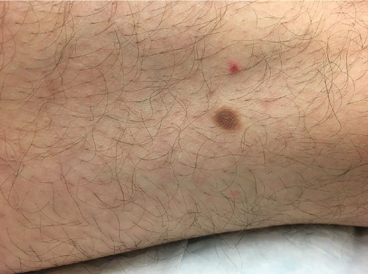
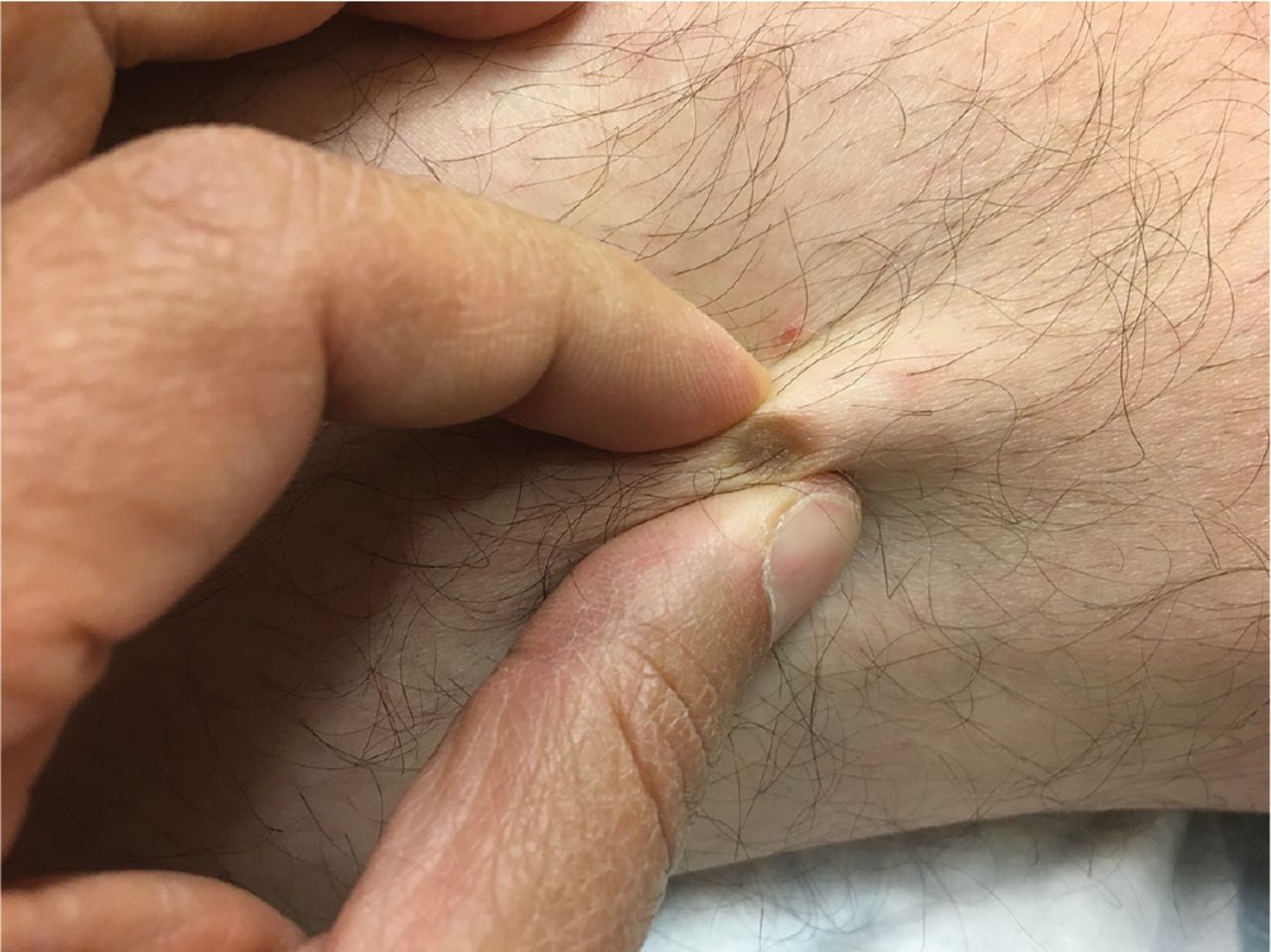
Physical examination revealed a firm, non-tender, hyperpigmented nodule (7 mm × 9 mm) on the lateral aspect of his right thigh (Figure 1). There was no surrounding erythema or warmth. Lateral pressure on the sides of the lesion produced a depression (“dimple” sign) (Figure 2).

A hyperpigmented nodule on the lateral thigh.

Retraction with lateral compression.
Based on the clinical picture and physical examination, the diagnosis of dermatofibroma was made.
THE DIFFERENTIAL DIAGNOSIS
Firm, hyperpigmented, macular or nodular skin lesions are prevalent and seen in a number of conditions (Table 1).
Differential diagnosis of a firm, hyperpigmented, macular skin lesion
Dermatofibroma (fibrous histiocytoma) is a benign proliferation of collagen fiber and other mesenchymal cell lines, likely in response to local inflammation or trauma.1
Dermatofibromas are more common in women and typically develop between ages 20 and 50.1,2 They often present as smooth, slow-growing, firm, tan to reddish brown papules or nodules less than 1 cm in diameter that classically dent on compression.1–3 They are mostly asymptomatic and may appear anywhere on the body, though 20% are on extremities.1–3 Lesions are typically darker in color in the center and lighter toward the perimeter.1–3
Lentigo maligna is a premalignant melanocytic nevus that may be considered melanoma in situ in its most advanced stages.4 It has a high risk of progression to invasive melanoma.4 Lesions typically present on sun-exposed skin such as the head or neck.4 They appear as heterogeneous asymmetric macules with irregular borders that grow centrifugally.4 Ultraviolet light examination with a Wood lamp can show extension of the lesion far beyond the pigmented borders.4
Treatment is typically by surgical excision with borders greater than 7 mm and histopathologic examination of the margins, though radiation and topical imiquimod may be used in specific circumstances.4
Dermatofibrosarcoma protuberans is a malignant neoplastic lesion, more common in women and darker-skinned individuals age 30 to 50.5 It can present as an asymptomatic, slow-growing, violaceous nodule or plaque, more often on the trunk or upper extremities.5 It is typically larger than a dermatofibroma, with an irregular border and deeper palpable skin invasion.3 Diagnosis is typically by excisional biopsy.5 Though it has a low metastatic potential, it can have a great capacity for local invasion and destruction.5
Treatment requires excision with exhaustive histopathologic examination of boundaries for tumor cells, either by Mohs micrographic surgery or wide local excision.5 Adjuvant and neoadjuvant therapies such as radiation and imatinib may also be used in cases refractory to excision or with extensive invasion.5
Seborrheic keratosis is a common benign skin tumor that becomes increasingly common with age, though lesions can present at any age.6 They can be pigmented and so may be mistaken for dermatofibroma, but they do not dimple to lateral compression.6 They typically present as sharply demarcated ovoid macules or papules 1 cm in diameter and with a shiny (“oily”) appearance.6 They classically appear raised and “stuck on” to the skin.1,6 Obvious seborrheic keratoses may be monitored, but questionable lesions should be diagnosed with shave excision or curettage and histopathology.6
Epidermoid inclusion (sebaceous) cyst is a common cystic lesion that can be flat or raised, with size ranging from a few millimeters to a few centimeters.1 They often have a dark central punctum, which occasionally drains.1 They are benign and should be removed only if they cause symptoms such as frequent infection or for cosmetic reasons.1
MANAGEMENT OF DERMATOFIBROMA
Most dermatofibromas do not require treatment unless they show signs of malignant progression such as a change in quality or rapid growth.1,2 It is essential to distinguish them from the far more malignant dermatofibro-sarcoma protuberans, as well as melanoma and other malignant lesions. Irregular borders or substantial palpable depth of invasion through skin should prompt excisional biopsy for definitive diagnosis.3
- Copyright © 2020 The Cleveland Clinic Foundation. All Rights Reserved.
In this issue


Jump to section
Related Articles
Cited By...
- No citing articles found.