


Based on the number of submissions we receive, The Clinical Picture is one of the most popular sections in CCJM—and also one of my favorites. Seemingly straightforward in approach, it is the section with our lowest acceptance rate, largely due to the fact that we have specific educational expectations for these pictures and their accompanying stories.
The Clinical Picture section was introduced in the Journal in 1997 by my preceding Editor in Chief Dr. John Clough. The very first article in this series was submitted by Dr. Gary Hoffman, who was at the time Chairman of Rheumatology at Cleveland Clinic. Dr. Hoffman, best known for his seminal work in the area of vasculitis, is an astute clinical educator who believes as I do in the power of visual imagery and the value of the clinical examination. His article depicted a patient with undertreated gout.1
In an ironic symmetry of content and message, the current issue of the Journal contains another Clinical Picture, this one from Drs. Hiroyuki Yano and Mitsuyo Kinjo,2 that depicts a patient with undertreated gout. They describe exactly the same management challenges Dr. Hoffman discussed 24 years ago.
Gout was extremely common 24 years ago, and is even more so now. These two articles demonstrate that, while clinical findings may be striking enough to be published, they may go unrecognized or underappreciated for their significance and be inadequately addressed in clinical practice.
We do not select images for publication in The Clinical Picture based solely on their uniqueness; in fact, it is often the opposite. We don’t generally accept the truly arcane one-of-a-kind image or illustrated unique case report. We look for images that reinforce the value of observation3 during the physical examination. We look for images that support what Nishigori et al4 have termed the “hypothesis-driven,” and that I have described in lectures and at the bedside as the “directed” physical examination. And when the images prompt us readers to be attentive and influence our clinical behavior when we recognize them in practice, it is a heuristic victory. Sometimes, as with the clinical picture provided by Drs. Yano and Kinjo, the story also provides a powerful message. These are the pictures we look to publish.
The man described by Drs. Yano and Kinjo2 suffered recurrent attacks of foot pain several times a year for 10 years, yet gout was apparently not diagnosed and was not treated until he developed chronic kidney disease along with palpable nodules and intradermal papules. Only then was urate-lowering treatment initiated. His attacks diminished, and some tophi regressed, although he never achieved a serum urate level of significantly less than 6.0 mg/dL, which is the generally accepted minimal treatment target for patients with demonstrable tophaceous gout.5 Although his treatment seemed to be successful as suggested by a decrease in the number of gout flares, it is unlikely that all tophi will resolve with an achieved level of serum urate higher than 6 mg/dL—thus, in the future, flares will likely resume, and further joint and bone damage ensue. This is likely to become an even more significant issue should he require renal transplant.6
Intradermal tophi have been repeatedly described in the literature. With a seeming preference for the cooler extremities, digital and ear locations are common, but diffuse miliary gout is well described.7,8 Interestingly, unlike in the patient described in this issue, these as well as other types of tophi (particularly those occurring around osteoarthritic finger joints) can be found on examination even before gout flares occur.9
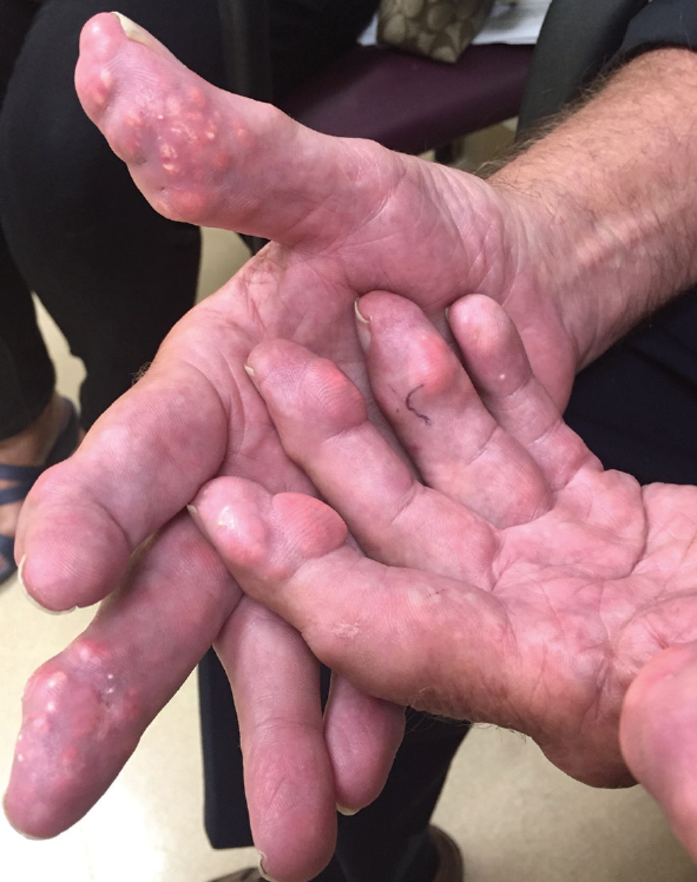
To dramatically illustrate the latter point, I attach 2 images of the hands of one of my patients, a 70-year-old international businessman. Despite having tophaceous nodulosis and intradermal tophi to the extent that he could not make a complete fist (Figure 1), he had not experienced any flares of arthritis. He had been told “it is only gout” and never received urate-lowering therapy. Figure 2 shows his hands after 6 months of very aggressive urate-lowering therapy to keep his serum urate level lower than 1 mg/dL. He regained virtually full use of his hands, and the urate-lowering therapy was subsequently changed to maintain a serum urate level of approximately 5 mg/dL.

Before treatment.

After treatment.
The teaching points here include that physical findings, once found, should be acted upon when appropriate. We can all benefit from being reminded of the value of looking for less-common findings and reacting to them in the appropriate clinical context. Gout remains a very common and very frequently undertreated clinical condition. A reminder of those facts every quarter-century seems appropriate.
- Copyright © 2021 The Cleveland Clinic Foundation. All Rights Reserved.


