
Main menu

  • Home
  • Content
    • Current Issue
    • Ahead of Print
    • Past Issues
    • Supplements
    • Article Type
  • Specialty
    • Articles by Specialty
  • CME/MOC
    • Articles
    • Calendar
  • Info For
    • Manuscript Submission
    • Authors & Reviewers
    • Subscriptions
    • About CCJM
    • Contact Us
    • Media Kit
  • Conversations with Leaders
  • Conference Coverage
    • Kidney Week 2024
    • CHEST 2024
    • ACR Convergence 2023
    • Kidney Week 2023
    • ObesityWeek 2023
    • IDWeek 2023
    • CHEST 2023
    • MDS 2023
    • IAS 2023
    • ACP 2023
    • AAN 2023
    • ACC / WCC 2023
    • AAAAI Meeting 2023
    • ACR Convergence 2022
    • Kidney Week 2022
    • AIDS 2022
  • Other Publications
    • www.clevelandclinic.org

User menu

  • Register
  • Log in

Search

  • Advanced search
Cleveland Clinic Journal of Medicine
  • Other Publications
    • www.clevelandclinic.org
  • Register
  • Log in
Cleveland Clinic Journal of Medicine

Advanced Search

  • Home
  • Content
    • Current Issue
    • Ahead of Print
    • Past Issues
    • Supplements
    • Article Type
  • Specialty
    • Articles by Specialty
  • CME/MOC
    • Articles
    • Calendar
  • Info For
    • Manuscript Submission
    • Authors & Reviewers
    • Subscriptions
    • About CCJM
    • Contact Us
    • Media Kit
  • Conversations with Leaders
  • Conference Coverage
    • Kidney Week 2024
    • CHEST 2024
    • ACR Convergence 2023
    • Kidney Week 2023
    • ObesityWeek 2023
    • IDWeek 2023
    • CHEST 2023
    • MDS 2023
    • IAS 2023
    • ACP 2023
    • AAN 2023
    • ACC / WCC 2023
    • AAAAI Meeting 2023
    • ACR Convergence 2022
    • Kidney Week 2022
    • AIDS 2022
The Clinical Picture

Fibrous epulis: A tumorlike gingival lesion

Keiichi Ohta, DDS and Hitoshi Yoshimura, DDS, PhD
Cleveland Clinic Journal of Medicine May 2021, 88 (5) 265-266; DOI: https://doi.org/10.3949/ccjm.88a.20127
Keiichi Ohta
Department of Dentistry and Oral Surgery, Unit of Sensory and Locomotor Medicine, Division of Medicine, Faculty of Medical Sciences, University of Fukui, Fukui, Japan
  • Find this author on Google Scholar
  • Find this author on PubMed
  • Search for this author on this site
  • For correspondence: [email protected]
Hitoshi Yoshimura
Department of Dentistry and Oral Surgery, Unit of Sensory and Locomotor Medicine, Division of Medicine, Faculty of Medical Sciences, University of Fukui, Fukui, Japan
  • Find this author on Google Scholar
  • Find this author on PubMed
  • Search for this author on this site
  • Article
  • Figures & Data
  • Info & Metrics
  • PDF
Loading

A 40-year-old woman presented with a 1-year history of an enlarging mass on the maxillary gingiva. The mass had been resected 4 years ago but had grown back. She said that she was otherwise in good health.

Examination revealed a nodule—well-circumscribed, smooth, elastic, hard, measuring 20 mm by 18 mm—on the left maxillary anterior gingiva (Figure 1). In addition, the left maxillary second incisor was hypermobile, with poor-quality dental restorations. Examination of the neck found no cervical lymphadenopathy.

Figure 1
  • Download figure
  • Open in new tab
  • Download powerpoint
Figure 1

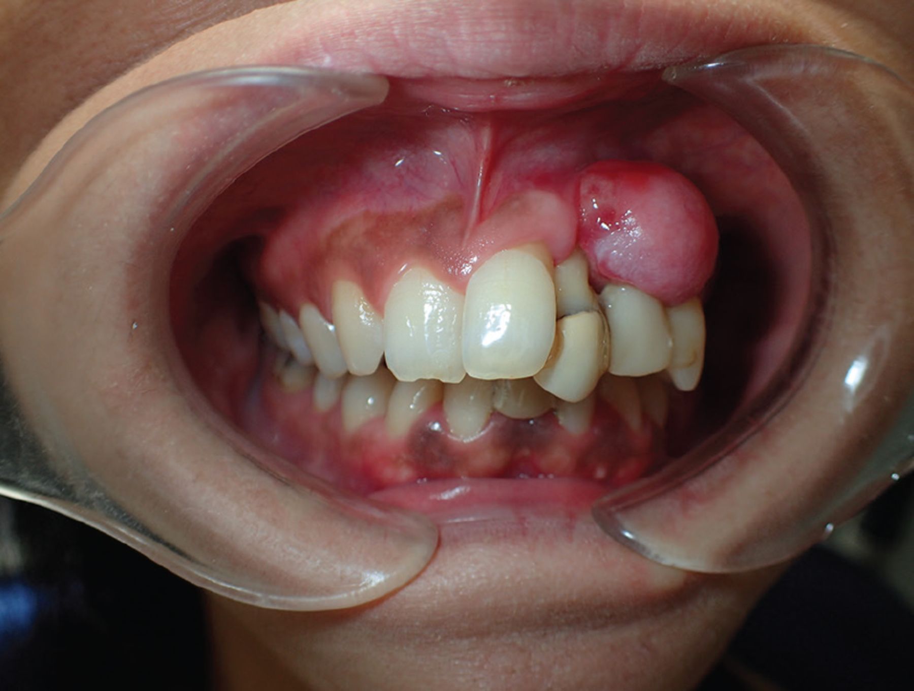
A well-circumscribed, smooth-surfaced, elastic, hard nodule, 20 mm by 18 mm, on the left maxillary anterior gingiva.

Computed tomography showed a well-demarcated, rim-enhanced soft-tissue mass in the left maxillary anterior gingiva with slight bone resorption at the left maxillary second incisor (Figure 2).

Figure 2
  • Download figure
  • Open in new tab
  • Download powerpoint
Figure 2

Computed tomography shows a well-demarcated, rim-enhanced soft tissue mass in the left maxillary anterior gingiva (A, arrow), with slight bone resorption at the left maxillary second incisor (B, arrow).

Biopsy was performed, and histopathologic study revealed keratinized epithelium overlying fibrous connective tissue with infiltration of inflammatory cells.

Based on these findings, we diagnosed recurrent fibrous epulis, resected the nodule, and extracted the loose tooth. At follow-up 20 months later there was no evidence of recurrence.

CLINICAL RECOGNITION AND DIAGNOSIS

Fibrous epulis, a type of inflammatory fibrous hyperplasia of the gingiva, is a relatively common tumorlike lesion.1,2 The possible origin is the periosteum and the periodontal ligament.1,2 Factors that lead to its development are local irritations such as poor-quality dental restorations, dental plaque, and calculus.2,3

The estimated prevalence of fibrous epulis is 0.09%. It occurs at a wide range of ages and in women more often than men.4 Most lesions occur on the maxillary anterior interdental papilla.1,2,4

Clinically, fibrous epulis is an asymptomatic, exophytic, smooth-surfaced or focally ulcerated, mucosal-colored mass with a variable growth rate.2,3 At presentation, most lesions are 10 mm to 20 mm in diameter; those that are large or grow rapidly tend to be misdiagnosed as neoplastic.2,3

On computed tomography, lesions appear as a soft-tissue mass in the gingiva with mild enhancement, and up to one-third contain calcifications that can be easily seen.1,3 These calcified lesions are termed mineralizing fibrous epulis or peripheral ossifying fibroma.1 Bone resorption is relatively uncommon.5

Histologically, fibrous epulis shows hyper-plastic epithelium that overlies fibrous connective tissue.1 Mineralized tissue, if present, consists of trabeculae or droplike metaplastic bone.1

The differential diagnosis includes pyogenic granuloma, peripheral giant cell granuloma, fibroma, peripheral odontogenic fibroma, fibrosarcoma, and squamous cell carcinoma.1,3,5 A slowly growing mass on the interdental papilla with local irritations and calcifications detected by computed tomography should raise suspicion of fibrous epulis.3 However, distinguishing fibrous epulis from the other conditions listed above may be difficult, and thus, histopathologic study is crucial.2,3

TREATMENT

Complete excision and curettage of the lesion is the preferred treatment because the recurrence rate is high, from 7% to 45%.3 Therefore, long-term follow-up is essential. Tooth extraction is not indicated unless there is underlying bone resorption.

DISCLOSURES

The authors report no relevant financial relationships which, in the context of their contributions, could be perceived as a potential conflict of interest.

  • Copyright © 2021 The Cleveland Clinic Foundation. All Rights Reserved.

REFERENCES

  1. ↵
    1. Brierley DJ,
    2. Crane H,
    3. Hunter KD
    . Lumps and bumps of the gingiva: a pathological miscellany. Head Neck Pathol 2019; 13(1):103–113. doi:10.1007/s12105-019-01000-w
    OpenUrlCrossRef
  2. ↵
    1. Fonseca GM,
    2. Fonseca RM,
    3. Cantín M
    . Massive fibrous epulis—a case report of a 10-year-old lesion. Int J Oral Sci 2014; 6(3):182–184. doi:10.1038/ijos.2013.75
    OpenUrlCrossRef
  3. ↵
    1. Moon WJ,
    2. Choi SY,
    3. Chung EC,
    4. Kwon KH,
    5. Chae SW
    . Peripheral ossifying fibroma in the oral cavity: CT and MR findings. Dentomaxillofac Radiol 2007; 36(3):180–182. doi:10.1259/dmfr/59377498
    OpenUrlAbstract/FREE Full Text
  4. ↵
    1. Shulman JD,
    2. Beach MM,
    3. Rivera-Hidalgo F
    . The preva lence of oral mucosal lesions in US adults: data from the Third National Health and Nutrition Examination Survey, 1988–1994. J Am Dent Assoc 2004; 135(9):1279–1286. doi:10.14219/jada.archive.2004.0403
    OpenUrlAbstract/FREE Full Text
  5. ↵
    1. Singh D,
    2. Pranab A,
    3. Mishra N,
    4. Sharma AK,
    5. Kumar S,
    6. Gupta P
    . Epulis—commonly misdiagnosed entity: a report of 2 cases. J Interdiscipl Med Dent Sci 2018, 6(2):2. doi:10.4172/2376-032X.1000230
    OpenUrlCrossRef
PreviousNext
Back to top

In this issue

Cleveland Clinic Journal of Medicine: 88 (5)
Cleveland Clinic Journal of Medicine
Vol. 88, Issue 5
1 May 2021
  • Table of Contents
  • Table of Contents (PDF)
  • Index by author
  • Complete Issue (PDF)
Print
Download PDF
Article Alerts
Sign In to Email Alerts with your Email Address
Email Article

Thank you for your interest in spreading the word on Cleveland Clinic Journal of Medicine.

NOTE: We only request your email address so that the person you are recommending the page to knows that you wanted them to see it, and that it is not junk mail. We do not capture any email address.

Enter multiple addresses on separate lines or separate them with commas.
Fibrous epulis: A tumorlike gingival lesion
(Your Name) has sent you a message from Cleveland Clinic Journal of Medicine
(Your Name) thought you would like to see the Cleveland Clinic Journal of Medicine web site.
CAPTCHA
This question is for testing whether or not you are a human visitor and to prevent automated spam submissions.
Citation Tools
Fibrous epulis: A tumorlike gingival lesion
Keiichi Ohta, Hitoshi Yoshimura
Cleveland Clinic Journal of Medicine May 2021, 88 (5) 265-266; DOI: 10.3949/ccjm.88a.20127

Citation Manager Formats

  • BibTeX
  • Bookends
  • EasyBib
  • EndNote (tagged)
  • EndNote 8 (xml)
  • Medlars
  • Mendeley
  • Papers
  • RefWorks Tagged
  • Ref Manager
  • RIS
  • Zotero
Share
Fibrous epulis: A tumorlike gingival lesion
Keiichi Ohta, Hitoshi Yoshimura
Cleveland Clinic Journal of Medicine May 2021, 88 (5) 265-266; DOI: 10.3949/ccjm.88a.20127
Twitter logo Facebook logo Mendeley logo
  • Tweet Widget
  • Facebook Like
  • Linkedin Share Button

Jump to section

  • Article
    • CLINICAL RECOGNITION AND DIAGNOSIS
    • TREATMENT
    • DISCLOSURES
    • REFERENCES
  • Figures & Data
  • Info & Metrics
  • PDF

Related Articles

  • No related articles found.
  • PubMed
  • Google Scholar

Cited By...

  • No citing articles found.
  • Google Scholar

More in this TOC Section

  • Tinea incognito
  • Prolonged venous filling time and dependent rubor in a patient with peripheral artery disease
  • Sarcoidosis with diffuse purplish erythematous plaques on the hands
Show more The Clinical Picture

Similar Articles

Subjects

  • Imaging
  • Oncology

Navigate

  • Current Issue
  • Past Issues
  • Supplements
  • Article Type
  • Specialty
  • CME/MOC Articles
  • CME/MOC Calendar
  • Media Kit

Authors & Reviewers

  • Manuscript Submission
  • Authors & Reviewers
  • Subscriptions
  • About CCJM
  • Contact Us
  • Cleveland Clinic Center for Continuing Education
  • Consult QD

Share your suggestions!

Copyright © 2025 The Cleveland Clinic Foundation. All rights reserved. The information provided is for educational purposes only. Use of this website is subject to the website terms of use and privacy policy. 

Powered by HighWire