
Main menu

  • Home
  • Content
    • Current Issue
    • Ahead of Print
    • Past Issues
    • Supplements
    • Article Type
  • Specialty
    • Articles by Specialty
  • CME/MOC
    • Articles
    • Calendar
  • Info For
    • Manuscript Submission
    • Authors & Reviewers
    • Subscriptions
    • About CCJM
    • Contact Us
    • Media Kit
  • Conversations with Leaders
  • Conference Coverage
    • Kidney Week 2024
    • CHEST 2024
    • ACR Convergence 2023
    • Kidney Week 2023
    • ObesityWeek 2023
    • IDWeek 2023
    • CHEST 2023
    • MDS 2023
    • IAS 2023
    • ACP 2023
    • AAN 2023
    • ACC / WCC 2023
    • AAAAI Meeting 2023
    • ACR Convergence 2022
    • Kidney Week 2022
    • AIDS 2022
  • Other Publications
    • www.clevelandclinic.org

User menu

  • Register
  • Log in

Search

  • Advanced search
Cleveland Clinic Journal of Medicine
  • Other Publications
    • www.clevelandclinic.org
  • Register
  • Log in
Cleveland Clinic Journal of Medicine

Advanced Search

  • Home
  • Content
    • Current Issue
    • Ahead of Print
    • Past Issues
    • Supplements
    • Article Type
  • Specialty
    • Articles by Specialty
  • CME/MOC
    • Articles
    • Calendar
  • Info For
    • Manuscript Submission
    • Authors & Reviewers
    • Subscriptions
    • About CCJM
    • Contact Us
    • Media Kit
  • Conversations with Leaders
  • Conference Coverage
    • Kidney Week 2024
    • CHEST 2024
    • ACR Convergence 2023
    • Kidney Week 2023
    • ObesityWeek 2023
    • IDWeek 2023
    • CHEST 2023
    • MDS 2023
    • IAS 2023
    • ACP 2023
    • AAN 2023
    • ACC / WCC 2023
    • AAAAI Meeting 2023
    • ACR Convergence 2022
    • Kidney Week 2022
    • AIDS 2022
The Clinical Picture

Pseudopathologic vertebral body enhancement

Yi Pan, MD, Peixin Tan, MD, Songxi Xie, MD, Jiewen Chen, MMed and Yi-Long Wu, MD
Cleveland Clinic Journal of Medicine August 2021, 88 (8) 424-425; DOI: https://doi.org/10.3949/ccjm.88a.20150
Yi Pan
Department of Radiation Oncology, Guangdong Lung Cancer Institute, Guangdong Provincial People’s Hospital, Guangdong Academy of Medical Sciences, School of Medicine, South China University of Technology, Guangzhou, PR China
  • Find this author on Google Scholar
  • Find this author on PubMed
  • Search for this author on this site
  • For correspondence: [email protected]
Peixin Tan
Department of Radiation Oncology, Guangdong Lung Cancer Institute, Guangdong Provincial People’s Hospital, Guangdong Academy of Medical Sciences, School of Medicine, South China University of Technology, Guangzhou, PR China
  • Find this author on Google Scholar
  • Find this author on PubMed
  • Search for this author on this site
Songxi Xie
Department of Radiation Oncology, Guangdong Provincial People’s Hospital, Guangdong Academy of Medical Sciences, School of Medicine, South China University of Technology, Guangzhou, PR China
  • Find this author on Google Scholar
  • Find this author on PubMed
  • Search for this author on this site
Jiewen Chen
Department of Radiation Oncology, Guangdong Provincial People’s Hospital, Guangdong Academy of Medical Sciences, School of Medicine, South China University of Technology, Guangzhou, PR China
  • Find this author on Google Scholar
  • Find this author on PubMed
  • Search for this author on this site
Yi-Long Wu
Guangdong Lung Cancer Institute, Guangdong Provincial People’s Hospital, Guangdong Academy of Medical Sciences, School of Medicine, South China University of Technology, Guangzhou, PR China
  • Find this author on Google Scholar
  • Find this author on PubMed
  • Search for this author on this site
  • Article
  • Figures & Data
  • Info & Metrics
  • PDF
Loading

A 45-year-old man presented to the emergency department with severe dyspnea and cough that had gradually worsened over the past 3 months. His medical history was notable for stage IIA esophageal squamous cell carcinoma that had been treated with esophagectomy 15 years ago; and 13 years ago, he had developed mediastinal lymph node metastasis and had received chemoradiotherapy. He had been a heavy smoker and drinker but had quit 15 years ago.

Emergency contrast-enhanced thoracic computed tomography (CT) revealed a tumor mass in the lower trachea, sclerotic enhancement of the T1 vertebral body (Figure 1) and from the C2 to T2 vertebral bodies, and thrombosis in the left brachiocephalic vein (Figure 2). The patient rapidly developed type 2 respiratory failure (defined as a Pao2 < 8.0 kPa and a Paco2 > 6.0 kPa) and underwent bedside fiberoptic bronchoscopy, which revealed an obstructive tumor in the lower trachea that had invaded the carina and both the left and right main bronchi. Bronchoscopy-guided radiofrequency ablation was performed to relieve airway obstruction. Moderately differentiated squamous cell carcinoma was confirmed by endobronchial biopsy study.

Figure 1
  • Download figure
  • Open in new tab
  • Download powerpoint
Figure 1

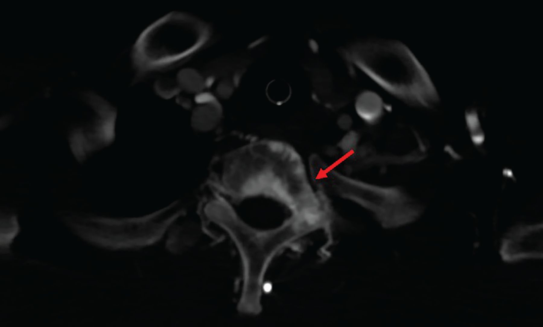
Contrast-enhanced thoracic computed tomography showed sclerotic enhancement of the T1 vertebral body (red arrow).

Figure 2
  • Download figure
  • Open in new tab
  • Download powerpoint
Figure 2

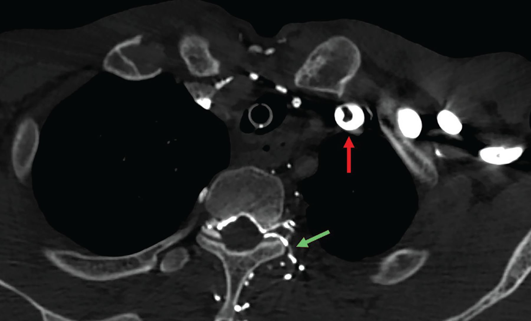
Contrast-enhanced computed tomography noted thrombosis in the left brachiocephalic vein (red arrow) and contrast filling of the paravertebral veins, usually occult on contrast enhancement (green arrow).

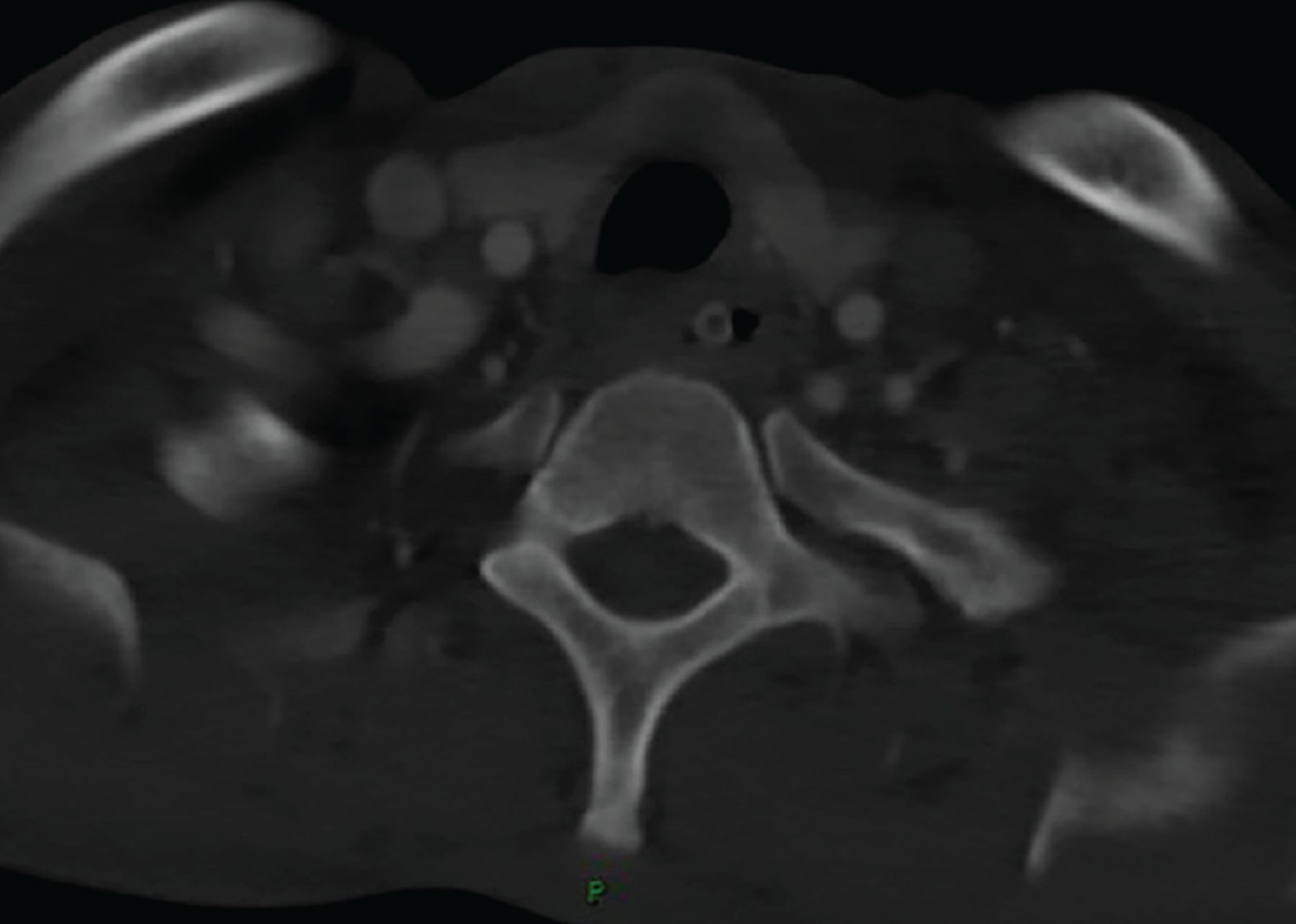
The patient’s dyspnea improved after bronchoscopic therapy. Repeat CT 5 days later showed no evidence of the vertebral body enhancement (Figure 3). Esophagoscopy showed an anastomotic stenosis 24 cm from the incisors and smooth mucosa in the esophagus. Anastomotic stenosis was consistent with the tumor location in the lower trachea and carina. Esophageal stenosis and smooth esophageal mucosa demonstrated external tumor compression of the esophagus, which did not support the diagnosis of esophageal carcinoma.

Figure 3
  • Download figure
  • Open in new tab
  • Download powerpoint
Figure 3

Repeat computed tomography showed the disappearance of the sclerotic vertebral body enhancement.

Based on the patient’s smoking and irradiation history and the long interval between the last anticancer treatment and the appearance of the second tumor, the obstructive tumor was diagnosed as a second primary bronchogenic carcinoma.

The patient underwent disease staging with positron emission tomography CT, which showed increased 18F-fluorodeoxyglucose uptake at the back wall of the trachea alone, indicating that the tumor was localized.

TREATMENT

The patient received 4 cycles of capecitabine and anlotinib, followed by 4 cycles of anlotinib. This achieved a partial response. At last follow-up, the patient was alive and without disease progression.

PSEUDOPATHOLOGIC VERTEBRAL BODY ENHANCEMENT

The differential diagnosis of sclerotic lesions on contrast-enhanced CT includes tumor metastasis, mastocytosis, sarcoidosis, osteomyelitis, lymphoma, Paget disease, and pseudopathologic vertebral body enhancement. For patients with a history of cancer, the leading cause is metastatic malignancy.

A limited number of case reports showed that pseudo pathologic vertebral body enhancement may exist in the presence of obstruction of the superior vena cava or brachiocephalic vein. This is uncommon and easily misdiagnosed as sclerotic osseous metastasis in clinical practice.1,2

The mechanism of pseudopathologic sclerotic enhancement of vertebral bodies is not well characterized. However, an elevated venous pressure that induced contrast agent reflux into the intravertebral venous plexus was proposed as a main reason.3

In our patient, narrowing of the left brachiocephalic vein proximal to the superior vena cava resulted in elevated venous pressure and blood flow into the vertebral venous plexus, causing reflux of contrast agent.1,4 Sclerotic enhancement of the vertebral bodies and vertebral venous collaterals, which are usually occult on CT, were seen. Eight consecutive vertebral bodies were involved, which is uncommon with metastasis. However, when the contrast agent was injected into the contralateral arm during the second CT, the vertebra enhancement disappeared (Figure 3).

Pseudopathologic vertebral body enhancement due to brachiocephalic vein narrowing is rare. However, sclerotic bone metastasis based on contrast-enhanced CT should prompt a careful evaluation when narrowing or obstruction of the brachiocephalic vein and paravertebral collateral veins is present on imaging. Spine magnetic resonance imaging or positron emission tomography CT should be performed to confirm the diagnosis.

DISCLOSURES

The authors report no relevant financial relationships which, in the context of their contributions, could be perceived as a potential conflict of interest.

  • Copyright © 2021 The Cleveland Clinic Foundation. All Rights Reserved.

REFERENCES

  1. ↵
    1. Berritto D,
    2. Abboud S,
    3. Kosmas C,
    4. Riherd D,
    5. Robbin M
    . Vertebral body enhancement mimicking sclerotic osseous lesions in the setting of bilateral brachiocephalic vein thrombosis. Skeletal Radiol 2015; 44(2):303–305. doi:10.1007/s00256-014-2037-9
    OpenUrlCrossRef
  2. ↵
    1. Thomas N,
    2. Oliver TB,
    3. Sudarshan T
    . Vanishing bone metastases—a pitfall in the interpretation of contrast enhanced CT in patients with superior vena cava obstruction. Br J Radiol 2011; 84(1005):e176–e178. doi:10.1259/bjr/50676625
    OpenUrlAbstract/FREE Full Text
  3. ↵
    1. Kim YK,
    2. Sung YM,
    3. Hwang KH,
    4. Cho EK,
    5. Choi HY
    . Pseudopathologic vertebral body enhancement in the presence of superior vena cava obstruction on computed tomography. Spine J 2015; 15(6):1295–1301. doi:10.1016/j.spinee.2013.07.440
    OpenUrlCrossRef
  4. ↵
    1. Chen JY,
    2. Mamourian AC,
    3. Messe SR,
    4. Wolf RL
    . Pseudopathologic brain parenchymal enhancement due to venous reflux from left-sided injection and brachiocephalic vein narrowing. AJNR Am J Neuroradiol 2010; 31(1):86–87. doi:10.3174/ajnr.A1688
    OpenUrlAbstract/FREE Full Text
PreviousNext
Back to top

In this issue

Cleveland Clinic Journal of Medicine: 88 (8)
Cleveland Clinic Journal of Medicine
Vol. 88, Issue 8
1 Aug 2021
  • Table of Contents
  • Table of Contents (PDF)
  • Index by author
  • Complete Issue (PDF)
Print
Download PDF
Article Alerts
Sign In to Email Alerts with your Email Address
Email Article

Thank you for your interest in spreading the word on Cleveland Clinic Journal of Medicine.

NOTE: We only request your email address so that the person you are recommending the page to knows that you wanted them to see it, and that it is not junk mail. We do not capture any email address.

Enter multiple addresses on separate lines or separate them with commas.
Pseudopathologic vertebral body enhancement
(Your Name) has sent you a message from Cleveland Clinic Journal of Medicine
(Your Name) thought you would like to see the Cleveland Clinic Journal of Medicine web site.
CAPTCHA
This question is for testing whether or not you are a human visitor and to prevent automated spam submissions.
Citation Tools
Pseudopathologic vertebral body enhancement
Yi Pan, Peixin Tan, Songxi Xie, Jiewen Chen, Yi-Long Wu
Cleveland Clinic Journal of Medicine Aug 2021, 88 (8) 424-425; DOI: 10.3949/ccjm.88a.20150

Citation Manager Formats

  • BibTeX
  • Bookends
  • EasyBib
  • EndNote (tagged)
  • EndNote 8 (xml)
  • Medlars
  • Mendeley
  • Papers
  • RefWorks Tagged
  • Ref Manager
  • RIS
  • Zotero
Share
Pseudopathologic vertebral body enhancement
Yi Pan, Peixin Tan, Songxi Xie, Jiewen Chen, Yi-Long Wu
Cleveland Clinic Journal of Medicine Aug 2021, 88 (8) 424-425; DOI: 10.3949/ccjm.88a.20150
Twitter logo Facebook logo Mendeley logo
  • Tweet Widget
  • Facebook Like
  • Linkedin Share Button

Jump to section

  • Article
    • TREATMENT
    • PSEUDOPATHOLOGIC VERTEBRAL BODY ENHANCEMENT
    • DISCLOSURES
    • REFERENCES
  • Figures & Data
  • Info & Metrics
  • PDF

Related Articles

  • No related articles found.
  • PubMed
  • Google Scholar

Cited By...

  • No citing articles found.
  • Google Scholar

More in this TOC Section

  • Tinea incognito
  • Prolonged venous filling time and dependent rubor in a patient with peripheral artery disease
  • Sarcoidosis with diffuse purplish erythematous plaques on the hands
Show more The Clinical Picture

Similar Articles

Subjects

  • Imaging
  • Oncology
  • Pulmonology

Navigate

  • Current Issue
  • Past Issues
  • Supplements
  • Article Type
  • Specialty
  • CME/MOC Articles
  • CME/MOC Calendar
  • Media Kit

Authors & Reviewers

  • Manuscript Submission
  • Authors & Reviewers
  • Subscriptions
  • About CCJM
  • Contact Us
  • Cleveland Clinic Center for Continuing Education
  • Consult QD

Share your suggestions!

Copyright © 2025 The Cleveland Clinic Foundation. All rights reserved. The information provided is for educational purposes only. Use of this website is subject to the website terms of use and privacy policy. 

Powered by HighWire