
Main menu

  • Home
  • Content
    • Current Issue
    • Ahead of Print
    • Past Issues
    • Supplements
    • Article Type
  • Specialty
    • Articles by Specialty
  • CME/MOC
    • Articles
    • Calendar
  • Info For
    • Manuscript Submission
    • Authors & Reviewers
    • Subscriptions
    • About CCJM
    • Contact Us
    • Media Kit
  • Conversations with Leaders
  • Conference Coverage
    • Kidney Week 2024
    • CHEST 2024
    • ACR Convergence 2023
    • Kidney Week 2023
    • ObesityWeek 2023
    • IDWeek 2023
    • CHEST 2023
    • MDS 2023
    • IAS 2023
    • ACP 2023
    • AAN 2023
    • ACC / WCC 2023
    • AAAAI Meeting 2023
    • ACR Convergence 2022
    • Kidney Week 2022
    • AIDS 2022
  • Other Publications
    • www.clevelandclinic.org

User menu

  • Register
  • Log in

Search

  • Advanced search
Cleveland Clinic Journal of Medicine
  • Other Publications
    • www.clevelandclinic.org
  • Register
  • Log in
Cleveland Clinic Journal of Medicine

Advanced Search

  • Home
  • Content
    • Current Issue
    • Ahead of Print
    • Past Issues
    • Supplements
    • Article Type
  • Specialty
    • Articles by Specialty
  • CME/MOC
    • Articles
    • Calendar
  • Info For
    • Manuscript Submission
    • Authors & Reviewers
    • Subscriptions
    • About CCJM
    • Contact Us
    • Media Kit
  • Conversations with Leaders
  • Conference Coverage
    • Kidney Week 2024
    • CHEST 2024
    • ACR Convergence 2023
    • Kidney Week 2023
    • ObesityWeek 2023
    • IDWeek 2023
    • CHEST 2023
    • MDS 2023
    • IAS 2023
    • ACP 2023
    • AAN 2023
    • ACC / WCC 2023
    • AAAAI Meeting 2023
    • ACR Convergence 2022
    • Kidney Week 2022
    • AIDS 2022
Review

Stress testing and noninvasive coronary imaging: What’s the best test for my patient?

Milad Matta, MD, Serge C. Harb, MD, Paul Cremer, MD, Rory Hachamovitch, MD, MSc and Chadi Ayoub, MD, PhD, FACC, FASE, FSCCT
Cleveland Clinic Journal of Medicine September 2021, 88 (9) 502-515; DOI: https://doi.org/10.3949/ccjm.88a.20068
Milad Matta
Department of Internal Medicine, Cleveland Clinic, Cleveland, OH
  • Find this author on Google Scholar
  • Find this author on PubMed
  • Search for this author on this site
  • For correspondence: [email protected]
Serge C. Harb
Section of Cardiovascular Imaging, Department of Cardiovascular Medicine, Heart, Vascular, and Thoracic Institute, Cleveland Clinic; Assistant Professor of Medicine, Cleveland Clinic Lerner College of Medicine of Case Western Reserve University, Cleveland, OH
  • Find this author on Google Scholar
  • Find this author on PubMed
  • Search for this author on this site
  • Find this author on Cleveland Clinic
Paul Cremer
Section of Cardiovascular Imaging, Department of Cardiovascular Medicine, Heart, Vascular, and Thoracic Institute, Cleveland Clinic, Cleveland, OH
  • Find this author on Google Scholar
  • Find this author on PubMed
  • Search for this author on this site
  • Find this author on Cleveland Clinic
Rory Hachamovitch
Section of Cardiovascular Imaging, Department of Cardiovascular Medicine, Heart, Vascular, and Thoracic Institute, Cleveland Clinic, Cleveland, OH
  • Find this author on Google Scholar
  • Find this author on PubMed
  • Search for this author on this site
Chadi Ayoub
Section of Cardiovascular Imaging, Department of Cardiovascular Medicine, Heart, Vascular, and Thoracic Institute, Cleveland Clinic; Assistant Professor of Medicine, Cleveland Clinic Lerner College of Medicine of Case Western Reserve University, Cleveland, OH
  • Find this author on Google Scholar
  • Find this author on PubMed
  • Search for this author on this site
  • Article
  • Figures & Data
  • Info & Metrics
  • PDF
Loading

Article Figures & Data

Figures

  • Tables
  • Figure 1
    • Download figure
    • Open in new tab
    • Download powerpoint
    Figure 1

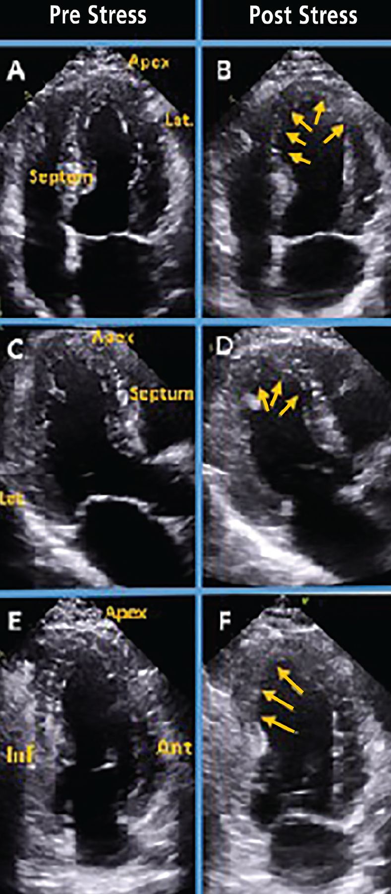
    Stress echocardiogram from a 54-year-old woman with chest pain shows ischemia in the left anterior descending artery and right coronary artery territories. Panels A and B show the 4-chamber view before and after the stress test. On the poststress image (B), the arrows point to hypokinesis at the apex and distal septum; enlargement of left ventricular cavity suggests significant ischemia. In the long-axis view (C, D), arrows point to hypo-kinesis at the apex and distal septum. In the 2-chamber view (E, F), arrows point to hypokinesis on the mid and apical inferior wall.

  • Figure 2
    • Download figure
    • Open in new tab
    • Download powerpoint
    Figure 2

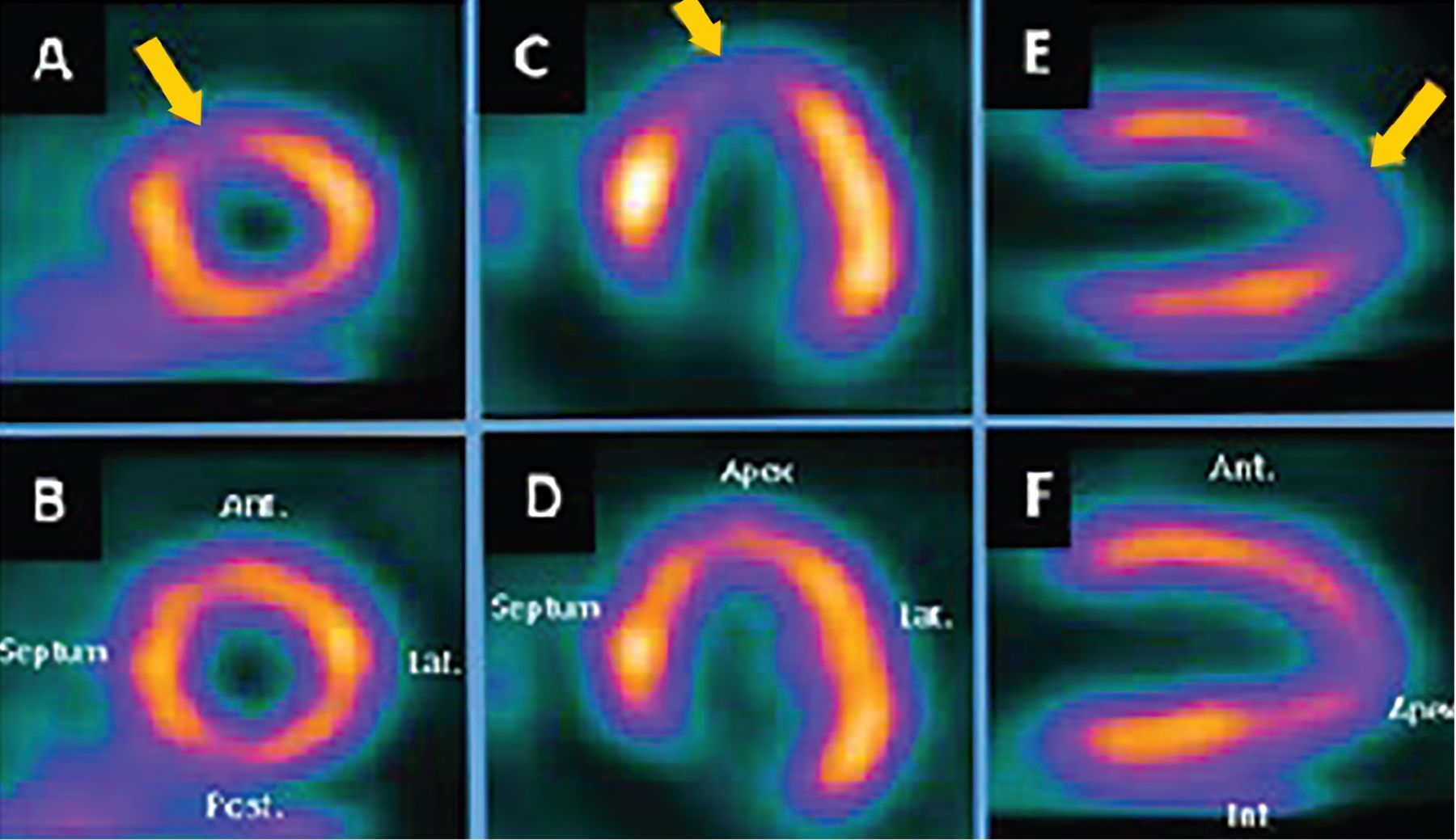
    Single-photon emission CT myocardial perfusion imaging in a 62-year-old man with diabetes and a 2-month history of dyspnea shows moderate left anterior descending coronary artery ischemia. Panels A, C, and E are poststress images that show perfusion defects in the apex, apical septum, and apical anterior wall (arrows). Panels B, D, and F show relatively normal perfusion at rest at the corresponding levels.

  • Figure 3
    • Download figure
    • Open in new tab
    • Download powerpoint
    Figure 3

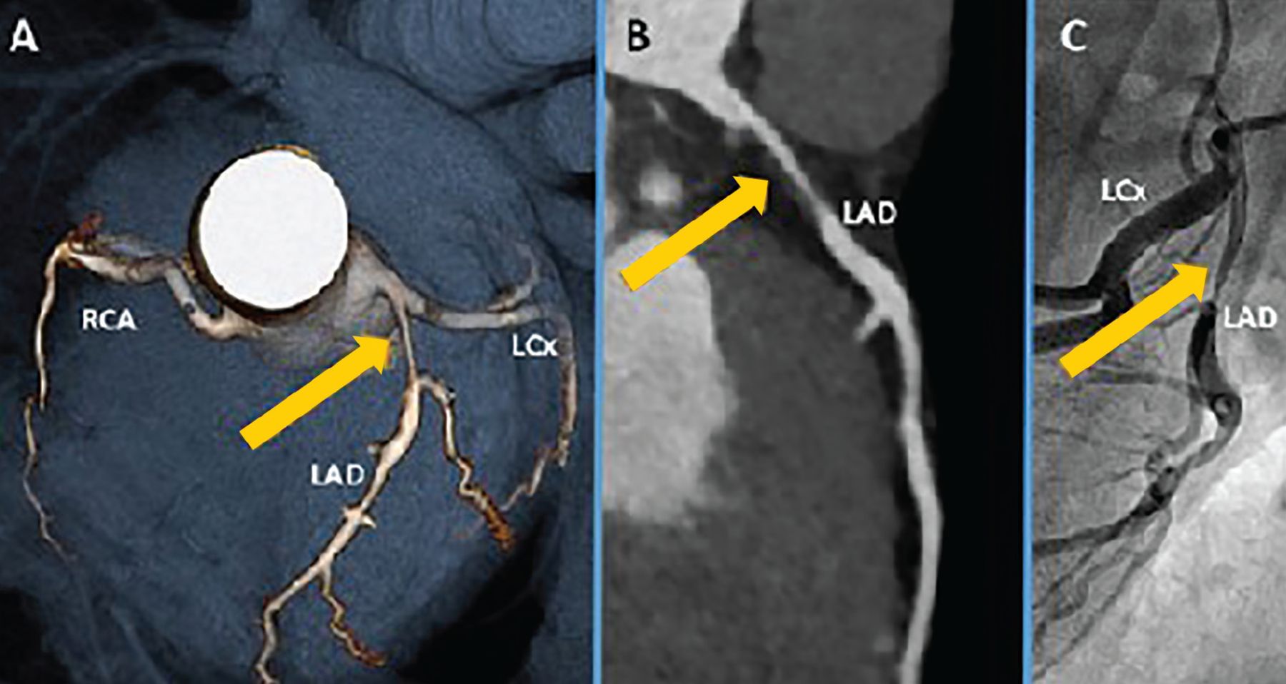
    Coronary computed tomography angiography in a 40-year-old man who smoked and had a family history of premature coronary artery disease. Panel A is a 3-D rendering showing proximal left anterior descending (LAD) coronary artery stenosis (arrow). Panel B is a multiplanar reconstruction showing proximal LAD coronary artery stenosis with predominantly soft (lipid-laden) noncalcified plaque (arrow). Panel C shows the corresponding LAD lesion (arrow) on coronary catheterization.

    LCx = left circumflex artery; RCA = right coronary artery

  • Figure 4
    • Download figure
    • Open in new tab
    • Download powerpoint
    Figure 4

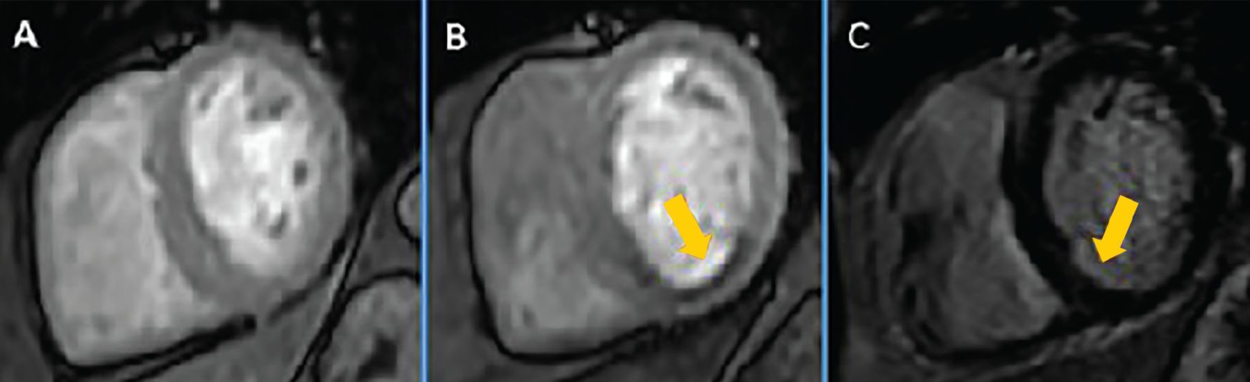
    Stress cardiac magnetic resonance imaging in a 67-year-old woman with diabetes and chest pain shows normal perfusion at rest (A). Panel B shows a poststress image with a perfusion defect in the inferior and inferoseptal segments (arrow), suggestive of ischemia in the right coronary artery territory. Panel C is a delayed gadolinium-enhanced image showing mild subendocardial enhancement (arrow) in the corresponding region, consistent with a small area of scar.

Tables

  • Figures
    • View popup
    TABLE 1

    Testing for coronary artery disease

    FunctionalElectrocardiography exercise stress test
    Stress echocardiography
    Nuclear medicine myocardial perfusion imaging techniques
     Single-photon emission computed tomography
     Positron emission tomography
    Cardiac magnetic resonance imaging with stress perfusion
    AnatomicCoronary computed tomography angiography
    Invasive coronary catheterization
    • View popup
    TABLE 2

    Common indications for stress testing and coronary computed tomography angiography

    Assessment for coronary artery disease
    Angina or anginal equivalent symptoms and negative cardiac enzymes
    Atypical symptoms in patients with diabetes or with high probability of diabetes
    New diagnosis of cardiomyopathy (to define whether the cause is ischemic or nonischemic)
    New or increasing heart failure symptoms despite adherence to medical therapy
    Re-evaluation of known heart failure (systolic or diastolic) in patients with a change in clinical status without a clear precipitating change in medication or diet
    Arrhythmias such as ventricular tachycardia or atrial fibrillation (to exclude ischemia as the cause) or new left bundle-branch block
    To exclude severe ischemia prior to noncardiac surgery in those with increased coronary artery disease risks, angina symptoms, or poor exercise capacity (< 4 metabolic equivalents)
    To define presence or absence of ischemia in those with moderate coronary stenosis (stress test or fractional flow reserve-computed tomography)
    Evaluation of anomalous coronary arteries
    Stress testing for indications other than coronary artery disease assessment
    Valve assessment
     Mitral valve stenosis or regurgitation severity (exercise stress echocardiography)
     Low-flow low gradient aortic stenosis (dobutamine stress echocardiography)
    Exercise-induced pulmonary hypertension or diastolic dysfunction (exercise stress echocardiography)
    Hypertrophic cardiomyopathy to demonstrate provocable left ventricular outflow tract obstruction (exercise stress echocardiography)
    Exercise-induced arrhythmia or chronotropic incompetence (exercise stress echocardiography)
    To define cardiopulmonary disease and aerobic exercise capacity (metabolic stress test)
    • Data from references 3 and 7–10.

    • View popup
    TABLE 3

    Prognostic indicators on electrocardiography stress testing

    IndicatorComments
    Functional capacityStrongest prognostic indicator, reported as metabolic equivalents.7,14
    ST-segment depression or elevation> 1-mm ST deviation is suggestive of ischemia.13
    Exercise-induced hypotensionDefined as systolic blood pressure that is lower during exercise than while standing at rest before exercise, reflecting failure of cardiac output to increase during exercise.
    Associated with severe coronary artery disease or left ventricular systolic dysfunction.15
    Chronotropic incompetenceFailure of heart rate to increase as expected during exercise, defined as achieving < 80% of predicted heart rate (or < 62% for patients taking beta-blockers).16
    Associated with increased all-cause and cardiovascular mortality.17
    Impaired heart rate recoveryHeart rate fails to decrease normally after cessation of exercise.
    Predicts all-cause mortality and cardiovascular events, including sudden death.18
    Ventricular arrhythmiaSustained ventricular tachychardia or ventricular fibrillation.
    Associated with significant coronary artery disease or left ventricular dysfunction.19
PreviousNext
Back to top

In this issue

Cleveland Clinic Journal of Medicine: 88 (9)
Cleveland Clinic Journal of Medicine
Vol. 88, Issue 9
1 Sep 2021
  • Table of Contents
  • Table of Contents (PDF)
  • Index by author
  • Complete Issue (PDF)
Print
Download PDF
Article Alerts
Sign In to Email Alerts with your Email Address
Email Article

Thank you for your interest in spreading the word on Cleveland Clinic Journal of Medicine.

NOTE: We only request your email address so that the person you are recommending the page to knows that you wanted them to see it, and that it is not junk mail. We do not capture any email address.

Enter multiple addresses on separate lines or separate them with commas.
Stress testing and noninvasive coronary imaging: What’s the best test for my patient?
(Your Name) has sent you a message from Cleveland Clinic Journal of Medicine
(Your Name) thought you would like to see the Cleveland Clinic Journal of Medicine web site.
CAPTCHA
This question is for testing whether or not you are a human visitor and to prevent automated spam submissions.
Citation Tools
Stress testing and noninvasive coronary imaging: What’s the best test for my patient?
Milad Matta, Serge C. Harb, Paul Cremer, Rory Hachamovitch, Chadi Ayoub
Cleveland Clinic Journal of Medicine Sep 2021, 88 (9) 502-515; DOI: 10.3949/ccjm.88a.20068

Citation Manager Formats

  • BibTeX
  • Bookends
  • EasyBib
  • EndNote (tagged)
  • EndNote 8 (xml)
  • Medlars
  • Mendeley
  • Papers
  • RefWorks Tagged
  • Ref Manager
  • RIS
  • Zotero
Share
Stress testing and noninvasive coronary imaging: What’s the best test for my patient?
Milad Matta, Serge C. Harb, Paul Cremer, Rory Hachamovitch, Chadi Ayoub
Cleveland Clinic Journal of Medicine Sep 2021, 88 (9) 502-515; DOI: 10.3949/ccjm.88a.20068
Twitter logo Facebook logo Mendeley logo
  • Tweet Widget
  • Facebook Like
  • Linkedin Share Button

Jump to section

  • Article
    • ABSTRACT
    • CORONARY ARTERY DISEASE
    • ELECTROCARDIOGRAPHY EXERCISE STRESS TESTING
    • STRESS ECHOCARDIOGRAPHY
    • NUCLEAR MEDICINE MYOCARDIAL PERFUSION IMAGING
    • CORONARY CT ANGIOGRAPHY
    • CORONARY ARTERY CALCIUM SCORE
    • STRESS CARDIAC MAGNETIC RESONANCE IMAGING
    • APPROPRIATE USE CRITERIA
    • TAKE-HOME MESSAGE
    • DISCLOSURES
    • REFERENCES
  • Figures & Data
  • Info & Metrics
  • PDF

Related Articles

  • No related articles found.
  • PubMed
  • Google Scholar

Cited By...

  • No citing articles found.
  • Google Scholar

More in this TOC Section

  • Alpha-gal syndrome: Recognizing and managing a tick-bite–related meat allergy
  • Managing right ventricular failure in the setting of pulmonary embolism
  • Psychedelics, spirituality, and existential distress in patients at the end of life
Show more Review

Similar Articles

Subjects

  • Cardiology
  • Imaging
  • Vascular Medicine

Navigate

  • Current Issue
  • Past Issues
  • Supplements
  • Article Type
  • Specialty
  • CME/MOC Articles
  • CME/MOC Calendar
  • Media Kit

Authors & Reviewers

  • Manuscript Submission
  • Authors & Reviewers
  • Subscriptions
  • About CCJM
  • Contact Us
  • Cleveland Clinic Center for Continuing Education
  • Consult QD

Share your suggestions!

Copyright © 2025 The Cleveland Clinic Foundation. All rights reserved. The information provided is for educational purposes only. Use of this website is subject to the website terms of use and privacy policy. 

Powered by HighWire