
Main menu

  • Home
  • Content
    • Current Issue
    • Ahead of Print
    • Past Issues
    • Supplements
    • Article Type
  • Specialty
    • Articles by Specialty
  • CME/MOC
    • Articles
    • Calendar
  • Info For
    • Manuscript Submission
    • Authors & Reviewers
    • Subscriptions
    • About CCJM
    • Contact Us
    • Media Kit
  • Conversations with Leaders
  • Conference Coverage
    • Kidney Week 2024
    • CHEST 2024
    • ACR Convergence 2023
    • Kidney Week 2023
    • ObesityWeek 2023
    • IDWeek 2023
    • CHEST 2023
    • MDS 2023
    • IAS 2023
    • ACP 2023
    • AAN 2023
    • ACC / WCC 2023
    • AAAAI Meeting 2023
    • ACR Convergence 2022
    • Kidney Week 2022
    • AIDS 2022
  • Other Publications
    • www.clevelandclinic.org

User menu

  • Register
  • Log in

Search

  • Advanced search
Cleveland Clinic Journal of Medicine
  • Other Publications
    • www.clevelandclinic.org
  • Register
  • Log in
Cleveland Clinic Journal of Medicine

Advanced Search

  • Home
  • Content
    • Current Issue
    • Ahead of Print
    • Past Issues
    • Supplements
    • Article Type
  • Specialty
    • Articles by Specialty
  • CME/MOC
    • Articles
    • Calendar
  • Info For
    • Manuscript Submission
    • Authors & Reviewers
    • Subscriptions
    • About CCJM
    • Contact Us
    • Media Kit
  • Conversations with Leaders
  • Conference Coverage
    • Kidney Week 2024
    • CHEST 2024
    • ACR Convergence 2023
    • Kidney Week 2023
    • ObesityWeek 2023
    • IDWeek 2023
    • CHEST 2023
    • MDS 2023
    • IAS 2023
    • ACP 2023
    • AAN 2023
    • ACC / WCC 2023
    • AAAAI Meeting 2023
    • ACR Convergence 2022
    • Kidney Week 2022
    • AIDS 2022
The Clinical Picture

Widespread skin-thickening and hyperpigmentation

India K. Poetzscher, Nikolai Klebanov, MD and Shinjita Das, MD, MPH
Cleveland Clinic Journal of Medicine September 2022, 89 (9) 485-487; DOI: https://doi.org/10.3949/ccjm.89a.21099
India K. Poetzscher
Department of Dermatology, Massachusetts General Hospital, Harvard Medical School, Boston, MA
  • Find this author on Google Scholar
  • Find this author on PubMed
  • Search for this author on this site
Nikolai Klebanov
Department of Dermatology, Massachusetts General Hospital, Harvard Medical School, Boston, MA
  • Find this author on Google Scholar
  • Find this author on PubMed
  • Search for this author on this site
Shinjita Das
Department of Dermatology, Massachusetts General Hospital, Harvard Medical School, Boston, MA
  • Find this author on Google Scholar
  • Find this author on PubMed
  • Search for this author on this site
  • For correspondence: [email protected]
  • Article
  • Figures & Data
  • Info & Metrics
  • PDF
Loading

A 37-year-old white male presented with skin-thickening and hyperpigmentation, the onset of which correlated with an increasingly sedentary lifestyle. He noted a subjective decrease in muscle density and a 40-pound weight gain in the decade following active military duty. His height was 6’1” (185.4 cm) and body mass index 32.3 kg/m2.

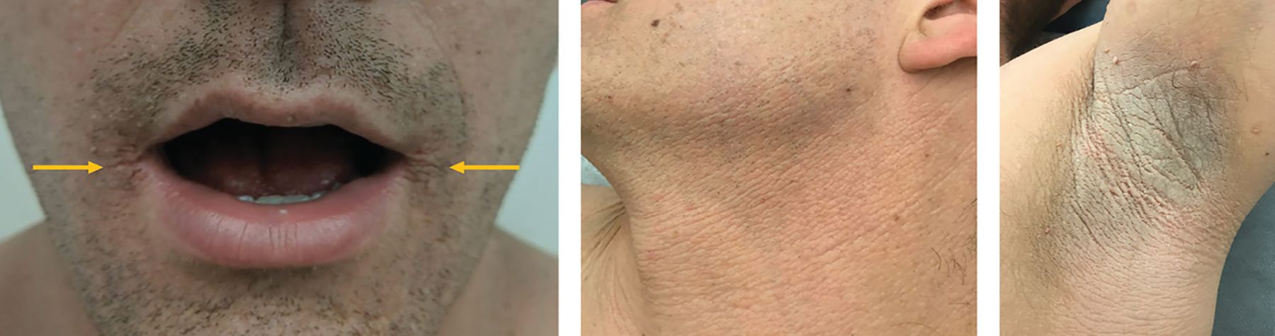
At presentation, the patient had extensive velvety hyperpigmented plaques on the forehead, corners of the lips, neck, axillae, trunk (including areolae), extremities, and groin, amounting to approximately 10% to 15% of the body surface area (Figure 1).

Figure 1
  • Download figure
  • Open in new tab
  • Download powerpoint
Figure 1

Plaques of acanthosis nigricans on areas of the lips, left neck, and left axilla at initial consultation.

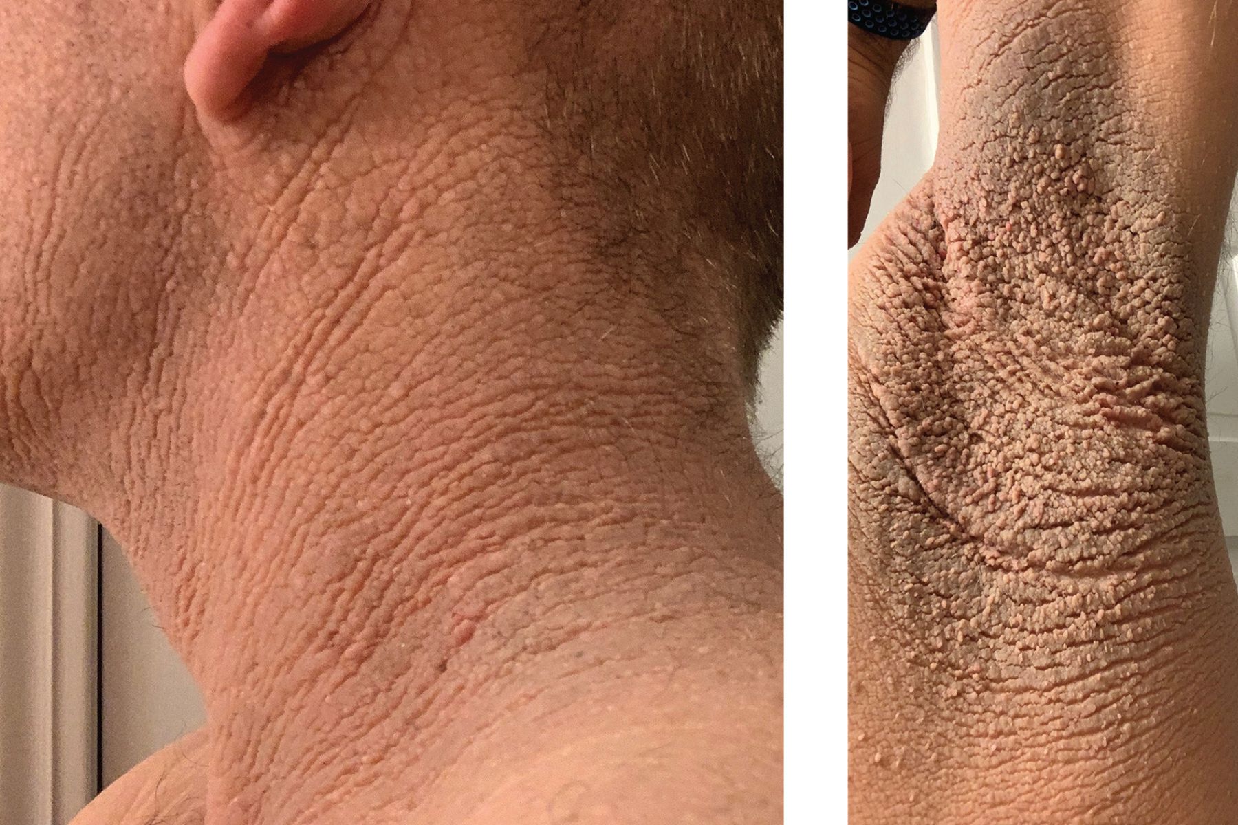
Clinical features and skin-shave biopsy (showing epidermal papillomatosis) at initial presentation supported the diagnosis of acanthosis nigricans. The distribution and body surface area involvement prompted an initial laboratory workup, with a complete blood cell count, lipid panel, renal panel, and endocrine panel, all of which were normal. Multiple management strategies were attempted, including metformin, isotretinoin, topical theapies (steroids and calcineurin inhibitors), and weight loss. The patient initially experienced mild improvement in visual appearance and pruritus from weight loss and tacrolimus ointment. Though he felt systemically well, his acanthosis nigricans progressively worsened over the next 2 to 3 years (Figure 2). This prompted a cancer screening workup (Table 1),1 but to date no major abnormalities have been identified except gradual elevation over 2 years of the total cholesterol level of 202 mg/dL.

Figure 2
  • Download figure
  • Open in new tab
  • Download powerpoint
Figure 2

Severe disease progression after 2 years.

View this table:
  • View inline
  • View popup
TABLE 1

Cancer screening workup for acanthosis nigricans with suspicious featuresa

PATHOPHYSIOLOGY AND CLINICAL VARIANTS

Acanthosis nigricans classically presents in skin folds (eg, neck, axillae) with symmetrically distributed hyperpigmented plaques with a velvety or verrucous texture.2 It is more common in Native Americans and African Americans.2 The precise pathophysiology of acanthosis nigricans is unknown but attributed to insulin-mediated stimulation of keratinocytes and fibroblasts via insulin-like growth factor receptors and epidermal growth factor receptors.2,3

Common causes of acanthosis nigricans include obesity, medications, and endocrine abnormalities, with rarer cases due to paraneoplastic phenomenon or a familial trait via germline fibroblast growth factor receptor 3 mutations.2–4 Obesity-associated acanthosis nigricans can present together with insulin resistance, high body mass index, diabetes, metabolic syndrome, or polycystic ovarian syndrome. Medications associated with acanthosis nigricans include nicotinic acid (niacin), oral corticosteroids, oral contraceptives, methyltestosterone.2 The paraneoplastic form is most often seen with gastric adenocarcinoma, though other causes have also been identified.2

Acanthosis nigricans with abrupt features such as severe and rapid onset, atypical sites (eg, palms or mucosa), widespread distribution at any age, or new onset in patients over age 40 should prompt evaluation for malignancy.2 A thorough history and physical examination followed by judicious use of laboratory and other investigations should help determine the underlying cause of acanthosis nigricans.

DIFFERENTIAL DIAGNOSIS OF SKIN-THICKENING AND HYPERPIGMENTATION

Other notable conditions can present with skin-thickening or hyperpigmentation:

  • Confluent and reticulated papillomatosis is more diffuse with a net-like appearance

  • Terra firma-forme dermatosis easily wipes off with rubbing alcohol

  • Hemochromatosis, Addison disease, and erythema dyschromicum perstans (ashy dermatosis) all present with flat hyperpigmentation only, without skin-thickening

  • Pemphigus vegetans is a rare form of pemphigus with warty (verrucous) ulcerated plaques.

MANAGEMENT

Acanthosis nigricans is first treated by addressing underlying causes with diet and exercise and oral metformin in obesity-associated acanthosis nigri-cans, surgery or chemotherapy for paraneoplastic acanthosis nigricans, and removal of the offending agent in drug-induced acanthosis nigricans.2–6 Skin treatments can help with the cosmetic appearance of plaques. Topical keratolytics (retinoids, salicylic acid, ammonium lactate, chemical peels) may help soften the appearance of acanthosis nigricans but carry a risk of skin irritation. Systemic isotretinoin may be considered in consultation with a dermatologist.

TAKE-HOME POINTS

While patients with milder acanthosis nigricans will not necessarily require a workup for malignancy, close clinical monitoring is recommended as the condition can be the first sign of diabetes or malignancy.6 Because approximately 17% of malignant acanthosis nigricans appears prior to tumor diagnosis, progression to severe acanthosis nigricans in this patient was the rationale for a more extensive clinical investigation.5–7

DISCLOSURES

Dr. Das has disclosed ownership interest (stock, stock ownership in a publicly owned company) in Bristol-Myers Squibb and Crisper Therapeutics, and work as advisor for Skin Analytics. The other authors report no relevant financial relationships which, in the context of their contributions, could be perceived as a potential conflict of interest.

  • Copyright © 2022 The Cleveland Clinic Foundation. All Rights Reserved.

REFERENCES

  1. ↵
    1. Department of Health and Human Services
    . US Preventive Services Task Force. A & B Recommendations. https://www.uspreventiveservicestaskforce.org/uspstf/recommendation-topics/uspstf-a-and-b-recommendations. Accessed August 18, 2022.
  2. ↵
    1. Phiske MM
    . An approach to acanthosis nigricans. Indian Dermatol Online J 2014; 5(3):239–249. doi:10.4103/2229-5178.137765
    OpenUrlCrossRefPubMed
  3. ↵
    1. Fukuchi K,
    2. Tatsuno K,
    3. Matsushita K,
    4. Kubo A,
    5. Ito T,
    6. Tokura Y
    . Familial acanthosis nigricans with p.K650T FGFR3 mutation. J Dermatol 2018; 45(2):207–210. doi:10.1111/1346-8138.14107
    OpenUrlCrossRef
  4. ↵
    1. Torley D,
    2. Bellus GA,
    3. Munro CS
    . Genes, growth factors and acanthosis nigricans. Br J Dermatol 2002; 147(6):1096–1101. doi:10.1046/j.1365-2133.2002.05150.x
    OpenUrlCrossRefPubMed
  5. ↵
    1. Talsania N,
    2. Harwood CA,
    3. Piras D,
    4. Cerio R
    . Paraneoplastic acanthosis nigricans: the importance of exhaustive and repeated malignancy screening. Dermatol Online J 2010; 16(8):8. pmid:20804685
    OpenUrlPubMed
  6. ↵
    1. Popa ML,
    2. Popa AC,
    3. Tanase C,
    4. Gheorghisan-Galateanu AA
    . Acanthosis nigricans: to be or not to be afraid. Oncol Lett 2019; 17(5):4133–4138. doi:10.3892/ol.2018.9736
    OpenUrlCrossRef
  7. ↵
    1. Krawczyk M,
    2. Mykała-Cieśla J,
    3. Kołodziej-Jaskuła A
    . Acanthosis nigricans as a paraneoplastic syndrome. Case reports and review of literature. Pol Arch Med Wewn 2009; 119(3):180–183. pmid:19514649
    OpenUrlPubMed
PreviousNext
Back to top

In this issue

Cleveland Clinic Journal of Medicine: 89 (9)
Cleveland Clinic Journal of Medicine
Vol. 89, Issue 9
1 Sep 2022
  • Table of Contents
  • Table of Contents (PDF)
  • Index by author
  • Complete Issue (PDF)
Print
Download PDF
Article Alerts
Sign In to Email Alerts with your Email Address
Email Article

Thank you for your interest in spreading the word on Cleveland Clinic Journal of Medicine.

NOTE: We only request your email address so that the person you are recommending the page to knows that you wanted them to see it, and that it is not junk mail. We do not capture any email address.

Enter multiple addresses on separate lines or separate them with commas.
Widespread skin-thickening and hyperpigmentation
(Your Name) has sent you a message from Cleveland Clinic Journal of Medicine
(Your Name) thought you would like to see the Cleveland Clinic Journal of Medicine web site.
CAPTCHA
This question is for testing whether or not you are a human visitor and to prevent automated spam submissions.
Citation Tools
Widespread skin-thickening and hyperpigmentation
India K. Poetzscher, Nikolai Klebanov, Shinjita Das
Cleveland Clinic Journal of Medicine Sep 2022, 89 (9) 485-487; DOI: 10.3949/ccjm.89a.21099

Citation Manager Formats

  • BibTeX
  • Bookends
  • EasyBib
  • EndNote (tagged)
  • EndNote 8 (xml)
  • Medlars
  • Mendeley
  • Papers
  • RefWorks Tagged
  • Ref Manager
  • RIS
  • Zotero
Share
Widespread skin-thickening and hyperpigmentation
India K. Poetzscher, Nikolai Klebanov, Shinjita Das
Cleveland Clinic Journal of Medicine Sep 2022, 89 (9) 485-487; DOI: 10.3949/ccjm.89a.21099
Twitter logo Facebook logo Mendeley logo
  • Tweet Widget
  • Facebook Like
  • Linkedin Share Button

Jump to section

  • Article
    • PATHOPHYSIOLOGY AND CLINICAL VARIANTS
    • DIFFERENTIAL DIAGNOSIS OF SKIN-THICKENING AND HYPERPIGMENTATION
    • MANAGEMENT
    • TAKE-HOME POINTS
    • DISCLOSURES
    • REFERENCES
  • Figures & Data
  • Info & Metrics
  • PDF

Related Articles

  • No related articles found.
  • PubMed
  • Google Scholar

Cited By...

  • No citing articles found.
  • Google Scholar

More in this TOC Section

  • Tinea incognito
  • Prolonged venous filling time and dependent rubor in a patient with peripheral artery disease
  • Sarcoidosis with diffuse purplish erythematous plaques on the hands
Show more The Clinical Picture

Similar Articles

Subjects

  • Dermatology
  • Physical Exam

Navigate

  • Current Issue
  • Past Issues
  • Supplements
  • Article Type
  • Specialty
  • CME/MOC Articles
  • CME/MOC Calendar
  • Media Kit

Authors & Reviewers

  • Manuscript Submission
  • Authors & Reviewers
  • Subscriptions
  • About CCJM
  • Contact Us
  • Cleveland Clinic Center for Continuing Education
  • Consult QD

Share your suggestions!

Copyright © 2025 The Cleveland Clinic Foundation. All rights reserved. The information provided is for educational purposes only. Use of this website is subject to the website terms of use and privacy policy. 

Powered by HighWire