
Main menu

  • Home
  • Content
    • Current Issue
    • Ahead of Print
    • Past Issues
    • Supplements
    • Article Type
  • Specialty
    • Articles by Specialty
  • CME/MOC
    • Articles
    • Calendar
  • Info For
    • Manuscript Submission
    • Authors & Reviewers
    • Subscriptions
    • About CCJM
    • Contact Us
    • Media Kit
  • Conversations with Leaders
  • Conference Coverage
    • Kidney Week 2024
    • CHEST 2024
    • ACR Convergence 2023
    • Kidney Week 2023
    • ObesityWeek 2023
    • IDWeek 2023
    • CHEST 2023
    • MDS 2023
    • IAS 2023
    • ACP 2023
    • AAN 2023
    • ACC / WCC 2023
    • AAAAI Meeting 2023
    • ACR Convergence 2022
    • Kidney Week 2022
    • AIDS 2022
  • Other Publications
    • www.clevelandclinic.org

User menu

  • Register
  • Log in

Search

  • Advanced search
Cleveland Clinic Journal of Medicine
  • Other Publications
    • www.clevelandclinic.org
  • Register
  • Log in
Cleveland Clinic Journal of Medicine

Advanced Search

  • Home
  • Content
    • Current Issue
    • Ahead of Print
    • Past Issues
    • Supplements
    • Article Type
  • Specialty
    • Articles by Specialty
  • CME/MOC
    • Articles
    • Calendar
  • Info For
    • Manuscript Submission
    • Authors & Reviewers
    • Subscriptions
    • About CCJM
    • Contact Us
    • Media Kit
  • Conversations with Leaders
  • Conference Coverage
    • Kidney Week 2024
    • CHEST 2024
    • ACR Convergence 2023
    • Kidney Week 2023
    • ObesityWeek 2023
    • IDWeek 2023
    • CHEST 2023
    • MDS 2023
    • IAS 2023
    • ACP 2023
    • AAN 2023
    • ACC / WCC 2023
    • AAAAI Meeting 2023
    • ACR Convergence 2022
    • Kidney Week 2022
    • AIDS 2022
The Clinical Picture

Hyperpigmentation as a clue to Addison disease

Yusuke Hirota, MD, PhD and Takaya Matsushita, MD, PhD
Cleveland Clinic Journal of Medicine September 2022, 89 (9) 498-499; DOI: https://doi.org/10.3949/ccjm.89a.21082
Yusuke Hirota
Department of Diabetology, Endocrinology and Metabolism, Tokyo Medical University Hachioji Medical Center, Tokyo, Japan
  • Find this author on Google Scholar
  • Find this author on PubMed
  • Search for this author on this site
  • For correspondence: [email protected]
Takaya Matsushita
Department of Diabetology, Endocrinology and Metabolism, Tokyo Medical University Hachioji Medical Center, Tokyo, Japan
  • Find this author on Google Scholar
  • Find this author on PubMed
  • Search for this author on this site
  • Article
  • Figures & Data
  • Info & Metrics
  • PDF
Loading

A 66-year-old woman presented to our department with generalized fatigue and loss of appetite.

A review of her medical record showed that at age 52 she had undergone kidney transplant for end-stage kidney disease, receiving treatment of tacrolimus and mycophenolate after induction therapy with methyl-prednisolone 1,000 mg/day tapered over 3 months. After that, without steroids or antihypertensive drugs, her blood pressure had remained within the normal range. Mild hyperkalemia had been noted, and her fasting blood glucose was in the normal range.

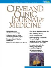
On admission, her heart rate was 74 beats per minute and her blood pressure was 117/80 mm Hg. Physical examination revealed hyperpigmentation of her tongue and soft palate (Figure 1), as well as the buccal mucosa, lower lip, fingers, and nail beds. The eyelids, neck, nipples, elbows, back, knees, and soles had very mild or no pigmentation. When asked about the hyperpigmentation, the patient said that she had first noticed it more than 20 years ago but that it had not interfered with her daily life, and she had never sought medical attention for it.

Figure 1
  • Download figure
  • Open in new tab
  • Download powerpoint
Figure 1

Skin pigmentation in the patient’s tongue (left) and on the soft palate (right).

INITIAL WORKUP

Laboratory testing at admission showed the following:

  • Potassium 5.7 mmol/L (reference range 3.6–4.8)

  • Adrenocorticotropic hormone (ACTH) of 372 pg/mL (7.2−63.3)

  • Dehydroepiandrosterone sulfate 15 μg/dL (12−133)

  • Estimated glomerular filtration rate of 33.2 mL/ min/1.73 m2 (≥ 60)

  • Morning cortisol 8.93 μg/dL (6.24−18.00)

  • 24-hour urinary free cortisol less than 12.5 μg/day (11.2−80.3)

  • Plasma renin activity and plasma aldosterone concentration were normal

  • Anti-adrenal antibody testing and cytomegalovirus antigenemia test were negative

  • Rapid ACTH stimulation test showed no significant increase in serum cortisol.

Computed tomography of the abdomen showed no adrenal hyperplasia, nodules, or malignant lesions.

These findings and the results of laboratory testing supported the diagnosis of Addison disease. The patient received glucocorticoid-replacement therapy without mineralocorticoid, which relieved the hyponatremia, hyperkalemia, and general symptoms. One month after the start of replacement therapy, the hyperpigmentation had improved slightly.

CAUSES AND SYMPTOMS OF PRIMARY ADRENAL INSUFFICIENCY

Addison disease, or primary adrenal insufficiency, is an uncommon disease caused by destruction of the adrenal cortex, usually owing to an autoimmune process. Other causes include infections, malignant infiltration, metabolic dysfunction, and granuloma-tous disease, as well as genetic, neonatal, vascular, pharmacologic, neoplastic, and surgical factors.1

Common clinical manifestations are fatigue, weight loss, orthostatic hypotension, skin and mucosal hyperpigmentation, nausea, vomiting, abdominal pain, libido reduction, depression, and salt-craving, depending on any deficiencies of glucocorticoids, mineralocorticoids, or androgens.1,2

Most manifestations are nonspecific and slowly progressive, so the diagnosis of Addison disease is often difficult or delayed. However, hyperpigmentation, caused by excess binding of ACTH and alpha-melanocyte stimulating hormone to the melanocortin 1 receptor,3 is a characteristic feature and a diagnostic clue for Addison disease.2

Diffuse hyperpigmentation is also caused by medications (eg, chemotherapeutic agents, antimalarials, oral contraceptives, prostaglandin agonists, amiodarone, minocycline) and by endocrine and metabolic diseases (hyperthyroidism, diabetes mellitus, hemochromatosis).4 This patient was taking none of these agents, and there was no endocrine or metabolic disease present to produce hyperpigmentation other than Addison disease.

FEATURES OF HYPERPIGMENTATION OFFER DIAGNOSTIC CLUES

The characteristics of skin hyperpigmentation can offer clues to the cause. For example, aminoquinoline drugs can induce gray-blue pigmentation on pretibial surfaces, oral mucosa, sclera, and subungual areas. Minocycline induces 3 patterns: generalized brown discoloration, dark blue discoloration confined to acne scars or areas of skin inflammation, and gray-blue discoloration on the front of the lower extremities or in exposed areas. In Addison disease, hyperpigmentation is muddy and diffuse on buccal, conjunctival, and genital mucosa and on nail beds, palmar creases, and nipples.4

TAKE-HOME MESSAGE

When Addison disease is considered in the differential diagnosis, it is important to review the patient’s medical history, to examine the skin and mucous membranes for hyperpigmentation, to measure early-morning plasma ACTH and serum cortisol, and to perform the ACTH stimulation test. Glucocorticoid and mineralocorticoid replacement therapy is essential.

DISCLOSURES

The authors report no relevant financial relationships which, in the context of their contributions, could be perceived as a potential conflict of interest.

  • Copyright © 2022 The Cleveland Clinic Foundation. All Rights Reserved.

REFERENCES

  1. ↵
    1. Betterle C,
    2. Presotto F,
    3. Furmaniak J
    . Epidemiology, pathogenesis, and diagnosis of Addison’s disease in adults. J Endocrinol Invest 2019; 42(12):1407–1433. doi:10.1007/s40618-019-01079-6
    OpenUrlCrossRefPubMed
  2. ↵
    1. Bancos I,
    2. Hahner S,
    3. Tomlinson J,
    4. Arlt W
    . Diagnosis and management of adrenal insufficiency. Lancet Diabetes Endocrinol 2015; 3(3):216–226. doi:10.1016/S2213-8587(14)70142-1
    OpenUrlCrossRefPubMed
  3. ↵
    1. Abdel-Malek ZA,
    2. Knittel J,
    3. Kadekaro AL,
    4. Swope VB,
    5. Starner R
    . The melanocortin 1 receptor and the UV response of human melanocytes—a shift in paradigm. Photochem Photobiol 2008; 84(2):501–508. doi:10.1111/j.1751-1097.2008.00294.x
    OpenUrlCrossRefPubMed
  4. ↵
    1. Stulberg DL,
    2. Clark N,
    3. Tovey D
    . Common hyperpigmentation disorders in adults: part I. Diagnostic approach, café au lait macules, diffuse hyperpigmentation, sun exposure, and phototoxic reactions. Am Fam Physician 2003; 68(10):1955–1960. pmid:14655804
    OpenUrlPubMed
PreviousNext
Back to top

In this issue

Cleveland Clinic Journal of Medicine: 89 (9)
Cleveland Clinic Journal of Medicine
Vol. 89, Issue 9
1 Sep 2022
  • Table of Contents
  • Table of Contents (PDF)
  • Index by author
  • Complete Issue (PDF)
Print
Download PDF
Article Alerts
Sign In to Email Alerts with your Email Address
Email Article

Thank you for your interest in spreading the word on Cleveland Clinic Journal of Medicine.

NOTE: We only request your email address so that the person you are recommending the page to knows that you wanted them to see it, and that it is not junk mail. We do not capture any email address.

Enter multiple addresses on separate lines or separate them with commas.
Hyperpigmentation as a clue to Addison disease
(Your Name) has sent you a message from Cleveland Clinic Journal of Medicine
(Your Name) thought you would like to see the Cleveland Clinic Journal of Medicine web site.
CAPTCHA
This question is for testing whether or not you are a human visitor and to prevent automated spam submissions.
Citation Tools
Hyperpigmentation as a clue to Addison disease
Yusuke Hirota, Takaya Matsushita
Cleveland Clinic Journal of Medicine Sep 2022, 89 (9) 498-499; DOI: 10.3949/ccjm.89a.21082

Citation Manager Formats

  • BibTeX
  • Bookends
  • EasyBib
  • EndNote (tagged)
  • EndNote 8 (xml)
  • Medlars
  • Mendeley
  • Papers
  • RefWorks Tagged
  • Ref Manager
  • RIS
  • Zotero
Share
Hyperpigmentation as a clue to Addison disease
Yusuke Hirota, Takaya Matsushita
Cleveland Clinic Journal of Medicine Sep 2022, 89 (9) 498-499; DOI: 10.3949/ccjm.89a.21082
Twitter logo Facebook logo Mendeley logo
  • Tweet Widget
  • Facebook Like
  • Linkedin Share Button

Jump to section

  • Article
    • INITIAL WORKUP
    • CAUSES AND SYMPTOMS OF PRIMARY ADRENAL INSUFFICIENCY
    • FEATURES OF HYPERPIGMENTATION OFFER DIAGNOSTIC CLUES
    • TAKE-HOME MESSAGE
    • DISCLOSURES
    • REFERENCES
  • Figures & Data
  • Info & Metrics
  • PDF

Related Articles

  • No related articles found.
  • PubMed
  • Google Scholar

Cited By...

  • No citing articles found.
  • Google Scholar

More in this TOC Section

  • Tinea incognito
  • Prolonged venous filling time and dependent rubor in a patient with peripheral artery disease
  • Sarcoidosis with diffuse purplish erythematous plaques on the hands
Show more The Clinical Picture

Similar Articles

Subjects

  • Dermatology
  • Endocrinology
  • Physical Exam

Navigate

  • Current Issue
  • Past Issues
  • Supplements
  • Article Type
  • Specialty
  • CME/MOC Articles
  • CME/MOC Calendar
  • Media Kit

Authors & Reviewers

  • Manuscript Submission
  • Authors & Reviewers
  • Subscriptions
  • About CCJM
  • Contact Us
  • Cleveland Clinic Center for Continuing Education
  • Consult QD

Share your suggestions!

Copyright © 2025 The Cleveland Clinic Foundation. All rights reserved. The information provided is for educational purposes only. Use of this website is subject to the website terms of use and privacy policy. 

Powered by HighWire