

Figure 2

Figure 2
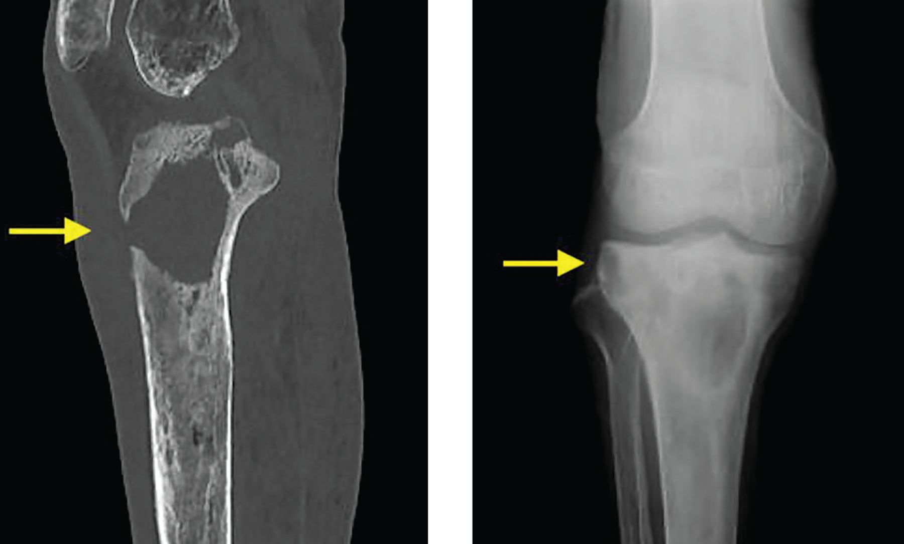
On hospital admission, computed tomography (left) demonstrated a well-visualized sinus tract connecting to subcutaneous tissue (arrow), and radiography (right) of the abscess showed well-circumscribed osteolysis with sclerotic margins (arrow), which had developed during the year since the initial presentation.
In this issue


Related Articles
Cited By...
- No citing articles found.