


A 45-year-old female presented with an 18-month history of skin discoloration spanning the nape of the neck, ears, and scalp that was accompanied by itching. There was no history of hair dye use. She denied experiencing skin tightness, Raynaud phenomenon, fingertip ulceration, dysphagia, retrosternal burning, difficulty opening her mouth, dyspnea, palpitations, pedal edema, or joint pain.
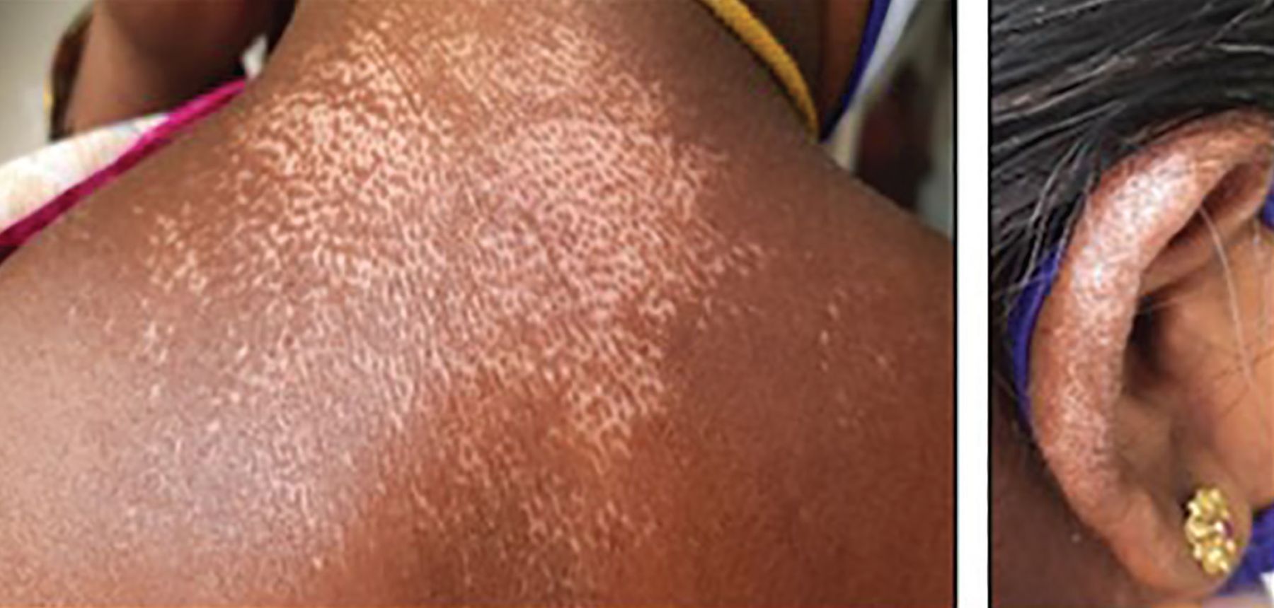
During physical examination, the patient exhibited salt-and-pepper pigmentation on the nape of the neck, pinna of each ear, and scalp (Figure 1). There was no restriction in opening her mouth, binding down of the skin, ragged cuticles, or abnormal chest expansion.

Salt-and-pepper skin pigmentation of the nape of the neck and pinna.
WORKUP AND DIAGNOSIS
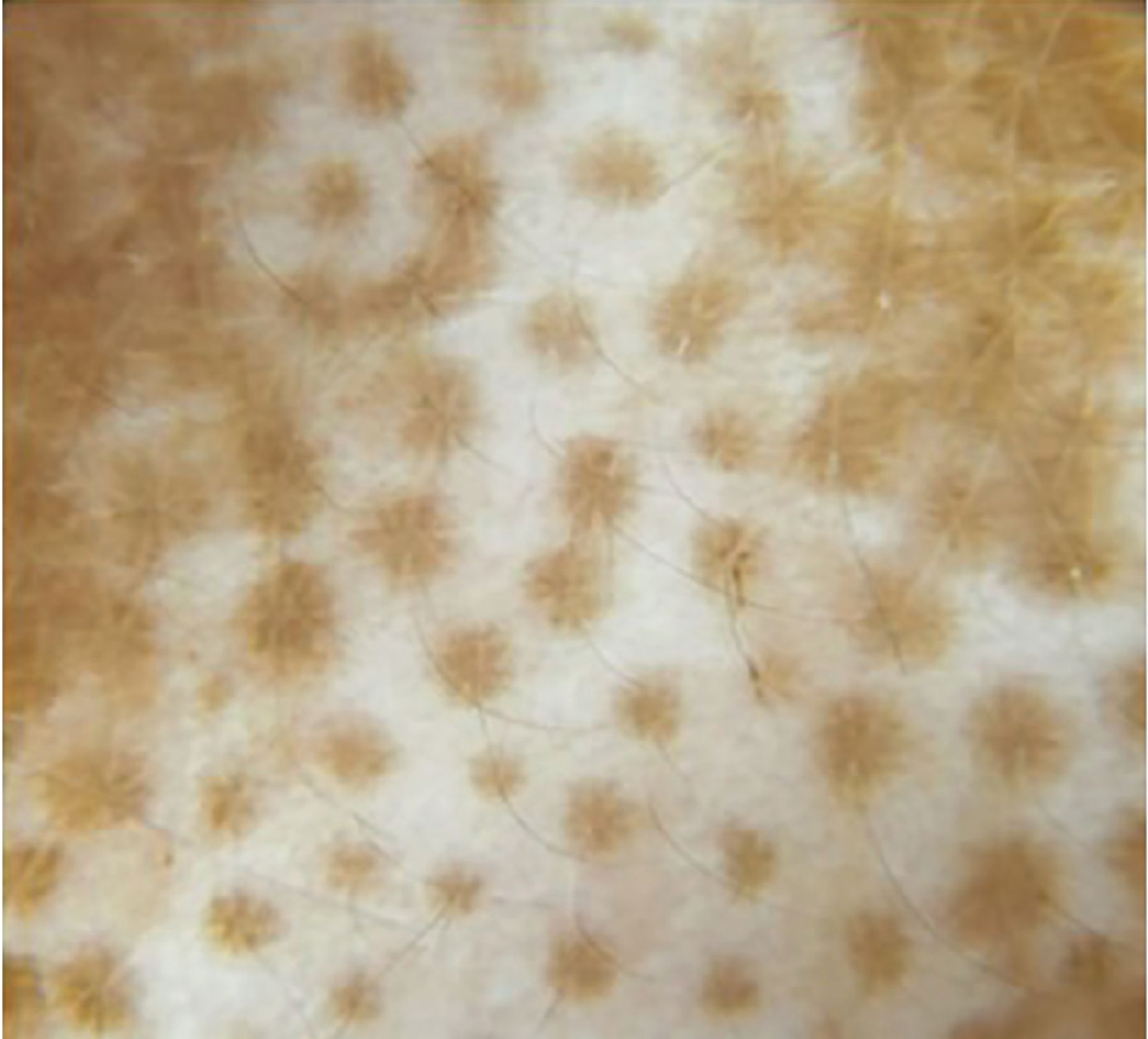
Dermoscopy of the skin lesions revealed homogeneous depigmented areas with perifollicular pigmentation (Figure 2). The differential diagnoses considered were vitiligo repigmentation and early scleroderma. The patient did not have other lesions elsewhere on the body, and the oral mucosa was normal. Results of a routine laboratory workup including complete blood cell count and metabolic panel were within normal limits. However, antinuclear antibody was detected (1:320) in a centromere pattern. Chest radiography and pulmonary function test results were normal. Biopsy of skin from the nape of the neck revealed a focally thinned out epidermis with loss of rete ridges. The superficial dermis exhibited mild perivascular lymphocytic infiltrate, with appendages appearing pulled up and bound down.

Homogeneous depigmented areas with perifollicular pigmentation.
Based on the classic skin lesion, absence of oral dyspigmentation, suggestive dermoscopic findings, and histopathologic findings consistent with scleroderma, a conclusive diagnosis of scleroderma was made.
The patient was started on methotrexate 0.3 mg/kg weekly to prevent cutaneous progression. On follow-up 4 months later, subjective improvement of the lesions was noted.
AN EARLY FEATURE OF SYSTEMIC SCLEROSIS
Systemic sclerosis is an autoimmune rheumatic disease characterized by chronic inflammation and fibrosis affecting the skin, gastrointestinal system, microvasculature, lungs, and heart.1 Presence of anticentromere antibody is usually associated with limited cutaneous disease and less chance of systemic involvement.2 Skin involvement manifests as areas of thickening, fibrosis, and, in some patients, a distinctive dyspigmentation known as salt-and-pepper pigmentation. Salt-and-pepper skin is characterized by vitiligo-like depigmentation, possibly triggered by trauma or immune dysfunction leading to destruction of melanocytes, with sparing of perifollicular areas. Perifollicular pigment retention has been attributed to the abundant capillary network around hair follicles, which can help preserve melanogenesis.3
There are several patterns of vitiligo repigmentation, the most common of which is perifollicular repigmentation.4 Vitiligo can involve the oral mucosa and exhibit features of Koebner phenomenon. Dermoscopy findings include diffuse whitening or alteration of the pigment network and perifollicular pigmentary changes.5
The primary goal of treatment is to prevent progression of the disease. Because salt-and-pepper pigmentation occurs in areas of cutaneous sclerosis, hypothetically, treating the underlying sclerosis could improve the overlying dyspigmentation. Topical Janus kinase inhibitors like ruxolitinib and tofacitinib are considered when only a few cutaneous lesions are present.6 Medications used to treat systemic sclerosis include methotrexate, mycophenolate mofetil, intravenous immunoglobulin, and biologic agents like rituximab, abatacept, and tocilizumab.7
Even in the absence of clinically detectable sclerosis, salt-and-pepper pigmentation can be an early indicator of systemic sclerosis and its presence should heighten suspicion for this diagnosis.
DISCLOSURES
The authors report no relevant financial relationships which, in the context of their contributions, could be perceived as a potential conflict of interest.
- Copyright © 2024 The Cleveland Clinic Foundation. All Rights Reserved.
In this issue


Jump to section
Related Articles
Cited By...
- No citing articles found.