
Main menu

  • Home
  • Content
    • Current Issue
    • Ahead of Print
    • Past Issues
    • Supplements
    • Article Type
  • Specialty
    • Articles by Specialty
  • CME/MOC
    • Articles
    • Calendar
  • Info For
    • Manuscript Submission
    • Authors & Reviewers
    • Subscriptions
    • About CCJM
    • Contact Us
    • Media Kit
  • Conversations with Leaders
  • Conference Coverage
    • Kidney Week 2024
    • CHEST 2024
    • ACR Convergence 2023
    • Kidney Week 2023
    • ObesityWeek 2023
    • IDWeek 2023
    • CHEST 2023
    • MDS 2023
    • IAS 2023
    • ACP 2023
    • AAN 2023
    • ACC / WCC 2023
    • AAAAI Meeting 2023
    • ACR Convergence 2022
    • Kidney Week 2022
    • AIDS 2022
  • Other Publications
    • www.clevelandclinic.org

User menu

  • Register
  • Log in

Search

  • Advanced search
Cleveland Clinic Journal of Medicine
  • Other Publications
    • www.clevelandclinic.org
  • Register
  • Log in
Cleveland Clinic Journal of Medicine

Advanced Search

  • Home
  • Content
    • Current Issue
    • Ahead of Print
    • Past Issues
    • Supplements
    • Article Type
  • Specialty
    • Articles by Specialty
  • CME/MOC
    • Articles
    • Calendar
  • Info For
    • Manuscript Submission
    • Authors & Reviewers
    • Subscriptions
    • About CCJM
    • Contact Us
    • Media Kit
  • Conversations with Leaders
  • Conference Coverage
    • Kidney Week 2024
    • CHEST 2024
    • ACR Convergence 2023
    • Kidney Week 2023
    • ObesityWeek 2023
    • IDWeek 2023
    • CHEST 2023
    • MDS 2023
    • IAS 2023
    • ACP 2023
    • AAN 2023
    • ACC / WCC 2023
    • AAAAI Meeting 2023
    • ACR Convergence 2022
    • Kidney Week 2022
    • AIDS 2022
The Clinical Picture

Acquired reactive perforating collagenosis in a patient with diabetes

Li-wen Zhang, MD, Juan Wu, MD, PhD, Rong-hua Xu, MD and Tao Chen, MD, PhD
Cleveland Clinic Journal of Medicine April 2024, 91 (4) 213-214; DOI: https://doi.org/10.3949/ccjm.91a.23074
Li-wen Zhang
Department of Dermatovenereology, Chengdu Second People’s Hospital, Chengdu, Sichuan, China
  • Find this author on Google Scholar
  • Find this author on PubMed
  • Search for this author on this site
Juan Wu
Sexually Transmitted Disease Institute, Shanghai Skin Disease Hospital, School of Medicine, Tongji University, Shanghai, China
  • Find this author on Google Scholar
  • Find this author on PubMed
  • Search for this author on this site
Rong-hua Xu
Institute of Dermatology, Chengdu Second People’s Hospital, Chengdu, Sichuan, China
  • Find this author on Google Scholar
  • Find this author on PubMed
  • Search for this author on this site
Tao Chen
Department of Dermatovenereology, Chengdu Second People’s Hospital, Chengdu, Sichuan, China
  • Find this author on Google Scholar
  • Find this author on PubMed
  • Search for this author on this site
  • For correspondence: [email protected] [email protected]
  • Article
  • Figures & Data
  • Info & Metrics
  • PDF
Loading

A 47-year-old woman presented with a 2-month history of pruritic eruptions on the left ankle and a complaint of thirst and polyuria for the past year. She was previously healthy and denied a history of insect bites.

Physical examination revealed multiple umbilicated hyperkeratotic papules and nodules with a central round crusted ulcer on the left ankle (Figure 1). Histologic study of the lesions revealed cup-shaped invagination of the epidermis plugged with necrotic inflammatory debris and transepidermal elimination of dermal collagen (Figure 2). Laboratory testing showed the following:

  • Fasting glucose 7.8 mmol/L [140.4 mg/dL] (reference range 3.9–5.6 [70–100])

  • Hemoglobin A1c 6.9%

  • Triglycerides 2.38 mmol/L [210.6 mg/dL] (< 1.7 [< 150])

  • Total cholesterol 7.21 mmol/L [278.4 mg/dL] (< 5.18 [< 200]).

Figure 1
  • Download figure
  • Open in new tab
  • Download powerpoint
Figure 1

Multiple umbilicated hyperkeratotic papules (black arrows) and nodules (blue arrows) with a central round crusted ulcer on the left ankle.

Figure 2
  • Download figure
  • Open in new tab
  • Download powerpoint
Figure 2

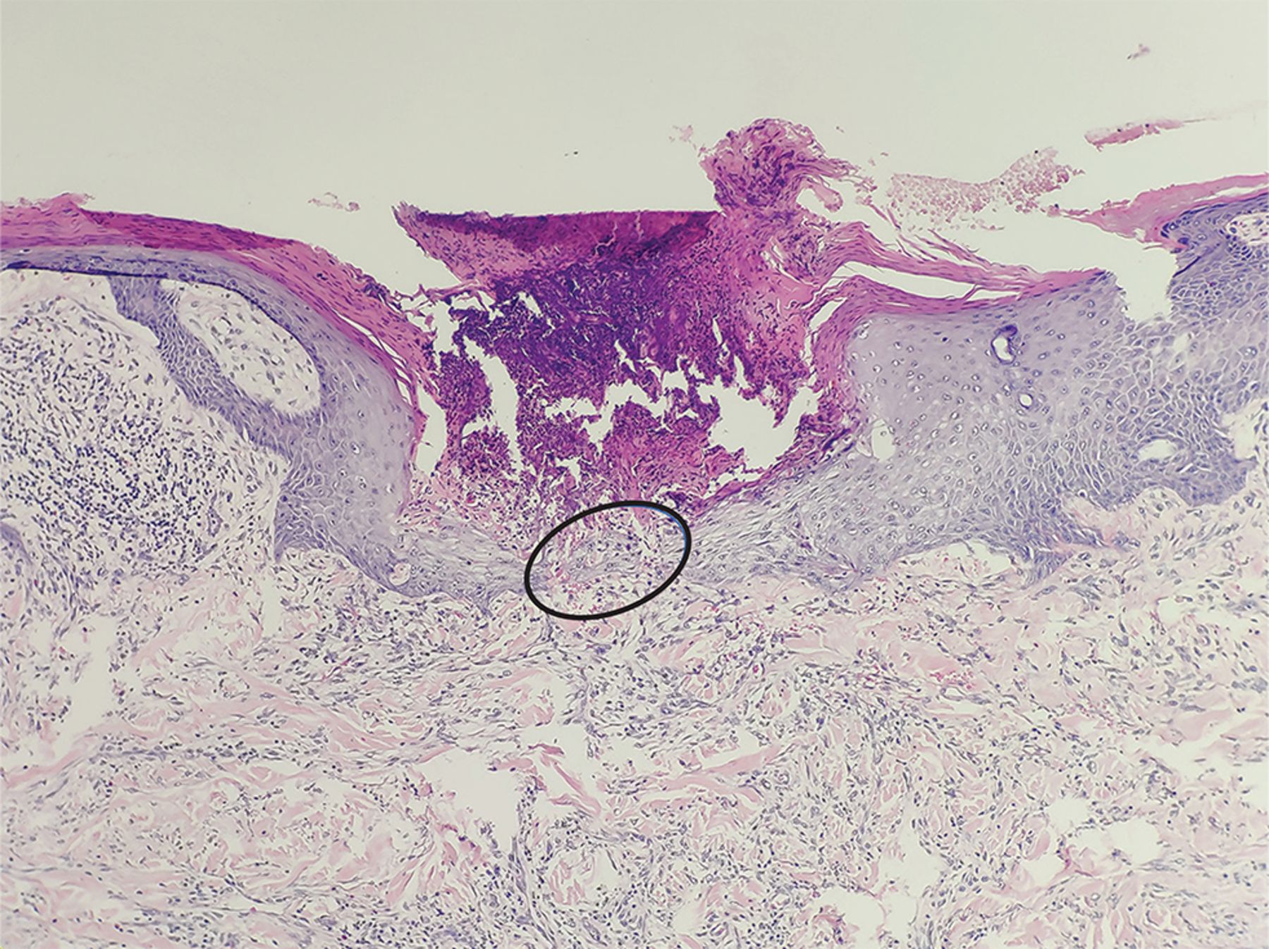
Histologic study of a papule revealed cup-shaped invagination of the epidermis, plugged with necrotic inflammatory debris, and transepidermal elimination of dermal collagen (circled area) (hematoxylin and eosin stain, magnification × 100).

Based on the clinical presentation and the results of histologic study and blood testing, we diagnosed acquired reactive perforating collagenosis and type 2 diabetes. The patient was referred to endocrinology for management, and her lesions improved after 4 weeks of treatment with loratadine and halometasone cream.

ACQUIRED PERFORATING DERMATOSIS

Perforating dermatoses are a group of chronic pruritic cutaneous disorders characterized histologically by transepidermal elimination of dermal components, ie, the components “perforate” the epidermis. They are divided into primary and secondary (ie, acquired) forms and, based on the dermal components of transepidermal elimination, can be further classifed into 4 subtypes: Kyrle disease, reactive perforating collagenosis (as in our patient), elastosis perforans serpiginosum, and perforating folliculitis.1

Acquired perforating dermatosis (APD) usually affects the extensor surfaces of the extremities and trunk and presents as multiple, nonfused, umbilicated, hyperkeratotic papules or nodules consisting of a central round crusted ulcer with a reddish brown raised border.

Symptoms and other features

Epidemiologic studies of APD are lacking. Seven retrospective case series studies have summarized a total of 282 cases of APD.1–7 The condition mainly affects patients in their 40s and 50s. Pruritus was the most common symptom, with a few complaining of pain. Koebner phenomenon was seen in 32% to 56% of patients.2–4,6,7

Patients with APD often have an underlying systemic disease, particularly diabetes (type 1 or type 2) or chronic kidney disease.1–7 Other underlying comorbidities include cardiovascular disease, infection (human immunodeficiency virus, hepatitis virus, and tuberculosis), rheumatic disease, pulmonary disease, malignancy, psychiatric disease, hypothyroidism, pregnancy, and dermatoses (atopic dermatitis, psoriasis, scabies).1–7

Pathogenesis still unclear

The pathogenesis of APD remains unknown. Possible theories include microtrauma caused by scratching prompted by pruritus, microangiopathy, and overloading of urinary metabolites due to renal insufficiency.4 In 1 study, pruritus was the most common symptom, and the presence of the Koebner phenomenon suggests that trauma plays a role in the pathogenesis of APD.4 Chronic pruritus is a symptom of many systemic diseases, including diabetes and chronic kidney disease. Diabetic microangiopathy may be involved in the formation of local hypoxia and necrosis.4

The differential diagnosis

The differential diagnosis includes insect bites, atopic dermatitis, and lichen planus. The lesions of APD typically have a crater-like structure, presenting as rounded, necrotic, dark brown crusts of variable size in the center of papulonodular lesions surrounded by a reddish brown halo. When lesions are atypical or the diagnosis is in doubt, histologic evidence of transepidermal elimination can confirm the diagnosis.

Treatment

Treatment of the underlying disease may help to improve APD.8 First-line therapies include antihistamines and topical emollients, corticosteroids, and keratolytics. Second-line options include intralesional corticosteroid injections and topical tretinoin, tazarotene, and imiquimod. Other potentially effective treatments include systemic drugs (retinoids, allopurinol, doxycycline, and corticosteroids), dupilumab,6 nemolizumab,9 narrow-band ultraviolet B light, and psoralen plus ultraviolet A light.1,2,8

DISCLOSURES

The authors report no relevant financial relationships which, in the context of their contributions, could be perceived as a potential conflict of interest.

  • Copyright © 2024 The Cleveland Clinic Foundation. All Rights Reserved.

REFERENCES

  1. ↵
    1. García-Malinis AJ,
    2. Del Valle Sánchez E,
    3. Sánchez-Salas MP,
    4. Del Prado E,
    5. Coscojuela C,
    6. Gilaberte Y
    . Acquired perforating dermatosis: clinicopathological study of 31 cases, emphasizing pathogenesis and treatment. J Eur Acad Dermatol Venereol 2017; 31(10):1757–1763. doi:10.1111/jdv.14220
    OpenUrlCrossRef
  2. ↵
    1. Saray Y,
    2. Seçkin D,
    3. Bilezikçi B
    . Acquired perforating dermatosis: clinicopathological features in twenty-two cases. J Eur Acad Dermatol Venereol 2006; 20(6):679–688. doi:10.1111/j.1468-3083.2006.01571.x
    OpenUrlCrossRefPubMed
    1. Kim SW,
    2. Kim MS,
    3. Lee JH, et al
    . A clinicopathologic study of thirty cases of acquired perforating dermatosis in Korea. Ann Dermatol 2014; 26(2):162–171. doi:10.5021/ad.2014.26.2.162
    OpenUrlCrossRef
  3. ↵
    1. Gore Karaali M,
    2. Erdil D,
    3. Erdemir VA,
    4. Gurel MS,
    5. Koku Aksu AE,
    6. Leblebici C
    . Evaluation of clinicopathological and treatment characteristics of 80 patients with acquired perforating dermatosis. Dermatol Ther 2020; 33(6):e14465. doi:10.1111/dth.14465
    OpenUrlCrossRef
    1. Garrido PM,
    2. Queirós C,
    3. Borges-Costa J,
    4. Soares-Almeida L,
    5. Filipe P
    . Acquired perforating dermatosis: clinicopathologic study of a 10-year period at a tertiary teaching hospital. Int J Dermatol 2020; 59(4):445–450. doi:10.1111/ijd.14760
    OpenUrlCrossRef
  4. ↵
    1. Gao Z,
    2. Lu SJ,
    3. Shan SJ
    . Acquired perforating dermatosis: a clinicopathologic study, and the features of dermoscopy and reflective confocal microscopy of 37 cases. Skin Res Technol 2023; 29(7):e13416. doi:10.1111/srt.13416
    OpenUrlCrossRef
  5. ↵
    1. Akoglu G,
    2. Emre S,
    3. Sungu N,
    4. Kurtoglu G,
    5. Metin A
    . Clinicopathological features of 25 patients with acquired perforating dermatosis. Eur J Dermatol 2013; 23(6):864–871. doi:10.1684/ejd.2013.2237
    OpenUrlCrossRef
  6. ↵
    1. Lukács J,
    2. Schliemann S,
    3. Elsner P
    . Treatment of acquired reactive perforating dermatosis—a systematic review. J Dtsch Dermatol Ges 2018; 16(7):825–842. doi:10.1111/ddg.13561
    OpenUrlCrossRef
  7. ↵
    1. Ohmori S,
    2. Sawada Y
    . Perforating dermatosis in a patient on haemodialysis successfully treated with nemolizumab. Clin Exp Dermatol 2023; 48(8):929–941. doi:10.1093/ced/llad119
    OpenUrlCrossRef
PreviousNext
Back to top

In this issue

Cleveland Clinic Journal of Medicine: 91 (4)
Cleveland Clinic Journal of Medicine
Vol. 91, Issue 4
1 Apr 2024
  • Table of Contents
  • Table of Contents (PDF)
  • Index by author
  • Complete Issue (PDF)
Print
Download PDF
Article Alerts
Sign In to Email Alerts with your Email Address
Email Article

Thank you for your interest in spreading the word on Cleveland Clinic Journal of Medicine.

NOTE: We only request your email address so that the person you are recommending the page to knows that you wanted them to see it, and that it is not junk mail. We do not capture any email address.

Enter multiple addresses on separate lines or separate them with commas.
Acquired reactive perforating collagenosis in a patient with diabetes
(Your Name) has sent you a message from Cleveland Clinic Journal of Medicine
(Your Name) thought you would like to see the Cleveland Clinic Journal of Medicine web site.
CAPTCHA
This question is for testing whether or not you are a human visitor and to prevent automated spam submissions.
Citation Tools
Acquired reactive perforating collagenosis in a patient with diabetes
Li-wen Zhang, Juan Wu, Rong-hua Xu, Tao Chen
Cleveland Clinic Journal of Medicine Apr 2024, 91 (4) 213-214; DOI: 10.3949/ccjm.91a.23074

Citation Manager Formats

  • BibTeX
  • Bookends
  • EasyBib
  • EndNote (tagged)
  • EndNote 8 (xml)
  • Medlars
  • Mendeley
  • Papers
  • RefWorks Tagged
  • Ref Manager
  • RIS
  • Zotero
Share
Acquired reactive perforating collagenosis in a patient with diabetes
Li-wen Zhang, Juan Wu, Rong-hua Xu, Tao Chen
Cleveland Clinic Journal of Medicine Apr 2024, 91 (4) 213-214; DOI: 10.3949/ccjm.91a.23074
Twitter logo Facebook logo Mendeley logo
  • Tweet Widget
  • Facebook Like
  • Linkedin Share Button

Jump to section

  • Article
    • ACQUIRED PERFORATING DERMATOSIS
    • DISCLOSURES
    • REFERENCES
  • Figures & Data
  • Info & Metrics
  • PDF

Related Articles

  • No related articles found.
  • PubMed
  • Google Scholar

Cited By...

  • No citing articles found.
  • Google Scholar

More in this TOC Section

  • Tinea incognito
  • Prolonged venous filling time and dependent rubor in a patient with peripheral artery disease
  • Sarcoidosis with diffuse purplish erythematous plaques on the hands
Show more The Clinical Picture

Similar Articles

Subjects

  • Dermatology
  • Diabetes
  • Physical Exam

Navigate

  • Current Issue
  • Past Issues
  • Supplements
  • Article Type
  • Specialty
  • CME/MOC Articles
  • CME/MOC Calendar
  • Media Kit

Authors & Reviewers

  • Manuscript Submission
  • Authors & Reviewers
  • Subscriptions
  • About CCJM
  • Contact Us
  • Cleveland Clinic Center for Continuing Education
  • Consult QD

Share your suggestions!

Copyright © 2025 The Cleveland Clinic Foundation. All rights reserved. The information provided is for educational purposes only. Use of this website is subject to the website terms of use and privacy policy. 

Powered by HighWire