
Main menu

  • Home
  • Content
    • Current Issue
    • Ahead of Print
    • Past Issues
    • Supplements
    • Article Type
  • Specialty
    • Articles by Specialty
  • CME/MOC
    • Articles
    • Calendar
  • Info For
    • Manuscript Submission
    • Authors & Reviewers
    • Subscriptions
    • About CCJM
    • Contact Us
    • Media Kit
  • Conversations with Leaders
  • Conference Coverage
    • Kidney Week 2024
    • CHEST 2024
    • ACR Convergence 2023
    • Kidney Week 2023
    • ObesityWeek 2023
    • IDWeek 2023
    • CHEST 2023
    • MDS 2023
    • IAS 2023
    • ACP 2023
    • AAN 2023
    • ACC / WCC 2023
    • AAAAI Meeting 2023
    • ACR Convergence 2022
    • Kidney Week 2022
    • AIDS 2022
  • Other Publications
    • www.clevelandclinic.org

User menu

  • Register
  • Log in

Search

  • Advanced search
Cleveland Clinic Journal of Medicine
  • Other Publications
    • www.clevelandclinic.org
  • Register
  • Log in
Cleveland Clinic Journal of Medicine

Advanced Search

  • Home
  • Content
    • Current Issue
    • Ahead of Print
    • Past Issues
    • Supplements
    • Article Type
  • Specialty
    • Articles by Specialty
  • CME/MOC
    • Articles
    • Calendar
  • Info For
    • Manuscript Submission
    • Authors & Reviewers
    • Subscriptions
    • About CCJM
    • Contact Us
    • Media Kit
  • Conversations with Leaders
  • Conference Coverage
    • Kidney Week 2024
    • CHEST 2024
    • ACR Convergence 2023
    • Kidney Week 2023
    • ObesityWeek 2023
    • IDWeek 2023
    • CHEST 2023
    • MDS 2023
    • IAS 2023
    • ACP 2023
    • AAN 2023
    • ACC / WCC 2023
    • AAAAI Meeting 2023
    • ACR Convergence 2022
    • Kidney Week 2022
    • AIDS 2022
Review

Wolff-Parkinson-White syndrome: Diagnostic and management strategies

Masihullah Barat, MD, David Torres Barba, MD, PhD and Gordon Ho, MD
Cleveland Clinic Journal of Medicine February 2025, 92 (2) 119-127; DOI: https://doi.org/10.3949/ccjm.92a.24059
Masihullah Barat
Clinical Fellow, Department of Cardiovacular Medicine, University of California San Diego, San Diego, CA
  • Find this author on Google Scholar
  • Find this author on PubMed
  • Search for this author on this site
David Torres Barba
Assistant Professor, Department of Cardiovascular Medicine, University of California San Diego, San Diego, CA
  • Find this author on Google Scholar
  • Find this author on PubMed
  • Search for this author on this site
  • For correspondence: [email protected]
Gordon Ho
Associate Professor, Department of Cardiovascular Medicine, University of California San Diego, San Diego, CA
  • Find this author on Google Scholar
  • Find this author on PubMed
  • Search for this author on this site
  • Article
  • Figures & Data
  • Info & Metrics
  • PDF
Loading

Article Figures & Data

Figures

  • Tables
  • Figure 1
    • Download figure
    • Open in new tab
    • Download powerpoint
    Figure 1

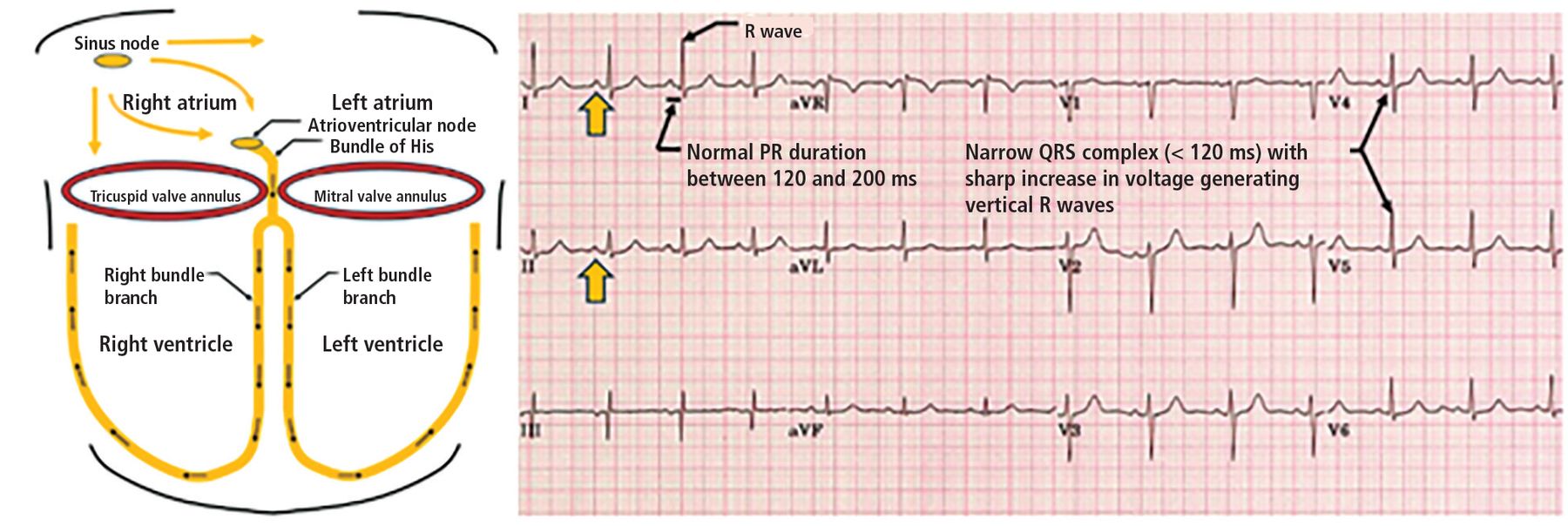
    Left, normal conduction pathway with normal sinus rhythm generated by the sinus node and conducted through the atrioventricular node, bundle of His, and subsequently through the left and right bundle branches, leading to normal PR duration (120–200 ms), normal QRS duration (< 120 ms), and no preexcitation. Right, normal electrocardiogram with clear upright P waves (yellow arrow), normal PR interval, and narrow QRS pattern.

  • Figure 2
    • Download figure
    • Open in new tab
    • Download powerpoint
    Figure 2

    Electrical conduction system with a left lateral accessory pathway. An accessory pathway provides an alternate atrioventricular (AV) conduction pathway, bypassing both the atrioventricular node and the His-Purkinje system, predisposing to malignant tachyarrhythmias. A left lateral accessory pathway leads to the characteristic type A Wolff-Parkinson-White pattern on electrocardiography (Figure 3).

    aAccessory pathways are capable of bidirectional flow, predisposing to retrograde conduction (conduction from ventricular to atrial tissue).

  • Figure 3
    • Download figure
    • Open in new tab
    • Download powerpoint
    Figure 3

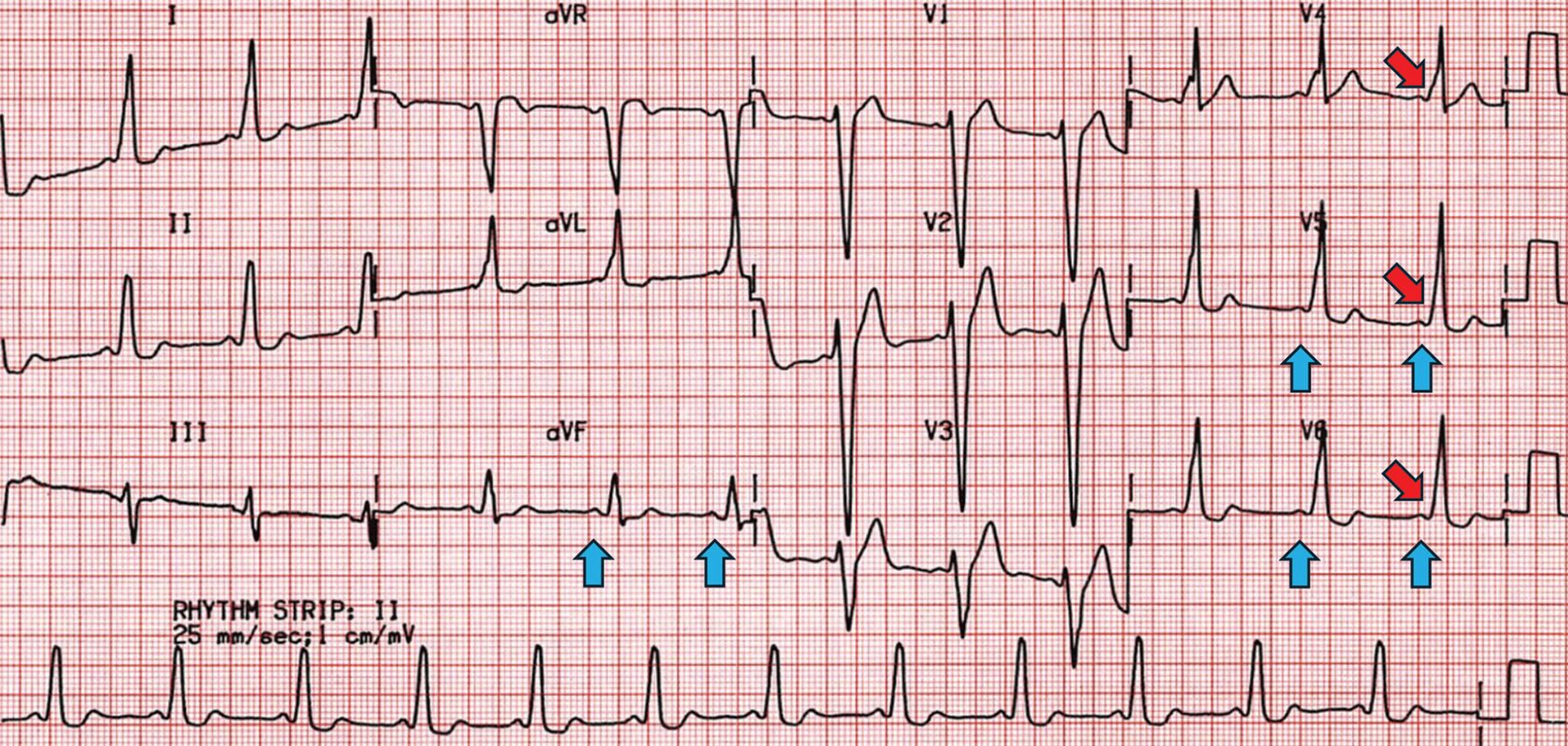
    Characteristic Wolff-Parkinson-White type A pattern, including a short PR interval (blue arrows), wide QRS, and delta waves (red arrows). Positive delta waves in V1–V3 suggest a left-sided accessory pathway (Figure 2).

  • Figure 4
    • Download figure
    • Open in new tab
    • Download powerpoint
    Figure 4

    Characteristic Wolff-Parkinson-White type B pattern, including a short PR interval (blue arrows), wide QRS, and delta waves (red arrows). Precordial transition after V2 suggests a right-sided accessory pathway.

  • Figure 5
    • Download figure
    • Open in new tab
    • Download powerpoint
    Figure 5

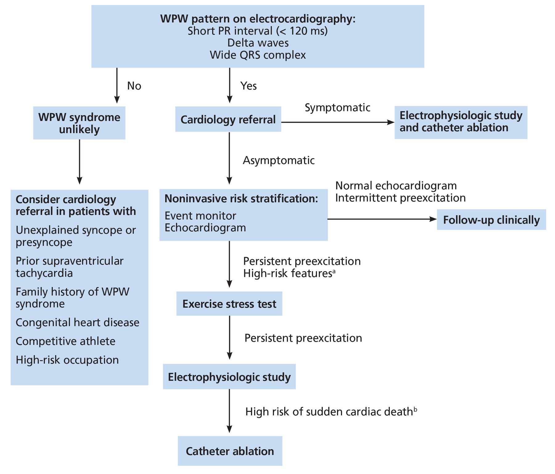
    Our proposed diagnostic and management guideline for patients with high clinical suspicion, Wolff-Parkinson-White (WPW) pattern, and WPW syndrome.

    aHigh-risk features: male sex, age less than 30, history of atrial fibrillation, family history of WPW syndrome, congenital heart disease, competitive athlete, high-risk occupation.

    bHigh risk of sudden cardiac death: multiple accessory pathways; preexcitation persists during induced atrial fibrillation; shortest RR interval < 250 ms during incremental atrial pacing, premature atrial contraction, or when in atrial fibrillation.

  • Figure 6
    • Download figure
    • Open in new tab
    • Download powerpoint
    Figure 6

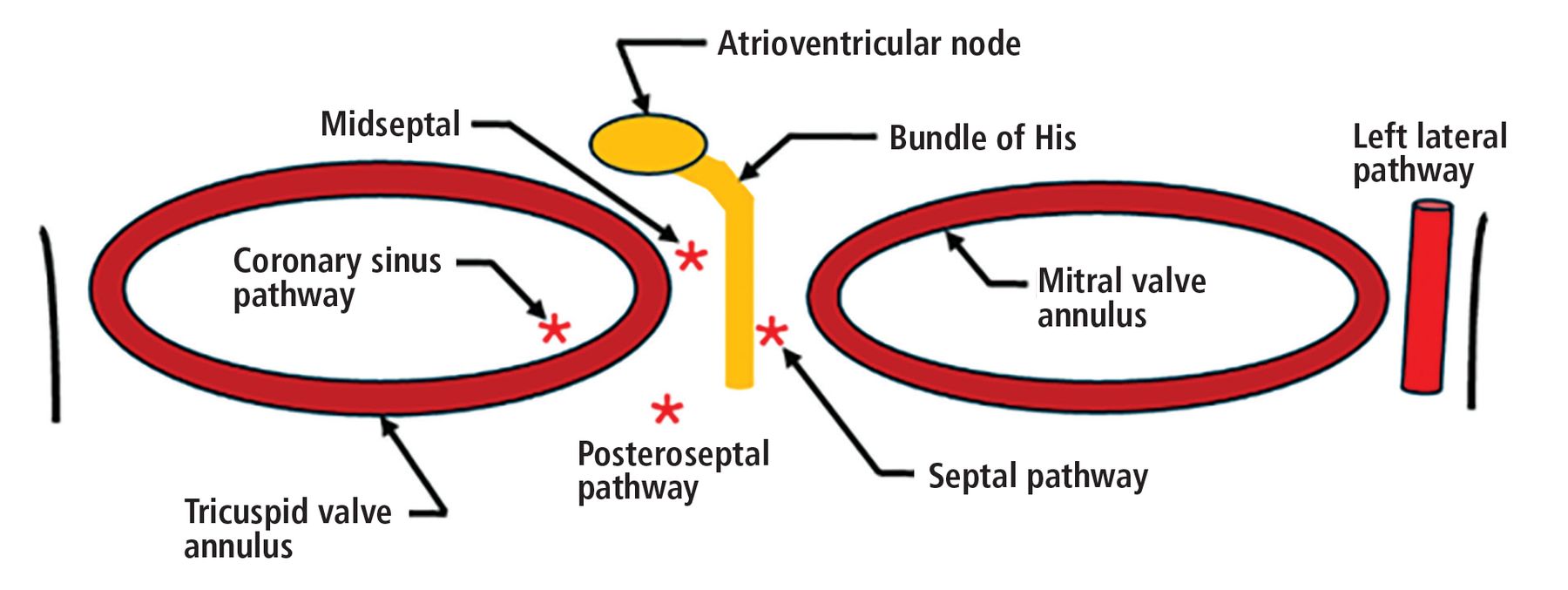
    The most reported accessory atrioventricular pathway location is the left lateral mitral valve annulus (30% to 58% of reported cases), followed by the posteroseptal region (about 25% of reported cases.25,26 The remainder of reported accessory pathway locations surround both the tricuspid annulus and the mitral valve annulus. Success rates of accessory pathway ablation vary by location, with highest success rates reported in those localized in the anterior, posterior, and lateral distribution (success rate of 90% and above), and lowest in those surrounding the coronary sinus (50%) and in the septal (50%) and midseptal distribution (73%).26
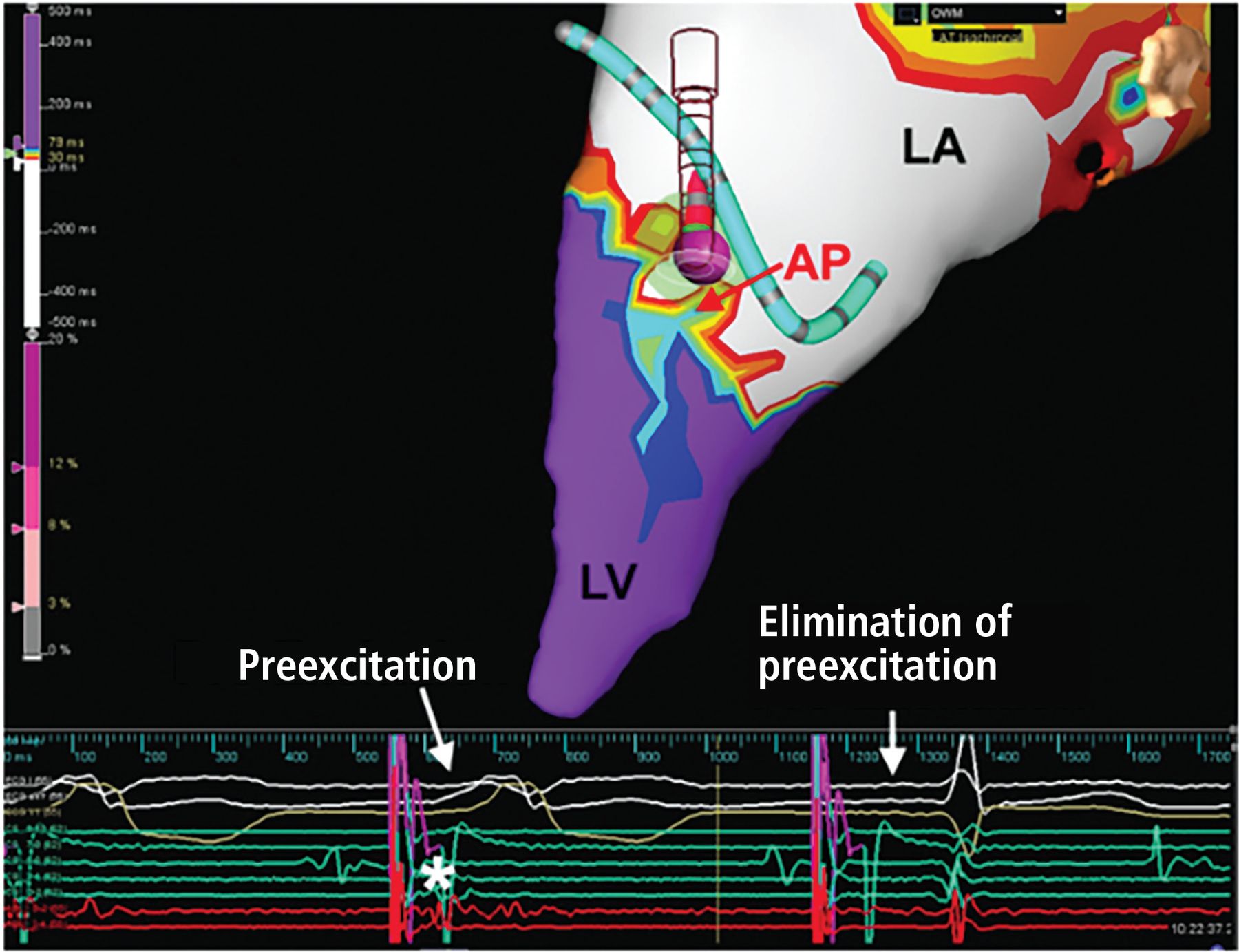
  • Figure 7
    • Download figure
    • Open in new tab
    • Download powerpoint
    Figure 7

    Catheter ablation of a left anterolateral accessory pathway in a 21-year-old patient. Open window mapping localized the accessory pathway (AP) to the anterolateral mitral annulus, where ablation (purple ball) eliminated the accessory pathway, with surface (white) and intracardiac electrocardiograms (red) showing loss of preexcitation and Kent potential (*) from the first to the second beat.

    LA = left atrium; LV = left ventricle

Tables

  • Figures
    • View popup
    TABLE 1

    Type A vs type B Wolff-Parkinson-White patterns

    Type A (associated with left-sided accessory pathway)Type B (associated with right-sided accessory pathway)
    Short PR interval (< 120 ms)Short PR interval (< 120 ms)
    Delta wave: broad QRS complex with slurred R waveDelta wave: broad QRS complex with slurred S wave
    May mimic right bundle branch blockMay mimic left bundle branch block
    Dominant R wave in V1Dominant S wave in V1
    Tall R waves and inverted T waves in leads V1–V3Tall S waves and inverted T waves in leads V4–V6
    • Based on information from reference 1.

PreviousNext
Back to top

In this issue

Cleveland Clinic Journal of Medicine: 92 (2)
Cleveland Clinic Journal of Medicine
Vol. 92, Issue 2
1 Feb 2025
  • Table of Contents
  • Table of Contents (PDF)
  • Index by author
  • Complete Issue (PDF)
Print
Download PDF
Article Alerts
Sign In to Email Alerts with your Email Address
Email Article

Thank you for your interest in spreading the word on Cleveland Clinic Journal of Medicine.

NOTE: We only request your email address so that the person you are recommending the page to knows that you wanted them to see it, and that it is not junk mail. We do not capture any email address.

Enter multiple addresses on separate lines or separate them with commas.
Wolff-Parkinson-White syndrome: Diagnostic and management strategies
(Your Name) has sent you a message from Cleveland Clinic Journal of Medicine
(Your Name) thought you would like to see the Cleveland Clinic Journal of Medicine web site.
CAPTCHA
This question is for testing whether or not you are a human visitor and to prevent automated spam submissions.
Citation Tools
Wolff-Parkinson-White syndrome: Diagnostic and management strategies
Masihullah Barat, David Torres Barba, Gordon Ho
Cleveland Clinic Journal of Medicine Feb 2025, 92 (2) 119-127; DOI: 10.3949/ccjm.92a.24059

Citation Manager Formats

  • BibTeX
  • Bookends
  • EasyBib
  • EndNote (tagged)
  • EndNote 8 (xml)
  • Medlars
  • Mendeley
  • Papers
  • RefWorks Tagged
  • Ref Manager
  • RIS
  • Zotero
Share
Wolff-Parkinson-White syndrome: Diagnostic and management strategies
Masihullah Barat, David Torres Barba, Gordon Ho
Cleveland Clinic Journal of Medicine Feb 2025, 92 (2) 119-127; DOI: 10.3949/ccjm.92a.24059
Twitter logo Facebook logo Mendeley logo
  • Tweet Widget
  • Facebook Like
  • Linkedin Share Button

Jump to section

  • Article
    • ABSTRACT
    • ACCESSORY PATHWAYS CAN LEAD TO MALIGNANT ARRHYTHMIAS
    • DIAGNOSIS: SYMPTOMS PLUS ELECTROCARDIOGRAPHIC SIGNS
    • WOLFF-PARKINSON-WHITE SYNDROME IN THE GENERAL POPULATION
    • MANAGEMENT STRATEGIES
    • DISCLOSURES
    • REFERENCES
  • Figures & Data
  • Info & Metrics
  • PDF

Related Articles

  • No related articles found.
  • PubMed
  • Google Scholar

Cited By...

  • No citing articles found.
  • Google Scholar

More in this TOC Section

  • High-output heart failure from arteriovenous dialysis access: A structured approach to diagnosis and management
  • Direct oral anticoagulants: Challenging prescribing scenarios in everyday practice
  • IgA nephropathy: Update on pathogenesis and treatment
Show more Review

Similar Articles

Subjects

  • Cardiology

Navigate

  • Current Issue
  • Past Issues
  • Supplements
  • Article Type
  • Specialty
  • CME/MOC Articles
  • CME/MOC Calendar
  • Media Kit

Authors & Reviewers

  • Manuscript Submission
  • Authors & Reviewers
  • Subscriptions
  • About CCJM
  • Contact Us
  • Cleveland Clinic Center for Continuing Education
  • Consult QD

Share your suggestions!

Copyright © 2025 The Cleveland Clinic Foundation. All rights reserved. The information provided is for educational purposes only. Use of this website is subject to the website terms of use and privacy policy. 

Powered by HighWire