
Main menu

  • Home
  • Content
    • Current Issue
    • Ahead of Print
    • Past Issues
    • Supplements
    • Article Type
  • Specialty
    • Articles by Specialty
  • CME/MOC
    • Articles
    • Calendar
  • Info For
    • Manuscript Submission
    • Authors & Reviewers
    • Subscriptions
    • About CCJM
    • Contact Us
    • Media Kit
  • Conversations with Leaders
  • Conference Coverage
    • Kidney Week 2024
    • CHEST 2024
    • ACR Convergence 2023
    • Kidney Week 2023
    • ObesityWeek 2023
    • IDWeek 2023
    • CHEST 2023
    • MDS 2023
    • IAS 2023
    • ACP 2023
    • AAN 2023
    • ACC / WCC 2023
    • AAAAI Meeting 2023
    • ACR Convergence 2022
    • Kidney Week 2022
    • AIDS 2022
  • Other Publications
    • www.clevelandclinic.org

User menu

  • Register
  • Log in
  • Log out

Search

  • Advanced search
Cleveland Clinic Journal of Medicine
  • Other Publications
    • www.clevelandclinic.org
  • Register
  • Log in
  • Log out
Cleveland Clinic Journal of Medicine

Advanced Search

  • Home
  • Content
    • Current Issue
    • Ahead of Print
    • Past Issues
    • Supplements
    • Article Type
  • Specialty
    • Articles by Specialty
  • CME/MOC
    • Articles
    • Calendar
  • Info For
    • Manuscript Submission
    • Authors & Reviewers
    • Subscriptions
    • About CCJM
    • Contact Us
    • Media Kit
  • Conversations with Leaders
  • Conference Coverage
    • Kidney Week 2024
    • CHEST 2024
    • ACR Convergence 2023
    • Kidney Week 2023
    • ObesityWeek 2023
    • IDWeek 2023
    • CHEST 2023
    • MDS 2023
    • IAS 2023
    • ACP 2023
    • AAN 2023
    • ACC / WCC 2023
    • AAAAI Meeting 2023
    • ACR Convergence 2022
    • Kidney Week 2022
    • AIDS 2022
The Clinical Picture

Not all wheezing is COPD

Vipul Patel, MD and Nikhil Madan, MD, FCCP
Cleveland Clinic Journal of Medicine March 2021, 88 (3) 147-149; DOI: https://doi.org/10.3949/ccjm.88a.20101
Vipul Patel
Department of Medicine, Division of Pulmonary and Critical Care Medicine, Newark Beth Israel Medical Center, Newark, NJ
  • Find this author on Google Scholar
  • Find this author on PubMed
  • Search for this author on this site
  • For correspondence: [email protected]
Nikhil Madan
Department of Medicine, Division of Pulmonary and Critical Care Medicine, Newark Beth Israel Medical Center, Newark, NJ
  • Find this author on Google Scholar
  • Find this author on PubMed
  • Search for this author on this site
  • Article
  • Figures & Data
  • Info & Metrics
  • PDF
Loading

A 53-year-old woman came to us because of shortness of breath. The problem had started about 3 years earlier, had slowly gotten worse, and was now limiting her activities of daily living. It was associated with wheezing, and her primary care physician had diagnosed it as asthma. Since that time, she had been hospitalized at least twice with “asthma exacerbations,” which were treated with systemic corticosteroids and intravenous antibiotics.

See related editorial, page 150

She had then been referred to a pulmonary physician for spirometry, which showed severe obstruction that did not reverse with bronchodilators. Her condition was diagnosed as chronic obstructive pulmonary disease (COPD) and was treated with montelukast and the combination of inhaled budesonine and formoterol at home.

These drugs did not relieve the symptoms, and she continued to have shortness of breath and wheezing, mainly on exertion. Her BODE index, which is used to predict mortality in COPD, was 5. BODE is an acronym that stands for body mass index, airflow obstruction [forced expiratory volume in 1 second (FEV1)], dyspnea, and exercise [6-minute walk distance]. The minimum score is 0 and the maximum score is 10. Lower scores are better than higher: a score of 5 indicates more than a 50% chance of death in 4 years.

The patient never smoked or used illicit drugs. She worked as a teacher most of her life and reported no environmental exposure to fumes or dust. She had never undergone endotracheal intubation. She was referred to our clinic for evaluation for a lung transplant.

PHYSICAL EXAMINATION AND WORKUP

The patient weighed 52 kg and her height was 155 cm. Her blood pressure was 132/84 mm Hg, heart rate 82 beats per minute, and respiratory rate 18 breaths per minute. General inspiratory and expiratory stridor sounds were detected in both lung fields and in the anterior neck area. The rest of the systemic examination was unremarkable.

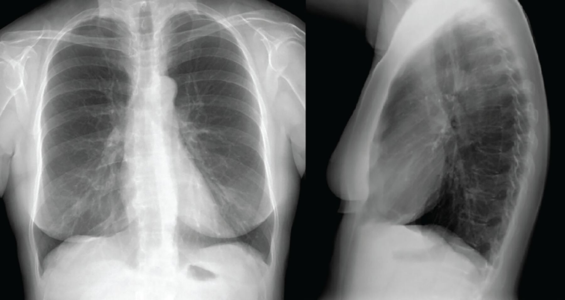
Chest radiography (Figure 1) and computed tomography of the chest were normal.

Figure 1
  • Download figure
  • Open in new tab
  • Download powerpoint
Figure 1

Chest radiography, posteroanterior and lateral views, showed normal findings.

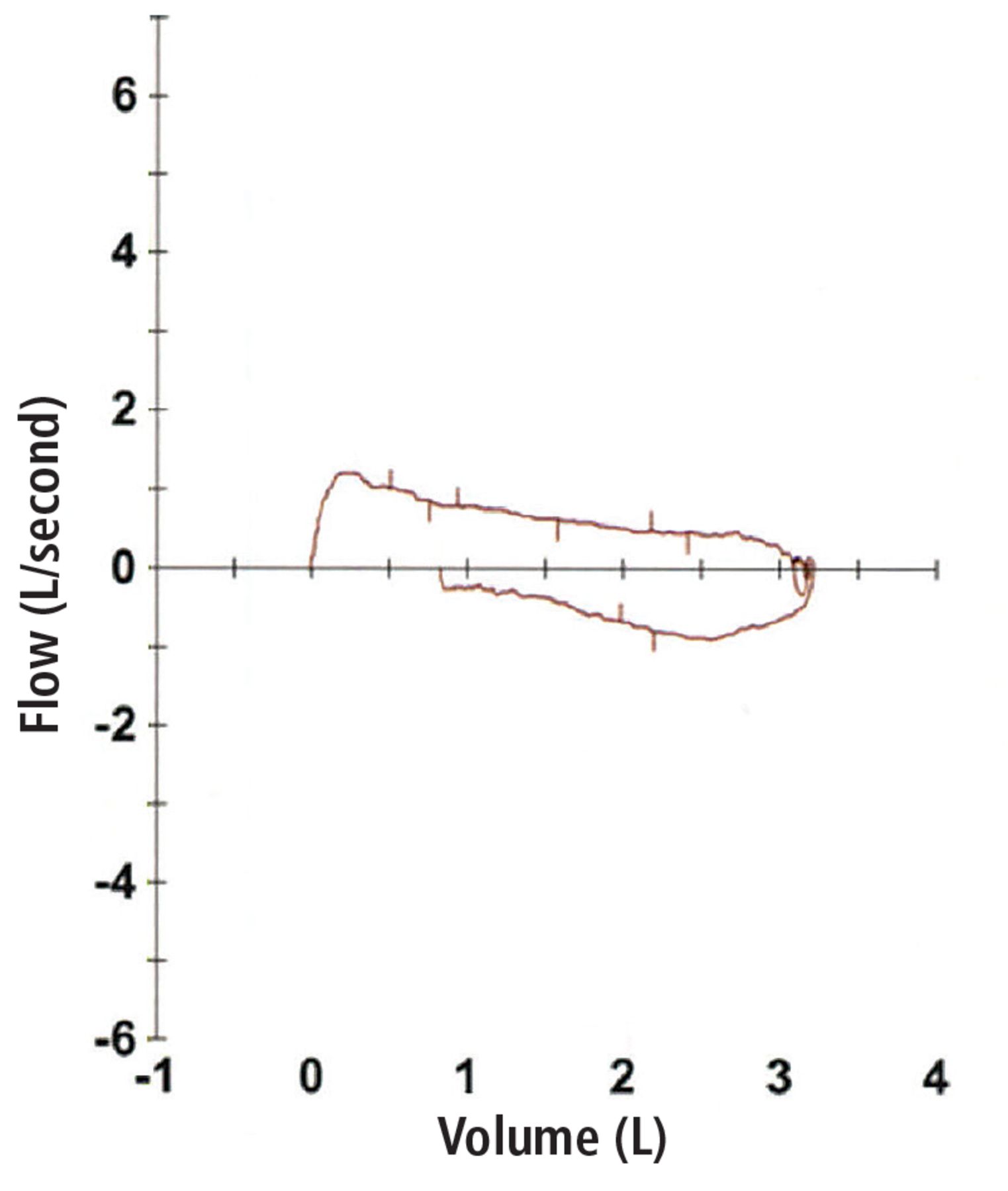
Spirometry showed severe obstructive pulmonary disease, and her forced vital capacity and FEV1 did not change when she was given a bronchodilator. Lung volume showed no evidence of air trapping or hyperinflation (Table 1). A flow-volume loop showed obstruction during inspiration and exhalation, suggesting a fixed extrathoracic airway obstruction (Figure 2).

View this table:
  • View inline
  • View popup
TABLE 1

Results of spirometry

Figure 2
  • Download figure
  • Open in new tab
  • Download powerpoint
Figure 2

Flow-volume loop showed a decrease in inspiratory and expiratory flow suggestive of fixed extrathoracic airway obstruction.

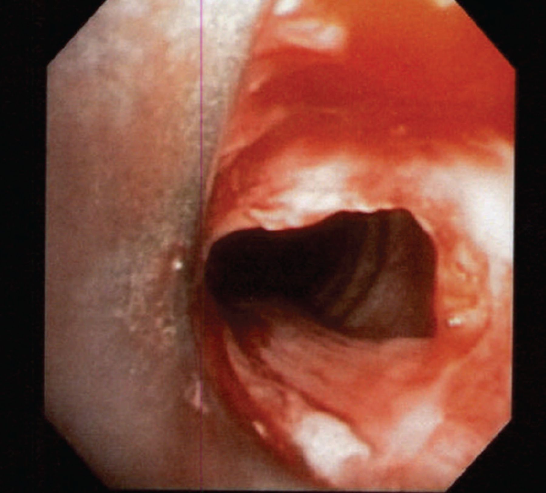
Flexible fiberoptic bronchoscopy was performed to evaluate the upper airway. The larynx was normal in shape, without laryngomalacia, and the vocal cords had synchronized movement with no abnormalities detected. However, an incomplete ring of tissue (web) was found about 1 cm below the vocal cords (Figure 3).

Figure 3
  • Download figure
  • Open in new tab
  • Download powerpoint
Figure 3

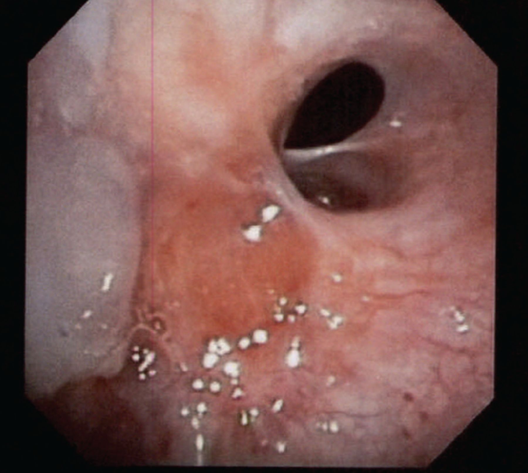
Tracheal web was seen below the vocal cords on flexible bronchoscopy.

The patient underwent resection of the tracheal web with endobronchial argon plasma coagulation. Afterward, bronchoscopy showed appropriate dilation of the tracheal stenosis (Figure 4), and her symptoms significantly improved. She was followed in the pulmonary clinic for 1 year, during which her symptoms completely resolved, and repeat pulmonary function testing showed normal findings.

Figure 4
  • Download figure
  • Open in new tab
  • Download powerpoint
Figure 4

Resection of tracheal web performed with successful endobronchial argon plasma coagulation dilation.

DISCUSSION

Tracheal web is a tissue layer covering the tracheal lumen. The web is usually incomplete and is not associated with a tracheal cartilage abnormality or deformity.

Congenital tracheal web is a rare anomaly. In children, the incidence has been estimated at 1 in 10,000 births.1 In adults, a few cases have been found incidentally during planned endotracheal intubation.1–5 In addition, tracheal stenosis can be a late complication of endotracheal intubation or tracheostomy in adults, and so can tracheal web, although it is rare.6,7

Tracheal web is usually misdiagnosed as asthma or COPD.2,6,8 In our patient, the correct diagnosis was missed by multiple physicians over many years, leading to inappropriate referral for lung transplant.

Bronchoscopy combined with computed tomography reconstruction is a reliable and sensitive method of evaluating tracheal web in suspected cases.6,9 It is also important to examine the flow-volume loop obtained during spirometry. Sometimes a rare diagnosis like tracheal web can be easily identified, and appropriate management can be rendered.

DISCLOSURES

The authors report no relevant financial relationships which, in the context of their contributions, could be perceived as a potential conflict of interest.

  • Copyright © 2021 The Cleveland Clinic Foundation. All Rights Reserved.

REFERENCES

  1. ↵
    1. Nguyen NK
    . Unexpected tracheal web encountered during difficult intubation in the operating room. Proc (Bayl Univ Med Cent) 2006; 19(3):224–225. doi:10.1080/08998280.2006.11928167
    OpenUrlCrossRefPubMed
  2. ↵
    1. Nakamura Y,
    2. Kurasako N,
    3. Iwaki T, et al
    . Unexpected difficult intubation caused by a laryngeal web. Masui 2000; 49(11):1278–1280. Japanese. pmid:11215243
    OpenUrlPubMed
    1. Yin Y,
    2. Zhang L
    . Successful diagnosis and treatment of congenital tracheal web using a fiberoptic bronchoscope. Pediatr Pulmonol 2010; 45(9):945–947. doi:10.1002/ppul.21235
    OpenUrlCrossRefPubMed
    1. Virmani S,
    2. Uppal R,
    3. Kiro K, et al
    . Successful double lumen tube insertion in a patient with unanticipated laryngeal and tracheal web. Ann Card Anaesth 2011; 14(3):211–213. doi:10.4103/0971-9784.84022
    OpenUrlCrossRefPubMed
  3. ↵
    1. Yamamoto S,
    2. Tetsuka K,
    3. Sato Y,
    4. Endo S
    . Unsuspected tracheal web inhibits endotracheal intubation: report of a case. J Anesth 2010; 24(1):132–133. doi:10.1007/s00540-009-0844-2
    OpenUrlCrossRefPubMed
  4. ↵
    1. Ozdulger A,
    2. Birbicer H,
    3. Duce MN
    . Tracheal web: presentation of a case with uncommon features. J Bronchology Interv Pulmonol 2009; 16(1):46–48. doi:10.1097/LBR.0b013e3181908c5d
    OpenUrlCrossRefPubMed
  5. ↵
    1. Kokkonouzis I,
    2. Mermigkis C,
    3. Psathakis K,
    4. Tsintiris K
    . Postintubation tracheal web. J Bronchol 2005; 12(4):271–272. doi:10.1097/01.laboratory.0000184836.74684.6d
    OpenUrlCrossRef
  6. ↵
    1. Lee JE,
    2. Chang MY,
    3. Kim KH,
    4. Jung YH
    . Post-intubation tracheoesophageal fistula with posterior glottic web. Clin Exp Otorhinolaryngol 2011; 4(2):105–108. doi:10.3342/ceo.2011.4.2.105
    OpenUrlCrossRefPubMed
  7. ↵
    1. Legasto AC,
    2. Haller JO,
    3. Giusti RJ
    . Tracheal web. Pediatr Radiol 2004; 34(3):256–258. doi:10.1007/s00247-003-1030-6
    OpenUrlCrossRefPubMed
PreviousNext
Back to top

In this issue

Cleveland Clinic Journal of Medicine: 88 (3)
Cleveland Clinic Journal of Medicine
Vol. 88, Issue 3
1 Mar 2021
  • Table of Contents
  • Table of Contents (PDF)
  • Index by author
  • Complete Issue (PDF)
Print
Download PDF
Article Alerts
Sign In to Email Alerts with your Email Address
Email Article

Thank you for your interest in spreading the word on Cleveland Clinic Journal of Medicine.

NOTE: We only request your email address so that the person you are recommending the page to knows that you wanted them to see it, and that it is not junk mail. We do not capture any email address.

Enter multiple addresses on separate lines or separate them with commas.
Not all wheezing is COPD
(Your Name) has sent you a message from Cleveland Clinic Journal of Medicine
(Your Name) thought you would like to see the Cleveland Clinic Journal of Medicine web site.
CAPTCHA
This question is for testing whether or not you are a human visitor and to prevent automated spam submissions.
Citation Tools
Not all wheezing is COPD
Vipul Patel, Nikhil Madan
Cleveland Clinic Journal of Medicine Mar 2021, 88 (3) 147-149; DOI: 10.3949/ccjm.88a.20101

Citation Manager Formats

  • BibTeX
  • Bookends
  • EasyBib
  • EndNote (tagged)
  • EndNote 8 (xml)
  • Medlars
  • Mendeley
  • Papers
  • RefWorks Tagged
  • Ref Manager
  • RIS
  • Zotero
Share
Not all wheezing is COPD
Vipul Patel, Nikhil Madan
Cleveland Clinic Journal of Medicine Mar 2021, 88 (3) 147-149; DOI: 10.3949/ccjm.88a.20101
Twitter logo Facebook logo Mendeley logo
  • Tweet Widget
  • Facebook Like
  • Linkedin Share Button

Jump to section

  • Article
    • PHYSICAL EXAMINATION AND WORKUP
    • DISCUSSION
    • DISCLOSURES
    • REFERENCES
  • Figures & Data
  • Info & Metrics
  • PDF

Related Articles

  • All that wheezes…
  • PubMed
  • Google Scholar

Cited By...

  • All that wheezes...
  • Google Scholar

More in this TOC Section

  • Tinea incognito
  • Prolonged venous filling time and dependent rubor in a patient with peripheral artery disease
  • Sarcoidosis with diffuse purplish erythematous plaques on the hands
Show more The Clinical Picture

Similar Articles

Subjects

  • Imaging
  • Pulmonology
  • Women's Health

Navigate

  • Current Issue
  • Past Issues
  • Supplements
  • Article Type
  • Specialty
  • CME/MOC Articles
  • CME/MOC Calendar
  • Media Kit

Authors & Reviewers

  • Manuscript Submission
  • Authors & Reviewers
  • Subscriptions
  • About CCJM
  • Contact Us
  • Cleveland Clinic Center for Continuing Education
  • Consult QD

Share your suggestions!

Copyright © 2025 The Cleveland Clinic Foundation. All rights reserved. The information provided is for educational purposes only. Use of this website is subject to the website terms of use and privacy policy. 

Powered by HighWire