


A 44-year-old male was brought to the emergency department after suffering a seizure while undergoing alcohol detoxification. His medical history was notable for liver cirrhosis secondary to excess alcohol consumption and hepatitis C infection.
The physical examination revealed signs of liver cirrhosis including palmar erythema, hepatomegaly, gynecomastia, paucity of chest hair, and red lesions on his arms, back, chest, and neck (Figure 1). The lesions blanched when pressure was applied centrally, and when pressure was released, they refilled from the center outward (Video 1).
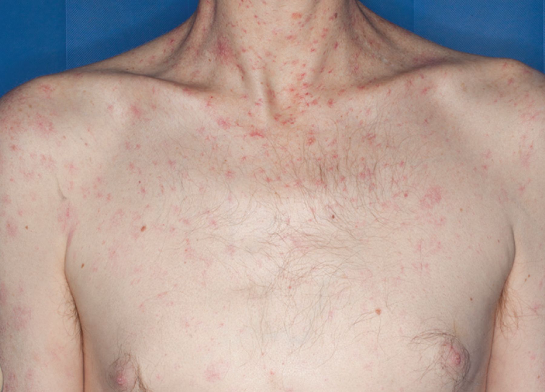
The patient presented with spider nevi on the chest, neck, and upper arms, as well as gynecomastia and paucity of chest hair.
Video 1. Spider nevus. When pressure is applied to the central arteriole, the whole lesion blanches. When pressure is released, the “legs” of the nevus refill from the center outward.
PATHOPHYSIOLOGY OF SPIDER NEVI
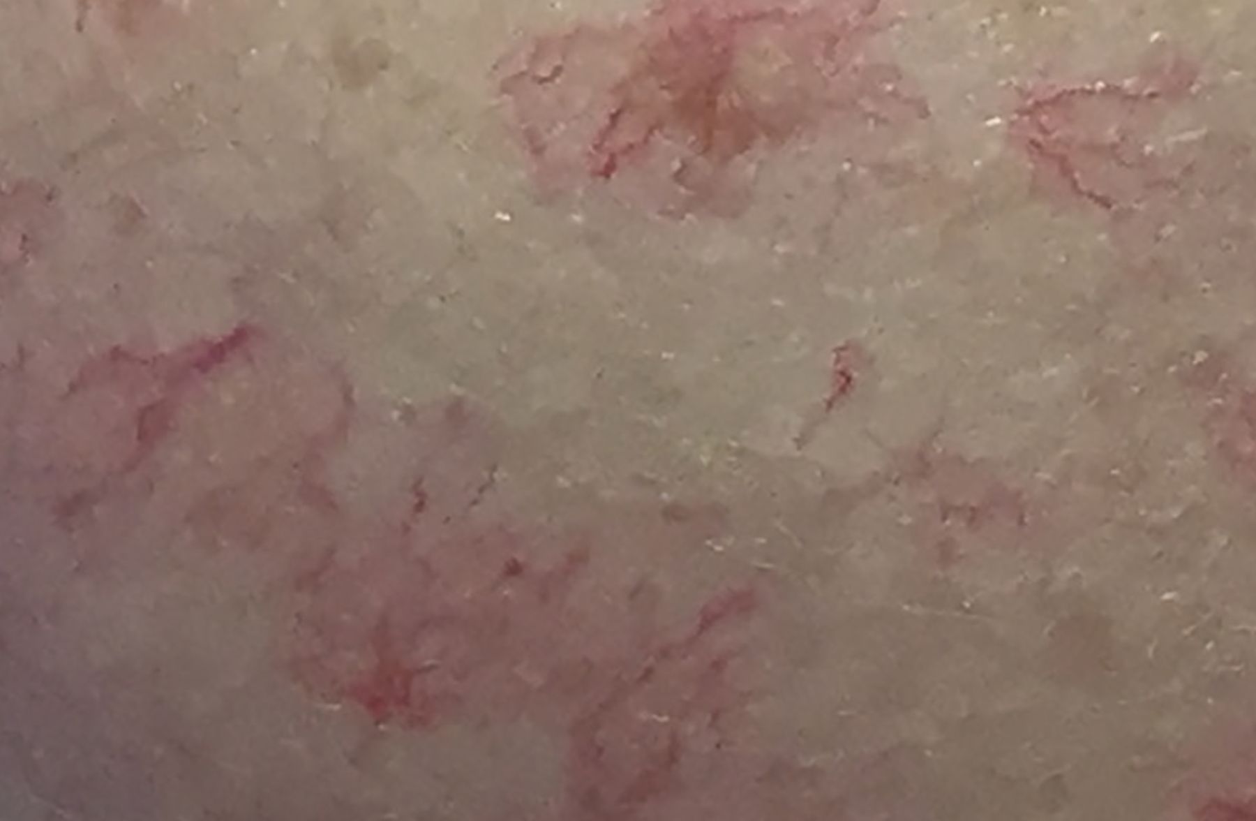
Spider nevi are the ends of arterioles from which capillaries radiate outward, resembling the legs of a spider (Figure 2). Patients with chronic liver disease from alcohol consumption commonly have many spider nevi. The nevi are present in up to one-third of patients with cirrhosis, and increasing numbers of lesions correlate with the severity of liver disease and the presence of esophageal and gastric varices.1,2

A close-up view of the spider nevi shows a central arteriole with vessels growing outward, resembling the legs of a spider.
As in this patient, the nevi are ordinarily distributed in blood vessels supplied by the superior vena cava (eg, in the face, neck, chest, and arms). They may also be found in patients with increased circulating estrogen, such as during pregnancy or when taking hormonal contraceptives. Occasionally, they can be seen in healthy patients with no underlying cause or pathology, and up to 3 lesions is considered normal.
Spider nevi occur due to the failure of sphincteric muscle surrounding a cutaneous arteriole.3 The mechanism of development is unclear, but they are thought to be caused by excess circulating estrogen due to pregnancy or insufficient hepatic metabolism, by inadequate metabolism of steroid hormones, by neovascularization from angiogenic growth factors, or by direct vasodilatory effects of alcohol.4
MANAGEMENT OF THIS PATIENT
The patient’s blood test results were within normal limits. He was admitted for alcohol detoxification regimen consisting of chlordiazepoxide and tapering doses of lorazepam, titrated to Clinical Institute Withdrawal Assessment for Alcohol-Revised score. His neurologic status improved over the course of an 8-day admission. He was discharged with follow-up with community-based alcohol services.
DISCLOSURES
The author reports no relevant financial relationships which, in the context of his contributions, could be perceived as a potential conflict of interest.
- Copyright © 2022 The Cleveland Clinic Foundation. All Rights Reserved.
In this issue


Jump to section
Related Articles
Cited By...
- No citing articles found.