
Main menu

  • Home
  • Content
    • Current Issue
    • Ahead of Print
    • Past Issues
    • Supplements
    • Article Type
  • Specialty
    • Articles by Specialty
  • CME/MOC
    • Articles
    • Calendar
  • Info For
    • Manuscript Submission
    • Authors & Reviewers
    • Subscriptions
    • About CCJM
    • Contact Us
    • Media Kit
  • Conversations with Leaders
  • Conference Coverage
    • Kidney Week 2024
    • CHEST 2024
    • ACR Convergence 2023
    • Kidney Week 2023
    • ObesityWeek 2023
    • IDWeek 2023
    • CHEST 2023
    • MDS 2023
    • IAS 2023
    • ACP 2023
    • AAN 2023
    • ACC / WCC 2023
    • AAAAI Meeting 2023
    • ACR Convergence 2022
    • Kidney Week 2022
    • AIDS 2022
  • Other Publications
    • www.clevelandclinic.org

User menu

  • Register
  • Log in

Search

  • Advanced search
Cleveland Clinic Journal of Medicine
  • Other Publications
    • www.clevelandclinic.org
  • Register
  • Log in
Cleveland Clinic Journal of Medicine

Advanced Search

  • Home
  • Content
    • Current Issue
    • Ahead of Print
    • Past Issues
    • Supplements
    • Article Type
  • Specialty
    • Articles by Specialty
  • CME/MOC
    • Articles
    • Calendar
  • Info For
    • Manuscript Submission
    • Authors & Reviewers
    • Subscriptions
    • About CCJM
    • Contact Us
    • Media Kit
  • Conversations with Leaders
  • Conference Coverage
    • Kidney Week 2024
    • CHEST 2024
    • ACR Convergence 2023
    • Kidney Week 2023
    • ObesityWeek 2023
    • IDWeek 2023
    • CHEST 2023
    • MDS 2023
    • IAS 2023
    • ACP 2023
    • AAN 2023
    • ACC / WCC 2023
    • AAAAI Meeting 2023
    • ACR Convergence 2022
    • Kidney Week 2022
    • AIDS 2022
The Clinical Picture

Ring-enhancing cerebral lesions

Aram Barbaryan, MD, Jignesh Modi, MD, Wajih Raqeem, MD, Michael I. Choi, MD, Alan Frigy, MD and Aibek E. Mirrakhimov, MD
Cleveland Clinic Journal of Medicine February 2017, 84 (2) 104-110; DOI: https://doi.org/10.3949/ccjm.84a.16037
Aram Barbaryan
Department of Medicine, HSHS Saint Mary’s Hospital, Decatur, IL
  • Find this author on Google Scholar
  • Find this author on PubMed
  • Search for this author on this site
  • For correspondence: [email protected]
Jignesh Modi
Department of Infectious Disease, HSHS Saint Mary’s Hospital, Decatur, IL
  • Find this author on Google Scholar
  • Find this author on PubMed
  • Search for this author on this site
Wajih Raqeem
Department of Medicine, HSHS Saint Mary’s Hospital, Decatur, IL
  • Find this author on Google Scholar
  • Find this author on PubMed
  • Search for this author on this site
Michael I. Choi
Department of Pathology, HSHS Saint Mary’s Hospital, Decatur, IL
  • Find this author on Google Scholar
  • Find this author on PubMed
  • Search for this author on this site
Alan Frigy
Department of Pathology, HSHS Saint Mary’s Hospital, Decatur, IL
  • Find this author on Google Scholar
  • Find this author on PubMed
  • Search for this author on this site
Aibek E. Mirrakhimov
Department of Medicine, University of Kentucky School of Medicine, Lexington, KY
  • Find this author on Google Scholar
  • Find this author on PubMed
  • Search for this author on this site
  • Article
  • Figures & Data
  • Info & Metrics
  • PDF
Loading

A 39-year-old woman with a history of human immunodeficiency virus (HIV) and hepatitis B virus infection was brought to the emergency department for evaluation of seizures, which had started a few days earlier. She was born and raised in a state bordering the Ohio River, an area where Histoplasma capsulatum is endemic. She denied any recent travel.

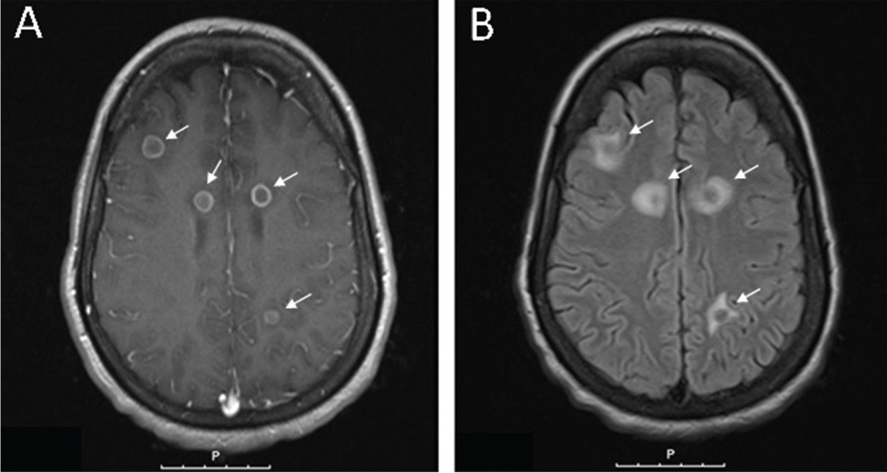
Her vital signs and neurologic examination were normal. Computed tomography of the head showed two areas of increased attenuation anterior to the frontal horns. To better characterize those lesions, magnetic resonance imaging (MRI) with contrast was done, which showed about a dozen 1-cm ring-enhancing lesions in the right cerebellum and both cerebral hemispheres (Figure 1).

FIGURE 1
  • Download figure
  • Open in new tab
  • Download powerpoint
FIGURE 1

(A) Axial contrast-enhanced T1-weighted magnetic resonance imaging showed ring-enhancing lesions (white arrows), while (B) axial T2-weighted images showed ring-enhancing lesions surrounding hyperintensity, consistent with vasogenic edema (white arrows).

Results of a complete blood cell count, metabolic profile, and chest radiography were normal. Her CD4 count was 428/μL (reference range 533–1,674) and 20% (60%–89%); her HIV viral load was 326,000 copies/mL.

She was initially treated empirically with sulfadiazine, pyrimethamine, and leukovorin for possible toxoplasmosis, which is the most common cause of ring-enhancing brain lesions in HIV patients. In the meantime, cerebrospinal fluid, blood, and urine were sent for a detailed workup for fungi, including Histoplasma. Results of the Histoplasma antibody and antigen studies of the serum, urine, and cerebrospinal fluid were positive, while cerebrospinal fluid testing for Toxoplasma by polymerase chain reaction testing was negative. Empirical treatment for toxoplasmosis was stopped and amphotericin B was started to treat disseminated histoplasmosis.

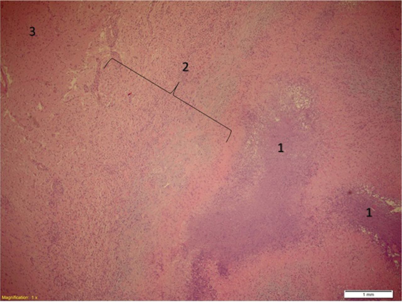
During her hospital course, she underwent brain biopsy via right frontotemporal craniotomy with resection of right frontal lesions. Pathologic study showed partially organizing abscesses with central necrosis (Figure 2), microscopy with Grocott-Gomori methenamine silver stain was positive for budding yeast forms consistent with H capsulatum (Figure 3), and special stain for acid-fast bacilli was negative for mycobacteria. Cultures of the brain biopsy specimen, blood, and cerebrospinal fluid for fungi, acid-fast bacilli, and bacteria did not reveal any growth after 28 days.

FIGURE 2
  • Download figure
  • Open in new tab
  • Download powerpoint
FIGURE 2

Partially organizing central nervous system abscess showing necrosis with acute inflammatory cells (1), fibrosis with acute and chronic inflammatory cells (2), and the normal-appearing brain tissue (3) (hematoxylin and eosin, × 4).

FIGURE 3
  • Download figure
  • Open in new tab
  • Download powerpoint
FIGURE 3

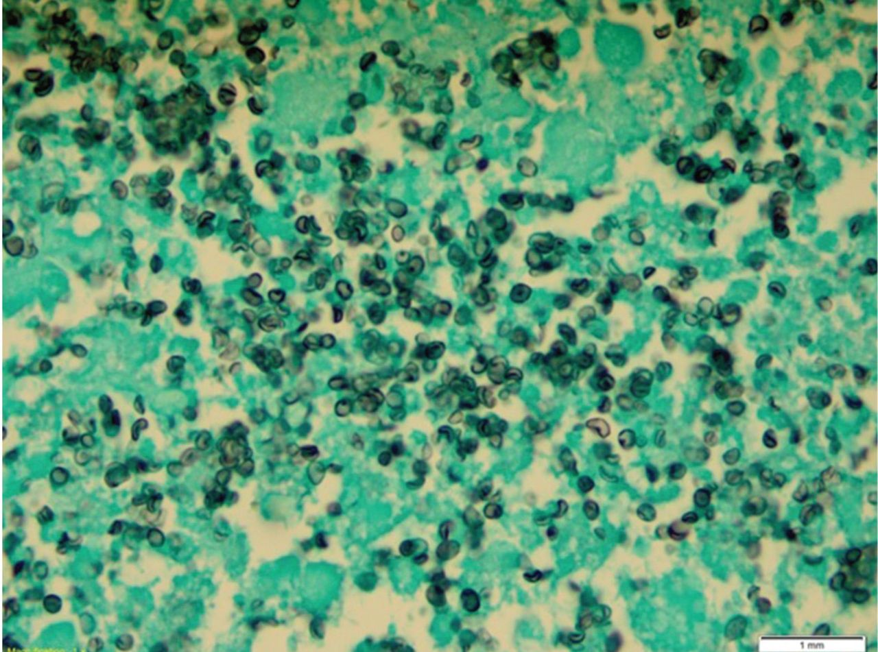
Grocott-Gomori methenamine silver staining of a biopsy specimen of a right frontal brain lesion showed budding yeast forms, consistent with Histoplasma capsulatum (× 100).

The patient was discharged home with instructions to take amphotericin B for a total of 6 weeks and then itraconazole. About 1 year later, she remained free of symptoms, although repeat MRI did not show any significant change in the size or number of histoplasmomas.

She did not comply well with her HIV treatment, and her immune status did not improve, so we decided to continue her itraconazole treatment for more than 1 year.

CEREBRAL HISTOPLASMOMA

The term “histoplasmoma” was introduced by Shapiro et al1 in 1955, when they first described numerous focal areas of softening, up to 1 cm in diameter, scattered throughout the brain at autopsy in a 41-year-old man who had died of disseminated histoplasmosis. They coined the word to describe these discrete areas of necrosis that might resemble tumors on the basis of their size, location, and capability of causing increased intracranial pressure.

Central nervous system involvement can either be a manifestation of disseminated disease or present as an isolated illness.2 It occurs in 5% to 10% of cases of disseminated histo-plasmosis.3 Histoplasmosis of the central nervous system can have different manifestations; the most common presentation is chronic meningitis.4

Laboratory diagnosis is based on detecting H capsulatum antigen and antibody in the urine, blood, and cerebrospinal fluid. Tissue biopsy (histopathology) as well as cultures of tissue samples or body fluids may also establish the diagnosis.4

Toxoplasmosis and primary central nervous system lymphoma are the most common causes of brain ring-enhancing lesions in HIV patients in developed countries, while in the developing world neurocysticercosis and tuberculomas are more common.5,6 Much less common causes include brain abscesses secondary to bacterial infections (pyogenic abscess),7 cryptococcomas,8 syphilitic cerebral gummata,9 primary brain tumors (gliomas), and metastases.10

Compared with other forms of the disease, histoplasmosis of the central nervous system has higher rates of treatment failure and relapse, so treatment should be prolonged and aggressive.2,3 The cure rate with amphotericin B ranges from 33% to 61%, and higher doses produce better response rates.3

Current treatment recommendations are based on 2007 guidelines of the Infectious Diseases Society of America.11 Liposomal amphotericin B is the drug of choice because it achieves higher concentrations in the central nervous system than other drugs and is less toxic. It is given for 4 to 6 weeks, followed by itraconazole for at least 1 year and until the cerebrospinal fluid Histoplasma antigen test is negative and other cerebrospinal fluid abnormalities are resolved.

In patients who have primary disseminated histoplasmosis that includes the central nervous system, itraconazole can be given for more than 1 year or until immune recovery is achieved—or lifelong if necessary.2,12 Long-term suppressive antifungal therapy also should be considered in patients for whom appropriate initial therapy fails.2

Nephrotoxicity (acute kidney injury, hypokalemia, and hypomagnesemia), infusion-related drug reactions, and rash are among the well-described side effects of amphotericin B. Maintenance of intravascular volume and replacement of electrolytes should be an integral part of the amphotericin B treatment regimen.13

TAKE-AWAY POINTS

  • Histoplasmomas should be considered in the differential diagnosis of ring-enhancing lesions of the central nervous system, along with toxoplasmosis and primary central nervous system lymphoma. This will allow timely initiation of the diagnostic workup, avoiding unnecessary and potentially risky interventions and delays in starting targeted antifungal therapy.

  • There is no single gold standard test for central nervous system histoplasmosis. Rather, the final diagnosis is based on the combination of clinical, laboratory, and radiologic findings.

ACKNOWLEDGMENT

Library research assistance provided by HSHS St. John’s Hospital Health Sciences Library staff.

  • Copyright © 2017 The Cleveland Clinic Foundation. All Rights Reserved.

REFERENCES

  1. ↵
    1. Shapiro JL,
    2. Lux JJ,
    3. Sprofkin BE
    . Histoplasmosis of the central nervous system. Am J Pathol 1955; 31:319–335.
    OpenUrlPubMed
  2. ↵
    1. Wheat LJ,
    2. Musial CE,
    3. Jenny-Avital E
    . Diagnosis and management of central nervous system histoplasmosis. Clin Infect Dis 2005; 40:844–852.
    OpenUrlCrossRefPubMed
  3. ↵
    1. Wheat LJ,
    2. Batteiger BE,
    3. Sathapatayavongs B
    . Histoplasma capsulatum infections of the central nervous system: a clinical review. Medicine (Baltimore) 1990; 69:244–260.
    OpenUrlCrossRefPubMed
  4. ↵
    1. Kauffman CA
    . Histoplasmosis: a clinical and laboratory update. Clin Microbiol Rev 2007; 20:115–132.
    OpenUrlAbstract/FREE Full Text
  5. ↵
    1. Modi M,
    2. Mochan A,
    3. Modi G
    . Management of HIV-associated focal brain lesions in developing countries. QJM 2004; 97:413–421.
    OpenUrlCrossRefPubMed
  6. ↵
    1. Miller RF,
    2. Hall-Craggs MA,
    3. Costa DC,
    4. et al
    . Magnetic resonance imaging, thallium-201 SPET scanning, and laboratory analyses for discrimination of cerebral lymphoma and toxoplasmosis in AIDS. Sex Transm Infect 1998; 74:258–264.
    OpenUrlAbstract
  7. ↵
    1. Cohen WA
    . Intracranial bacterial infections in patients with AIDS. Neuroimaging Clin N Am 1997; 7:223–229.
    OpenUrlPubMed
  8. ↵
    1. Troncoso A,
    2. Fumagalli J,
    3. Shinzato R,
    4. Gulotta H,
    5. Toller M,
    6. Bava J
    . CNS cryptococcoma in an HIV-positive patient. J Int Assoc Physicians AIDS Care (Chic) 2002; 1:131–133.
    OpenUrlCrossRefPubMed
  9. ↵
    1. Land AM,
    2. Nelson GA,
    3. Bell SG,
    4. Denby KJ,
    5. Estrada CA,
    6. Willett LL
    . Widening the differential for brain masses in human immunodeficiency virus-positive patients: syphilitic cerebral gummata. Am J Med Sci 2013; 346:253–255.
    OpenUrl
  10. ↵
    1. Balsys R,
    2. Janousek JE,
    3. Batnitzky S,
    4. Templeton AW
    . Peripheral enhancement in computerized cranial tomography: a non-specific finding. Surg Neurol 1979; 11:207–216.
    OpenUrlPubMed
  11. ↵
    1. Wheat LJ,
    2. Freifeld AG,
    3. Kleiman MB,
    4. et al
    5. Infectious Diseases Society of America
    . Clinical practice guidelines for the management of patients with histoplasmosis: 2007 update by the Infectious Diseases Society of America. Clin Infect Dis 2007; 45:807–825.
    OpenUrlCrossRefPubMed
  12. ↵
    1. Wheat J,
    2. Hafner R,
    3. Wulfsohn M,
    4. et al
    5. National Institute of Allergy and Infectious Diseases Clinical Trials and Mycoses Study Group Collaborators
    . Prevention of relapse of histoplasmosis with itraconazole in patients with the acquired immunodeficiency syndrome. Ann Intern Med 1993; 118:610–616.
    OpenUrlCrossRefPubMed
  13. ↵
    1. Saccente M
    . Central nervous system histoplasmosis. Curr Treat Options Neurol 2008; 10:161–167.
    OpenUrlCrossRefPubMed
PreviousNext
Back to top

In this issue

Cleveland Clinic Journal of Medicine: 84 (2)
Cleveland Clinic Journal of Medicine
Vol. 84, Issue 2
1 Feb 2017
  • Table of Contents
  • Table of Contents (PDF)
  • Index by author
Print
Download PDF
Article Alerts
Sign In to Email Alerts with your Email Address
Email Article

Thank you for your interest in spreading the word on Cleveland Clinic Journal of Medicine.

NOTE: We only request your email address so that the person you are recommending the page to knows that you wanted them to see it, and that it is not junk mail. We do not capture any email address.

Enter multiple addresses on separate lines or separate them with commas.
Ring-enhancing cerebral lesions
(Your Name) has sent you a message from Cleveland Clinic Journal of Medicine
(Your Name) thought you would like to see the Cleveland Clinic Journal of Medicine web site.
CAPTCHA
This question is for testing whether or not you are a human visitor and to prevent automated spam submissions.
Citation Tools
Ring-enhancing cerebral lesions
Aram Barbaryan, Jignesh Modi, Wajih Raqeem, Michael I. Choi, Alan Frigy, Aibek E. Mirrakhimov
Cleveland Clinic Journal of Medicine Feb 2017, 84 (2) 104-110; DOI: 10.3949/ccjm.84a.16037

Citation Manager Formats

  • BibTeX
  • Bookends
  • EasyBib
  • EndNote (tagged)
  • EndNote 8 (xml)
  • Medlars
  • Mendeley
  • Papers
  • RefWorks Tagged
  • Ref Manager
  • RIS
  • Zotero
Share
Ring-enhancing cerebral lesions
Aram Barbaryan, Jignesh Modi, Wajih Raqeem, Michael I. Choi, Alan Frigy, Aibek E. Mirrakhimov
Cleveland Clinic Journal of Medicine Feb 2017, 84 (2) 104-110; DOI: 10.3949/ccjm.84a.16037
Twitter logo Facebook logo Mendeley logo
  • Tweet Widget
  • Facebook Like
  • Linkedin Share Button

Jump to section

  • Article
    • CEREBRAL HISTOPLASMOMA
    • TAKE-AWAY POINTS
    • ACKNOWLEDGMENT
    • REFERENCES
  • Figures & Data
  • Info & Metrics
  • PDF

Related Articles

  • No related articles found.
  • PubMed
  • Google Scholar

Cited By...

  • Fungal organisms in the brain (February 2017)
  • Google Scholar

More in this TOC Section

  • Tinea incognito
  • Prolonged venous filling time and dependent rubor in a patient with peripheral artery disease
  • Sarcoidosis with diffuse purplish erythematous plaques on the hands
Show more The Clinical Picture

Similar Articles

Subjects

  • Emergency Medicine
  • Hospital Medicine
  • Imaging
  • Infectious Diseases
  • Neurology

Navigate

  • Current Issue
  • Past Issues
  • Supplements
  • Article Type
  • Specialty
  • CME/MOC Articles
  • CME/MOC Calendar
  • Media Kit

Authors & Reviewers

  • Manuscript Submission
  • Authors & Reviewers
  • Subscriptions
  • About CCJM
  • Contact Us
  • Cleveland Clinic Center for Continuing Education
  • Consult QD

Share your suggestions!

Copyright © 2025 The Cleveland Clinic Foundation. All rights reserved. The information provided is for educational purposes only. Use of this website is subject to the website terms of use and privacy policy. 

Powered by HighWire