


Article Figures & Data
Figures
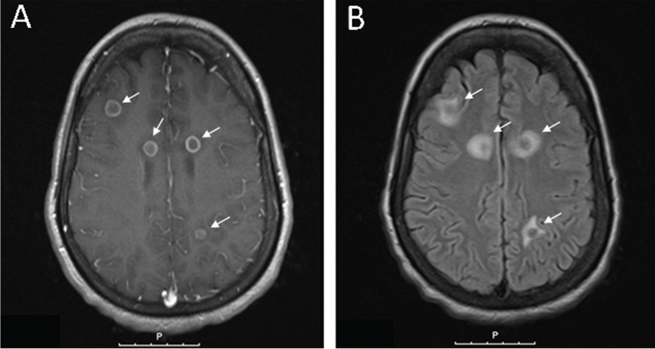
- FIGURE 1
(A) Axial contrast-enhanced T1-weighted magnetic resonance imaging showed ring-enhancing lesions (white arrows), while (B) axial T2-weighted images showed ring-enhancing lesions surrounding hyperintensity, consistent with vasogenic edema (white arrows).
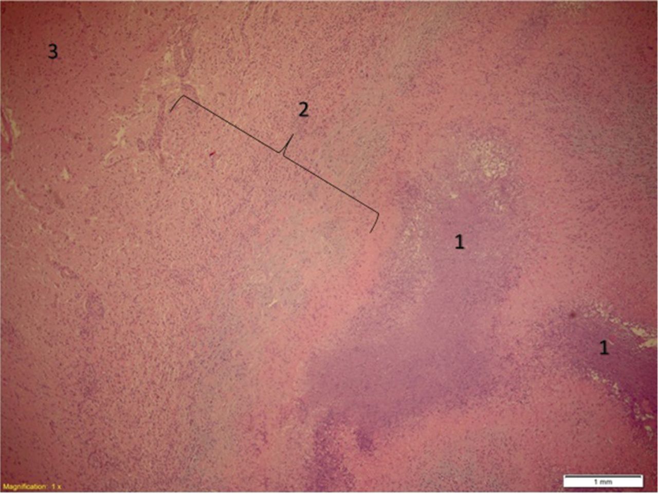
- FIGURE 2
Partially organizing central nervous system abscess showing necrosis with acute inflammatory cells (1), fibrosis with acute and chronic inflammatory cells (2), and the normal-appearing brain tissue (3) (hematoxylin and eosin, × 4).
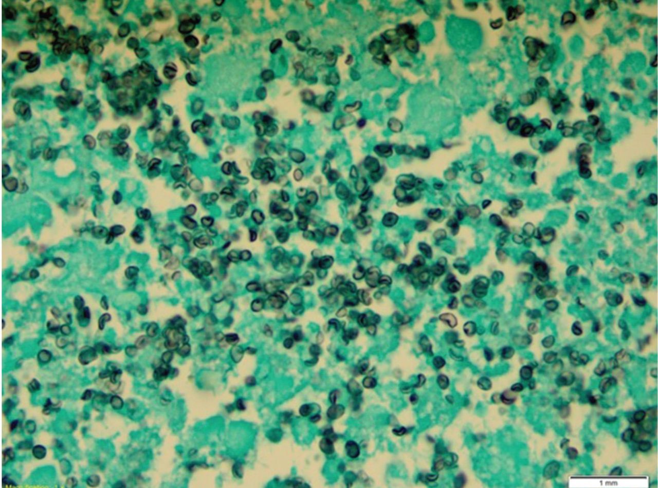
- FIGURE 3
Grocott-Gomori methenamine silver staining of a biopsy specimen of a right frontal brain lesion showed budding yeast forms, consistent with Histoplasma capsulatum (× 100).


