
Main menu

  • Home
  • Content
    • Current Issue
    • Ahead of Print
    • Past Issues
    • Supplements
    • Article Type
  • Specialty
    • Articles by Specialty
  • CME/MOC
    • Articles
    • Calendar
  • Info For
    • Manuscript Submission
    • Authors & Reviewers
    • Subscriptions
    • About CCJM
    • Contact Us
    • Media Kit
  • Conversations with Leaders
  • Conference Coverage
    • Kidney Week 2024
    • CHEST 2024
    • ACR Convergence 2023
    • Kidney Week 2023
    • ObesityWeek 2023
    • IDWeek 2023
    • CHEST 2023
    • MDS 2023
    • IAS 2023
    • ACP 2023
    • AAN 2023
    • ACC / WCC 2023
    • AAAAI Meeting 2023
    • ACR Convergence 2022
    • Kidney Week 2022
    • AIDS 2022
  • Other Publications
    • www.clevelandclinic.org

User menu

  • Register
  • Log in

Search

  • Advanced search
Cleveland Clinic Journal of Medicine
  • Other Publications
    • www.clevelandclinic.org
  • Register
  • Log in
Cleveland Clinic Journal of Medicine

Advanced Search

  • Home
  • Content
    • Current Issue
    • Ahead of Print
    • Past Issues
    • Supplements
    • Article Type
  • Specialty
    • Articles by Specialty
  • CME/MOC
    • Articles
    • Calendar
  • Info For
    • Manuscript Submission
    • Authors & Reviewers
    • Subscriptions
    • About CCJM
    • Contact Us
    • Media Kit
  • Conversations with Leaders
  • Conference Coverage
    • Kidney Week 2024
    • CHEST 2024
    • ACR Convergence 2023
    • Kidney Week 2023
    • ObesityWeek 2023
    • IDWeek 2023
    • CHEST 2023
    • MDS 2023
    • IAS 2023
    • ACP 2023
    • AAN 2023
    • ACC / WCC 2023
    • AAAAI Meeting 2023
    • ACR Convergence 2022
    • Kidney Week 2022
    • AIDS 2022
The Clinical Picture

Porcelain heart in a uremic patient

Hsiu-Chien Yang, MD, Chang-Han Lo, MD, Chun-Chi Chen, MD and Chia-Chao Wu, MD, PhD
Cleveland Clinic Journal of Medicine March 2017, 84 (3) 184-185; DOI: https://doi.org/10.3949/ccjm.84a.15177
Hsiu-Chien Yang
Department of Internal Medicine, Division of Nephrology, Tri-Service General Hospital, National Defense Medical Center, Taipei, Taiwan; and Division of Nephrology, Department of Internal Medicine, Zuoying Branch of Kaohsiung Armed Forces General Hospital, Kaohsiung, Taiwan
  • Find this author on Google Scholar
  • Find this author on PubMed
  • Search for this author on this site
Chang-Han Lo
Department of Internal Medicine, Division of Nephrology, Tri-Service General Hospital, National Defense Medical Center, Taipei, Taiwan
  • Find this author on Google Scholar
  • Find this author on PubMed
  • Search for this author on this site
Chun-Chi Chen
Department of Internal Medicine, Division of Nephrology, Tri-Service General Hospital, National Defense Medical Center, Taipei, Taiwan
  • Find this author on Google Scholar
  • Find this author on PubMed
  • Search for this author on this site
Chia-Chao Wu
Department of Internal Medicine, Division of Nephrology, Tri-Service General Hospital, National Defense Medical Center, Taipei, Taiwan
  • Find this author on Google Scholar
  • Find this author on PubMed
  • Search for this author on this site
  • For correspondence: [email protected]
  • Article
  • Figures & Data
  • Info & Metrics
  • PDF
Loading

A 58-year-old man with end-stage renal disease due to diabetic nephropathy was admitted with aggravated exertional dyspnea and intermittent chest pain for 1 week. He had been on hemodialysis for 15 years.

His blood pressure was 124/69 mm Hg, pulse 96 beats per minute, and temperature 35.8°C. On physical examination, he had bilateral diffuse crackles, elevated jugular venous pressure (9.5 cm H2O) with positive hepatojugular reflux, and apparent dependent pedal edema. The Kussmaul sign was not observed.

Cardiac enzymes were in the normal range (creatine kinase 73 U/L, troponin I 0.032 ng/mL), but the brain-natriuretic peptide level was elevated at 340 pg/mL. Other laboratory findings included calcium 9 mg/dL (reference range 8.4–10.2 mg/dL), inorganic phosphate 5 mg/dL (2.5–4.5 mg/dL), and intact parathyroid hormone 1,457 pg/mL (10–69 pg/mL).

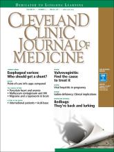
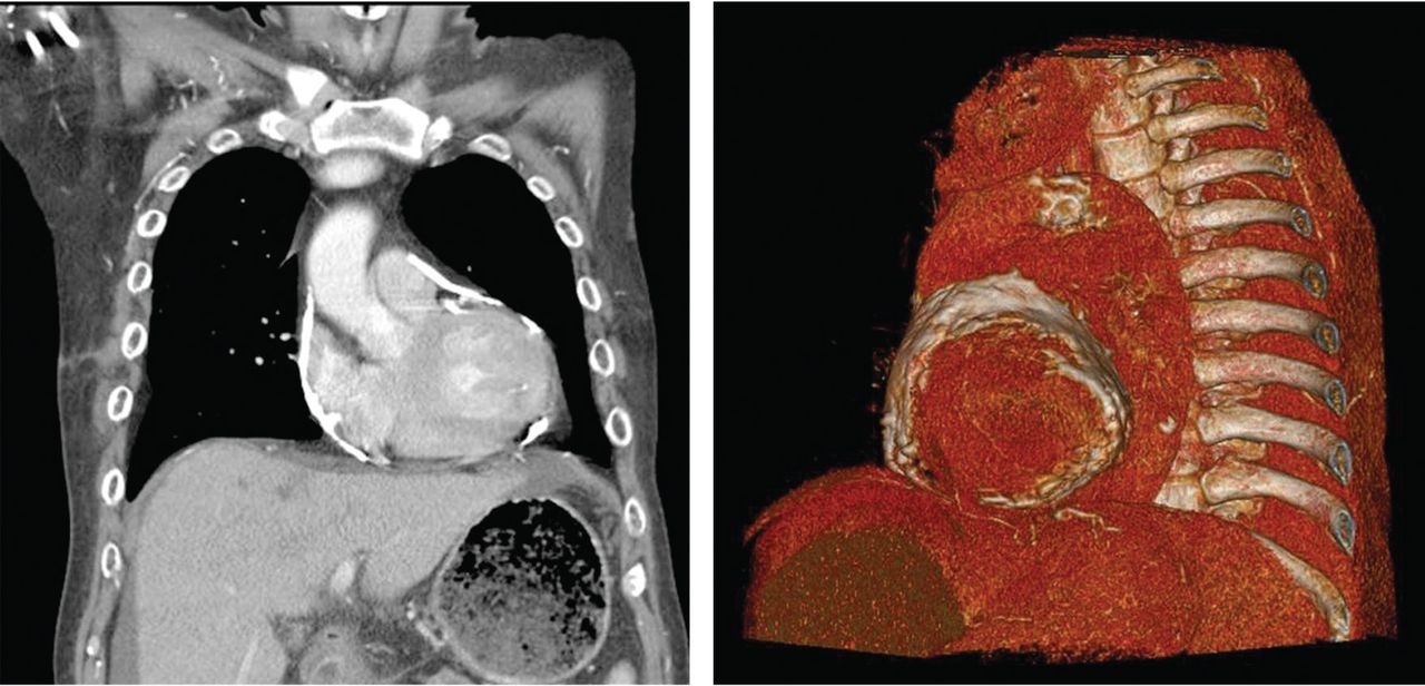
Electrocardiography showed sinus tachycardia with low voltage in diffuse leads and generalized flattening of the T wave. Chest radiography showed a bilateral reticulo nodular pattern, mild costo phrenic angle obliteration, and notable calcifications along the cardiac contour. Thoracic computed tomography showed a porcelain-like encasement of the heart (Figure 1). Transthoracic echocardiography showed thickened pericardium, pericardial calcification, and mild interventricular septal bounce in diastole, with no dyskinesia of ventricular wall motion. We decided not to perform an invasive hemodynamic assessment.

FIGURE 1
  • Download figure
  • Open in new tab
  • Download powerpoint
FIGURE 1

Thoracic computed tomography revealed calcified pericardium with heart encasement in the coronal view (left) and sagittal view (right).

CAUSES OF PERICARDIAL CALCIFICATION

Pericardial calcification, abnormal calcium deposits in response to inflammation,1 has become more widely reported as the use of chest computed tomography has become more widespread. The common identifiable causes of pericardial calcification include recurrent or chronic pericarditis, radiation therapy for Hodgkin lymphoma or breast cancer, tuberculosis, and end-stage kidney disease.2,3 Other possible causes are retention of uremic metabolites, metastatic calcification induced by secondary hyperparathyroidism, and calcium-phosphate deposition induced by hyperphosphatemia.4

In chronic kidney disease, the amount of pericardial fluid and fibrinous pericardial deposition is thought to contribute to increased pericardial thickness and constriction. In some patients, pericardial calcification and thickening would lead to constrictive pericarditis, which could be confirmed by echocardiography and cardiac catheterization. About 25% to 50% of cases of pericardial calcification are complicated by constrictive pericarditis.5,6 Constrictive pericarditis occurs in up to 4% of patients with end-stage renal disease, even with successful dialysis.7

Partial clinical improvement may be obtained with intensive hemodialysis, strict volume control, and decreased catabolism in patients with multiple comorbidities.8 However, the definite treatment is total pericardiectomy, which reduces symptoms substantially and offers a favorable long-term outcome.7

SECONDARY HYPERPARATHYROIDISM

Secondary hyperparathyroidism is a common complication in patients with end-stage renal disease and is characterized by derangements in the homeostasis of calcium, phosphorus, and vitamin D.9

Because renal function is decreased, phosphate is retained and calcitriol synthesis is reduced, resulting in hypocalcemia, which induces parathyroid gland hyperplasia and parathyroid hormone secretion.10 Moreover, some patents with long-standing secondary hyperparathyroidism may develop tertiary hyperparathyroidism associated with autonomous parathyroid hormone secretion, hypercalcemia, and hyperphosphatemia.11

The Kidney Disease: Improving Global Outcomes (KDIGO) Work Group recommends screening for and managing secondary hyperparathyroidism in all patients with stage 3 chronic kidney disease (estimated glomerular filtration rate < 60 mL/min). In patients with stage 5 chronic kidney disease or on dialysis, the serum calcium and phosphorus levels should be monitored every 1 to 3 months and the parathyroid hormone levels every 3 to 6 months.12

According to KDIGO guidelines, the target level of calcium is less than 10.2 mg/dL, and the target phosphorus level is less than 4.6 mg/dL. The level of parathyroid hormone should be maintained at 2 to 9 times the upper limit of normal for the assay.

The management of secondary hyperparathyroidism includes a low-phosphorus diet, calcium-containing or calcium-free phosphate binders, a calcitriol supplement, and calcimimetics. If medical treatment fails and manifestations are significant, parathyroidectomy may be indicated.13

  • Copyright © 2017 The Cleveland Clinic Foundation. All Rights Reserved.

References

  1. ↵
    1. Alpert MA,
    2. Ravenscraft MD
    . Pericardial involvement in end-stage renal disease. Am J Med Sci 2003; 325:228–236.
    OpenUrlCrossRefPubMed
  2. ↵
    1. Gowda RM,
    2. Boxt LM
    . Calcifications of the heart. Radiol Clin North Am 2004; 42:603–617.
    OpenUrlCrossRefPubMed
  3. ↵
    1. Kleynberg RL,
    2. Kleynberg VM,
    3. Kleynberg LM,
    4. Farahmandian D
    . Chronic constrictive pericarditis in association with end-stage renal disease. Int J Nephrol 2011; 2011:469602.
    OpenUrlPubMed
  4. ↵
    1. Rao N,
    2. Crail S
    . Metastatic calcification and long-term hemodialysis. N Engl J Med 2013; 368:2415.
    OpenUrl
  5. ↵
    1. Ling LH,
    2. Oh JK,
    3. Schaff HV,
    4. et al
    . Constrictive pericarditis in the modern era: evolving clinical spectrum and impact on outcome after pericardiectomy. Circulation 1999; 100:1380–1386.
    OpenUrlAbstract/FREE Full Text
  6. ↵
    1. Bergman M,
    2. Vitrai J,
    3. Salman H
    . Constrictive pericarditis: a reminder of a not so rare disease. Eur J Intern Med 2006; 17:457–464.
    OpenUrlCrossRefPubMed
  7. ↵
    1. Szabó G,
    2. Schmack B,
    3. Bulut C,
    4. et al
    . Constrictive pericarditis: risks, aetiologies and outcomes after total pericardiectomy: 24 years of experience. Eur J Cardiothorac Surg 2013; 44:1023–1028.
    OpenUrlCrossRefPubMed
  8. ↵
    1. Feldman V,
    2. Dovrish Z,
    3. Weisenberg N,
    4. Neuman Y,
    5. Amital H
    . Uremic pericarditis. Isr Med Assoc J 2011; 13:256–257.
    OpenUrlPubMed
  9. ↵
    1. Levin A,
    2. Bakris GL,
    3. Molitch M,
    4. et al
    . Prevalence of abnormal serum vitamin D, PTH, calcium, and phosphorus in patients with chronic kidney disease: results of the study to evaluate early kidney disease. Kidney Int 2007; 71:31–38.
    OpenUrlCrossRefPubMed
  10. ↵
    1. Martin KJ,
    2. Gonzalez EA
    . Metabolic bone disease in chronic kidney disease. J Am Soc Nephrol 2007; 18:875–885.
    OpenUrlAbstract/FREE Full Text
  11. ↵
    1. Kerby J,
    2. Rue LW,
    3. Blair H,
    4. Hudson S,
    5. Sellers MT,
    6. Diethelm AG
    . Operative treatment of tertiary hyperparathyroidism: a single-center experience. Ann Surg 1998; 227:878–886.
    OpenUrlCrossRefPubMed
  12. ↵
    1. Kidney Disease: Improving Global Outcomes (KDIGO) CKD MBD Work Group
    . KDIGO clinical practice guideline for the diagnosis, evaluation, prevention, and treatment of chronic kidney disease—mineral and bone disorder (CKD-MBD). Kidney Int Suppl 2009; 76:S1–130.
    OpenUrlPubMed
  13. ↵
    1. National Kidney Foundation
    . K/DOQI clinical practice guidelines for bone metabolism and disease in chronic kidney disease. Am J Kidney Dis 2003; 42(4 suppl 3):S1–201.
    OpenUrlPubMed
PreviousNext
Back to top

In this issue

Cleveland Clinic Journal of Medicine: 84 (3)
Cleveland Clinic Journal of Medicine
Vol. 84, Issue 3
1 Mar 2017
  • Table of Contents
  • Table of Contents (PDF)
  • Index by author
Print
Download PDF
Article Alerts
Sign In to Email Alerts with your Email Address
Email Article

Thank you for your interest in spreading the word on Cleveland Clinic Journal of Medicine.

NOTE: We only request your email address so that the person you are recommending the page to knows that you wanted them to see it, and that it is not junk mail. We do not capture any email address.

Enter multiple addresses on separate lines or separate them with commas.
Porcelain heart in a uremic patient
(Your Name) has sent you a message from Cleveland Clinic Journal of Medicine
(Your Name) thought you would like to see the Cleveland Clinic Journal of Medicine web site.
CAPTCHA
This question is for testing whether or not you are a human visitor and to prevent automated spam submissions.
Citation Tools
Porcelain heart in a uremic patient
Hsiu-Chien Yang, Chang-Han Lo, Chun-Chi Chen, Chia-Chao Wu
Cleveland Clinic Journal of Medicine Mar 2017, 84 (3) 184-185; DOI: 10.3949/ccjm.84a.15177

Citation Manager Formats

  • BibTeX
  • Bookends
  • EasyBib
  • EndNote (tagged)
  • EndNote 8 (xml)
  • Medlars
  • Mendeley
  • Papers
  • RefWorks Tagged
  • Ref Manager
  • RIS
  • Zotero
Share
Porcelain heart in a uremic patient
Hsiu-Chien Yang, Chang-Han Lo, Chun-Chi Chen, Chia-Chao Wu
Cleveland Clinic Journal of Medicine Mar 2017, 84 (3) 184-185; DOI: 10.3949/ccjm.84a.15177
Twitter logo Facebook logo Mendeley logo
  • Tweet Widget
  • Facebook Like
  • Linkedin Share Button

Jump to section

  • Article
    • CAUSES OF PERICARDIAL CALCIFICATION
    • SECONDARY HYPERPARATHYROIDISM
    • References
  • Figures & Data
  • Info & Metrics
  • PDF

Related Articles

  • No related articles found.
  • PubMed
  • Google Scholar

Cited By...

  • No citing articles found.
  • Google Scholar

More in this TOC Section

  • Severe cutaneous reaction induced by allopurinol
  • Amoxicillin rash in infectious mononucleosis
  • Tinea incognito
Show more The Clinical Picture

Similar Articles

Subjects

  • Cardiology
  • Endocrinology
  • Imaging
  • Nephrology

Navigate

  • Current Issue
  • Past Issues
  • Supplements
  • Article Type
  • Specialty
  • CME/MOC Articles
  • CME/MOC Calendar
  • Media Kit

Authors & Reviewers

  • Manuscript Submission
  • Authors & Reviewers
  • Subscriptions
  • About CCJM
  • Contact Us
  • Cleveland Clinic Center for Continuing Education
  • Consult QD

Share your suggestions!

Copyright © 2025 The Cleveland Clinic Foundation. All rights reserved. The information provided is for educational purposes only. Use of this website is subject to the website terms of use and privacy policy. 

Powered by HighWire