


Article Figures & Data
Figures
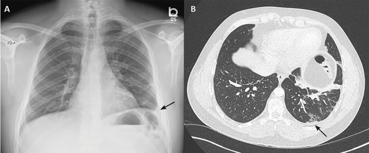
- Figure 1
Chest radiography in the emergency department (A) showed a mild left-sided pleural reaction (arrow). Computed tomography (B) showed a mild pleural reaction (arrow) and parenchymal atelectatic and fi brotic changes.
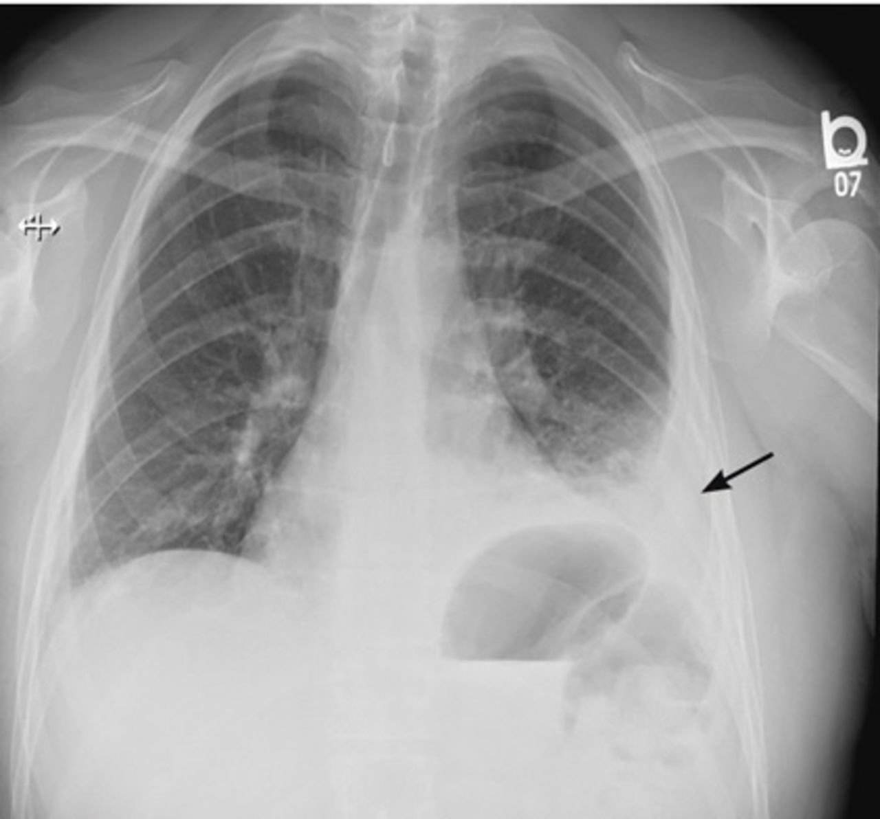
- Figure 2
Chest radiography 5 days after the emergency department presentation showed development of a left-sided pleural effusion.
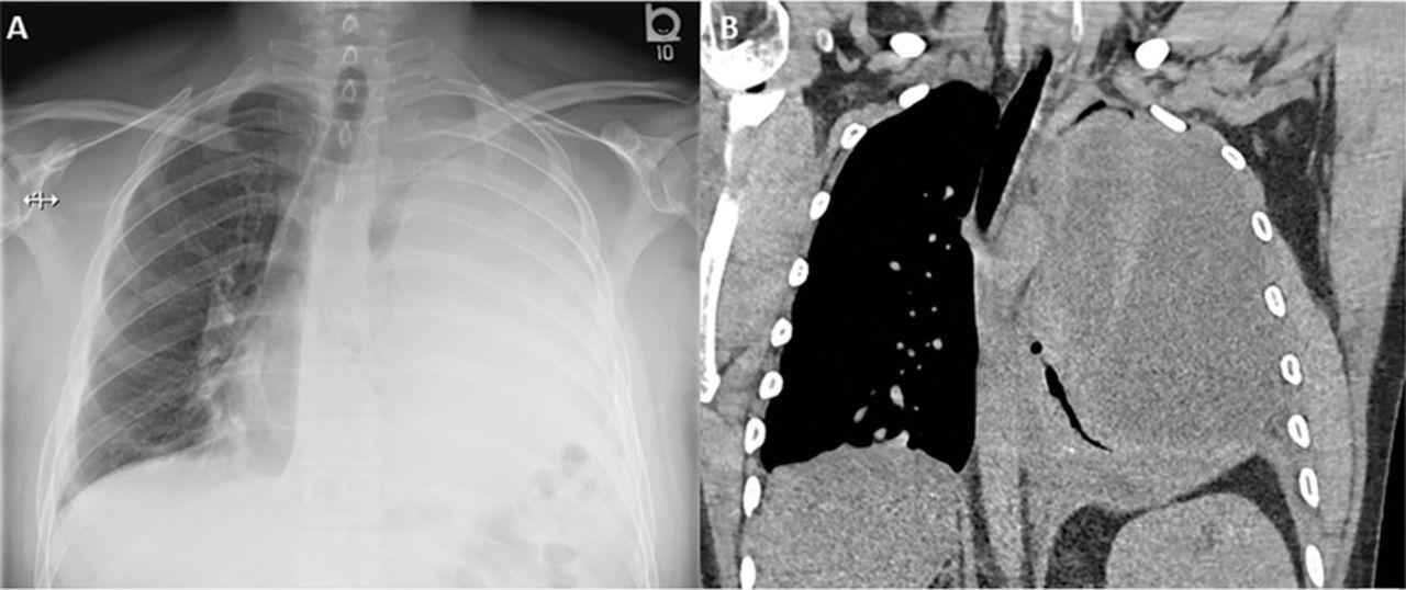
- Figure 3
Complete opacification of the left hemothorax on chest radiography (A) and massive pleural effusion causing mediastinal shift to the right on computed tomography (B).
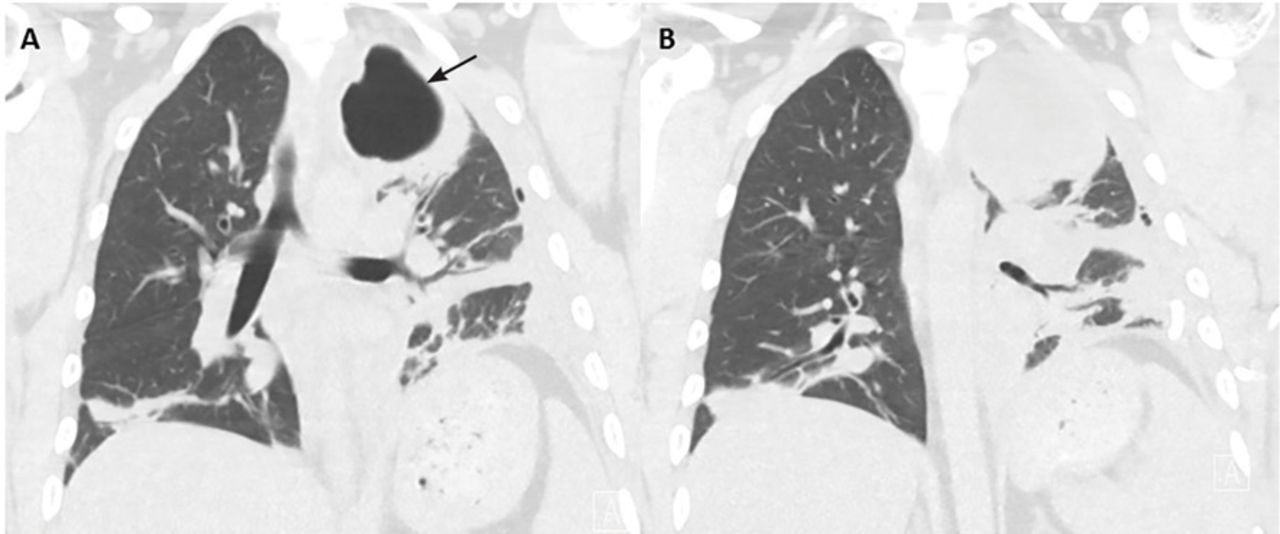
- Figure 4
Computed tomography 2 days after initial chest tube placement showed a non-communicating apical pocket.


Tables
- TABLE 1
Prognostic assessment of pleural effusion: the American College of Chest Physicians guidelines
Pleural space anatomy Pleural fluid bacteriology Pleural fluid chemistry Category Risk of poor outcome Drainage Minimal, free-flowing effusion (< 10 mm on lateral decubitus image) and Culture and Gram stain results unknown and pH unknown 1 Very low No Small to moderate free- flowing effusion (> 10 mm and < 1/2 hemithorax) and Negative culture and Gram stain and pH ≥ 7.20 2 Low No Large, free-flowing effusion (≥ 1/2 hemothorax), loculated effusion, or effusion with thickened parietal pleura or Positive culture and Gram stain or pH < 7.20 3 Moderate Yes Pus 4 High Yes Reprinted from Coulice et al,1 with permission from Elsevier; www.sciencedirect.com/science/journal/chest.
Test Pleural fluid analysis Value Glucose 0.8 mmol/L Total protein 53 g/L Lactate dehydrogenase 687 IU/L Triglyceride 0.75 mmol/L Cholesterol 2.41 mmol/L pH 7.00 Gram stain No organism seen Culture (bacterial, fungal, acid-fast bacilli) No growth Serum levels Lactate dehydrogenase 228 IU/L Protein 71 g/L The fluid is defined as an exudate if at least 1 of the following 3 criteria is met: Ratio of pleural fluid protein to serum protein > 0.5
Ratio of pleural fluid lactate dehydrogenase (LDH) to serum LDH > 0.6
Pleural fluid LDH more than 2/3 the upper limits of the laboratory normal serum LDH
Information from Light et al, reference 7.


