
Main menu

  • Home
  • Content
    • Current Issue
    • Ahead of Print
    • Past Issues
    • Supplements
    • Article Type
  • Specialty
    • Articles by Specialty
  • CME/MOC
    • Articles
    • Calendar
  • Info For
    • Manuscript Submission
    • Authors & Reviewers
    • Subscriptions
    • About CCJM
    • Contact Us
    • Media Kit
  • Conversations with Leaders
  • Conference Coverage
    • Kidney Week 2024
    • CHEST 2024
    • ACR Convergence 2023
    • Kidney Week 2023
    • ObesityWeek 2023
    • IDWeek 2023
    • CHEST 2023
    • MDS 2023
    • IAS 2023
    • ACP 2023
    • AAN 2023
    • ACC / WCC 2023
    • AAAAI Meeting 2023
    • ACR Convergence 2022
    • Kidney Week 2022
    • AIDS 2022
  • Other Publications
    • www.clevelandclinic.org

User menu

  • Register
  • Log in

Search

  • Advanced search
Cleveland Clinic Journal of Medicine
  • Other Publications
    • www.clevelandclinic.org
  • Register
  • Log in
Cleveland Clinic Journal of Medicine

Advanced Search

  • Home
  • Content
    • Current Issue
    • Ahead of Print
    • Past Issues
    • Supplements
    • Article Type
  • Specialty
    • Articles by Specialty
  • CME/MOC
    • Articles
    • Calendar
  • Info For
    • Manuscript Submission
    • Authors & Reviewers
    • Subscriptions
    • About CCJM
    • Contact Us
    • Media Kit
  • Conversations with Leaders
  • Conference Coverage
    • Kidney Week 2024
    • CHEST 2024
    • ACR Convergence 2023
    • Kidney Week 2023
    • ObesityWeek 2023
    • IDWeek 2023
    • CHEST 2023
    • MDS 2023
    • IAS 2023
    • ACP 2023
    • AAN 2023
    • ACC / WCC 2023
    • AAAAI Meeting 2023
    • ACR Convergence 2022
    • Kidney Week 2022
    • AIDS 2022
Review

Managing malignant pleural effusion

Ali Saeed Wahla, MBBS, Mateen Uzbeck, MBBS, Yaser Abu El Sameed, MBBS and Zaid Zoumot, MBBS, MRCP, MSC, PHD
Cleveland Clinic Journal of Medicine February 2019, 86 (2) 95-99; DOI: https://doi.org/10.3949/ccjm.86a.17095
Ali Saeed Wahla
Respiratory and Critical Care Institute, Cleveland Clinic Abu Dhabi, Abu Dhabi, UAE
  • Find this author on Google Scholar
  • Find this author on PubMed
  • Search for this author on this site
  • For correspondence: [email protected]
Mateen Uzbeck
Respiratory and Critical Care Institute, Cleveland Clinic Abu Dhabi, Abu Dhabi, UAE
  • Find this author on Google Scholar
  • Find this author on PubMed
  • Search for this author on this site
Yaser Abu El Sameed
Respiratory and Critical Care Institute, Cleveland Clinic Abu Dhabi, Abu Dhabi, UAE
  • Find this author on Google Scholar
  • Find this author on PubMed
  • Search for this author on this site
Zaid Zoumot
Respiratory and Critical Care Institute, Cleveland Clinic Abu Dhabi, Abu Dhabi, UAE
  • Find this author on Google Scholar
  • Find this author on PubMed
  • Search for this author on this site
  • Article
  • Figures & Data
  • Info & Metrics
  • PDF
Loading

ABSTRACT

Malignant pleural effusion can be managed in different ways, including clinical observation, thoracentesis, placement of an indwelling pleural catheter, and chemical pleurodesis. The optimal strategy depends on a variety of clinical factors. This article uses cases to illustrate the rationale for determining the best approach in different situations.

KEY POINTS
  • Asymptomatic pleural effusion in patients currently on chemotherapy does not require treatment but should be monitored for progression.

  • Indwelling pleural catheters are best used to treat effusion with lung collapse and are increasingly used as first-line therapy in other settings.

  • Chemical or mechanical pleurodesis results in filling the pleural space to prevent further fluid accumulation and can be accomplished by one of several methods.

  • For patients near the end of life, simple thoracentesis, repeated as needed, is a reasonable strategy.

Managing patients with malignant pleural effusion can be challenging. Symptoms are often distressing, and its presence signifies advanced disease. Median survival after diagnosis is 4 to 9 months,1–3 although prognosis varies considerably depending on the type and stage of the malignancy.

How patients are best managed depends on clinical circumstances. Physicians should consider the risks and benefits of each option while keeping in mind realistic goals of care.

This article uses brief case presentations to review management strategies for malignant pleural effusion.

CANCER IS A COMMON CAUSE OF PLEURAL EFFUSION

Physicians and surgeons, especially in tertiary care hospitals, must often manage malignant pleural effusion.4 Malignancy is the third leading cause of pleural effusion after heart failure and pneumonia, accounting for 44% to 77% of exudates.5 Although pleural effusion can arise secondary to many different malignancies, the most common causes are lung cancer in men and breast cancer in women; these cancers account for about 75% of all cases of malignant pleural effusion.6,7

A WOMAN ON CHEMOTHERAPY WITH ASYMPTOMATIC PLEURAL EFFUSION

An 18-year-old woman with non-Hodgkin lymphoma has received her first cycle of chemotherapy and is now admitted to the hospital for diarrhea. A routine chest radiograph reveals a left-sided pleural effusion covering one-third of the thoracic cavity. She is asymptomatic and reports no shortness of breath at rest or with exertion. Her oxygen saturation level is above 92% on room air without supplemental oxygen.

Thoracentesis reveals an exudative effusion, and cytologic study shows malignant lymphoid cells, consistent with a malignant pleural effusion. Cultures are negative.

What is the appropriate next step to manage this patient’s effusion?

Observation is reasonable

This patient is experiencing no symptoms and has just begun chemotherapy for her lymphoma. Malignant pleural effusion associated with lymphoma, small-cell lung cancer, and breast cancer is most sensitive to chemotherapy.5 For patients who do not have symptoms from the pleural effusion and who are scheduled to receive further chemotherapy, a watch-and-wait approach is reasonable.

It is important to follow the patient for developing symptoms and obtain serial imaging to evaluate for an increase in the effusion size. We recommend repeat imaging at 2- to 4-week intervals, and sooner if symptoms develop.

If progression is evident or if the patient’s oncologist indicates that the cancer is unresponsive to systemic therapy, further intervention may be necessary with one of the options discussed below.

A MAN WITH LUNG CANCER WITH PLEURAL EFFUSION, LUNG COLLAPSE

A 42-year-old man with a history of lung cancer is admitted for worsening shortness of breath. Chest radiography reveals a large left-sided pleural effusion with complete collapse of the left lung and contralateral shift of midline structures (Figure 1). Large-volume thoracentesis improves his symptoms. Pleural fluid cytology is positive for malignant cells. A repeat chest radiograph shows incomplete expansion of the left lung, thick pleura, and pneumothorax, indicating a trapped lung (ie, one unable to expand fully). Two weeks later, his symptoms recur, and chest radiography reveals a recurrent effusion.

Figure 1
  • Download figure
  • Open in new tab
  • Download powerpoint
Figure 1

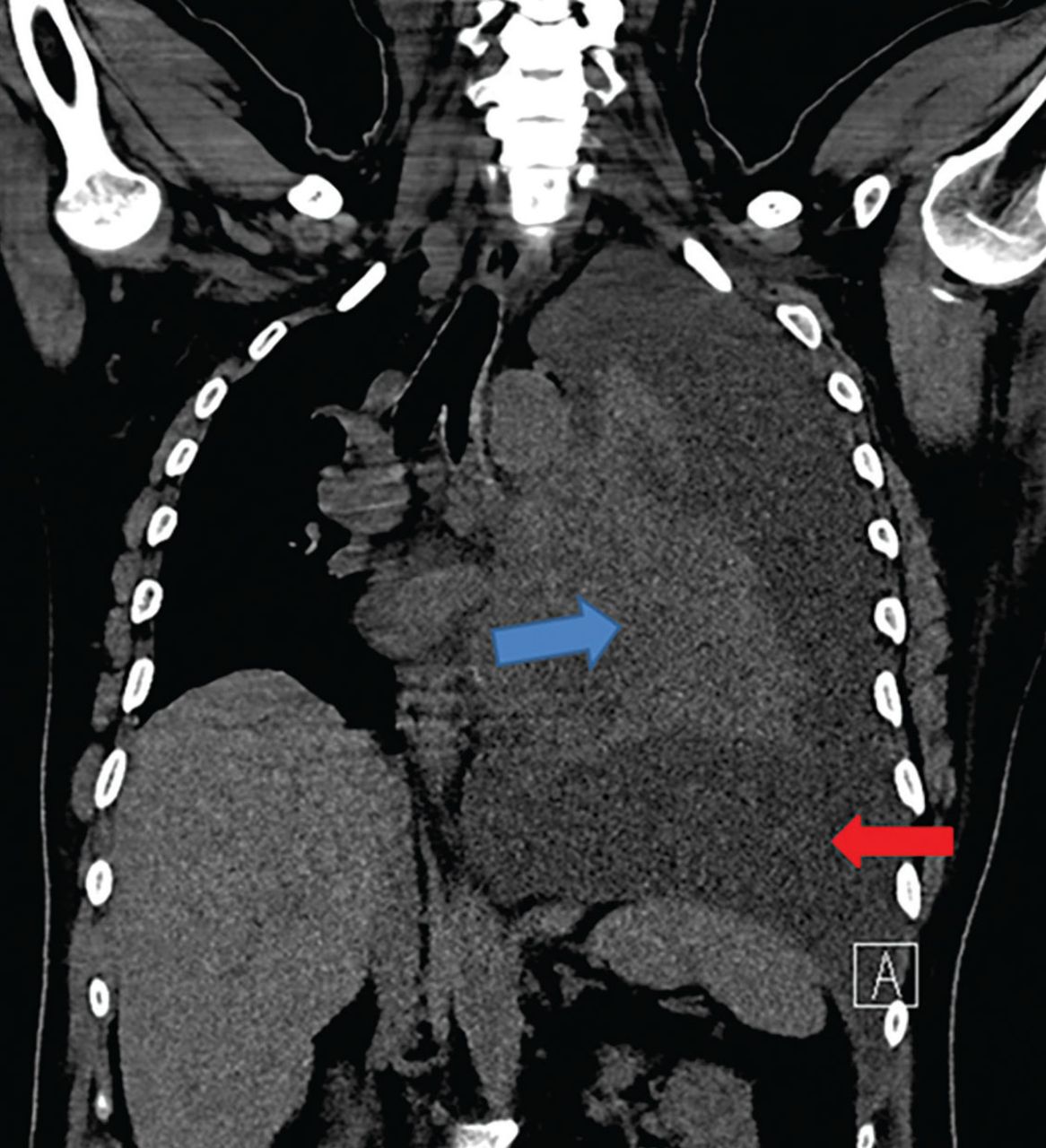
Coronal computed tomography shows left-sided pleural effusion (red arrow) and collapsed lung (blue arrow), along with midline shift.

How should this effusion be managed?

Indwelling pleural catheter placement

In a retrospective cohort study,8 malignant pleural effusion recurred in 97% of patients within 1 month (mean, 4.2 days) of therapeutic aspiration, highlighting the need for definitive treatment.

In the absence of lung expansion, pleurodesis is rarely successful, and placing an indwelling pleural catheter in symptomatic patients is the preferred strategy. The US Food and Drug Administration approved this use in 1997.9

Indwelling pleural catheters are narrow (15.5 French, or about 5 mm in diameter) and soft (made of silicone), with distal fenestrations. The distal end remains positioned in the pleural cavity to enable drainage of pleural fluid. The middle portion passes through subcutaneous tissue, where a polyester cuff prevents dislodgement and infection. The proximal end of the catheter remains outside the patient’s skin and is connected to a 1-way valve that prevents air or fluid flow into the pleural cavity.

Pleural fluid is typically drained every 2 or 3 days for palliation. Patients must be educated about home drainage and proper catheter care.

Indwelling pleural catheters are now initial therapy for many

Although indwelling pleural catheters were first used for patients who were not candidates for pleurodesis, they are now increasingly used as first-line therapy.

Since these devices were introduced, several clinical series including more than 800 patients have found that their use for malignant pleural infusion led to symptomatic improvement in 89% to 100% of cases, with 90% of patients needing no subsequent pleural procedures after catheter insertion.10–13

Davies et al14 randomized 106 patients with malignant pleural effusion to either receive an indwelling pleural catheter or undergo pleurodesis. In the first 6 weeks, the 2 groups had about the same incidence of dyspnea, but the catheter group had less dyspnea at 6 months, shorter index hospitalization (0 vs 4 days), fewer hospital days in the first year for treatment-related complications (1 vs 4.5 days), and fewer patients needing follow-up pleural procedures (6% vs 22%). On the other hand, adverse events were more frequent in the indwelling pleural catheter group (40% vs 13%). The most frequent events were pleural infection, cellulitis, and catheter blockage.

Fysh et al15 also compared indwelling pleural catheter insertion and pleurodesis (based on patient choice) in patients with malignant pleural effusion. As in the previous trial, those who received a catheter required significantly fewer days in the hospital and fewer additional pleural procedures than those who received pleurodesis. Safety profiles and symptom control were comparable.

Indwelling pleural catheters have several other advantages. They have been found to be more cost-effective than talc pleurodesis in patients not expected to live long (survival < 14 weeks).16 Patients with an indwelling pleural catheter can receive chemotherapy, and concurrent treatment does not increase risk of infection.17 And a systematic review18 found a 46% rate of autopleurodesis at a median of 52 days after insertion of an indwelling pleural catheter.

Drainage rate may need to be moderated

Chest pain has been reported with the use of indwelling pleural catheters, related to rapid drainage of the effusion in the setting of failed reexpansion of the trapped lung due to thickened pleura. Drainage schedules may need to be adjusted, with more frequent draining of smaller volumes, to control dyspnea without causing significant pain.

A WOMAN WITH RECURRENT PLEURAL EFFUSION, GOOD PROGNOSIS

A 55-year-old woman with a history of breast cancer presents with shortness of breath. Chest radiography reveals a right-sided effusion, which on thoracentesis is found to be malignant. After fluid removal, repeat chest radiography shows complete lung expansion.

One month later, she returns with symptoms and recurrence of the effusion. Ultrasonography does not reveal any adhesions in the pleural space. Her oncologist informs you that her expected survival is in years.

What is the next step?

Chemical pleurodesis

Chemical pleurodesis involves introducing a sclerosant into the pleural space to provoke an intense inflammatory response, creating adhesions and fibrosis that will obliterate the space. The sclerosing agent (typically talc) can be delivered by tube thoracostomy, video-assisted thoracic surgery (VATS), or medical pleuroscopy. Although the latter 2 methods allow direct visualization of the pleural space and, in theory, a more even distribution of the sclerosing agent, current evidence does not favor 1 option over the other,19 and practice patterns vary between institutions.

Tube thoracostomy. Typically, the sclerosing agent is administered once a chest radiograph shows lung reexpansion, and tube output of pleural fluid is less than 150 mL/day.19 However, some studies indicate that if pleural apposition can be confirmed using ultrasonography, then sclerosant administration at that time leads to optimal pleurodesis efficacy and shorter hospitalization.20,21

VATS is usually done in the operating room with the patient under general anesthesia. A double-lumen endotracheal tube allows for single-lung ventilation; a camera is then inserted into the pleural space of the collapsed lung. Multiple ports of entry are usually employed, and the entire pleural space can be visualized and the sclerosing agent instilled uniformly. The surgeon may alternatively choose to perform mechanical pleurodesis, which entails abrading the visceral and parietal pleura with dry gauze to provoke diffuse petechial hemorrhage and an inflammatory reaction. VATS can also be used to perform biopsy, lobectomy, and pneumonectomy.

Medical pleuroscopy. Medical pleuroscopy is usually done using local anesthesia with the patient awake, moderately sedated, and not intubated. Because no double-lumen endotracheal tube is used, lung collapse may not be complete, making it difficult to completely visualize the entire pleural surfaces.

Although no randomized study of VATS vs medical pleuroscopy exists, a retrospective case-matched study22 comparing VATS (under general anesthesia) to single-port VATS (under local anesthesia) noted equivalent rates of pleurodesis. However, the local anesthesia group had a lower perioperative mortality rate (0% vs 2.3%), a lower postoperative major morbidity rate (5.2% vs 9%), earlier improvement in quality of life, and shorter hospitalization (3 vs 5 days).22 In general, the diagnostic sensitivity of pleuroscopy for pleural malignancy is similar to that of Vats (93% vs 97%).23,24

A MAN WITH PLEURAL EFFUSION AND A POOR PROGNOSIS

A 60-year-old man with metastatic pancreatic cancer is brought to the clinic for worsening shortness of breath over the past 2 months. During that time, he has lost 6 kg and has become bedridden.

On examination, he has severe cachexia and is significantly short of breath at rest with associated hypoxia. His oncologist expects him to survive less than 3 months.

His laboratory investigations reveal hypoalbuminemia and leukocytosis. A chest radiograph shows a large left-sided pleural effusion that was not present 2 months ago.

What should be done for him?

Thoracentesis, repeat as needed

Malignant pleural effusion causing dyspnea is not uncommon in certain advanced malignancies and may contribute to significant suffering at the end of life. A study of 298 patients with malignant pleural effusion noted that the presence of leukocytosis, hypoalbuminemia, and hypoxemia was associated with a poorer prognosis. Patients having all 3 factors had a median survival of 42 days.25

Thoracentesis, the least invasive option that may improve dyspnea, can be done in the clinic setting and is a reasonable strategy for patients with advanced cancer and an expected survival of less than 3 months.26 Although recurrence is expected, it may take up to a few weeks, and repeat thoracentesis can be performed as needed.

  • Copyright © 2019 The Cleveland Clinic Foundation. All Rights Reserved.
View Abstract

REFERENCES

  1. ↵
    1. Roberts ME,
    2. Neville E,
    3. Berrisford RG,
    4. Antunes G,
    5. Ali NJ,
    6. BTS Pleural Disease Guideline Group
    . Management of a malignant pleural effusion: British Thoracic Society pleural disease guideline 2010. Thorax 2010; 65(suppl 2):ii32–ii40. doi:10.1136/thx.2010.136994
    OpenUrlFREE Full Text
    1. Ruckdeschel JC
    . Management of malignant pleural effusions. Semin Oncol 1995; 22(2 suppl 3):58–63. pmid:7740322
    OpenUrlPubMed
  2. ↵
    1. Bielsa S,
    2. Martín-Juan J,
    3. Porcel JM,
    4. Rodríguez-Panadero F
    . Diagnostic and prognostic implications of pleural adhesions in malignant effusions. J Thorac Oncol 2008; 3(11):1251–1256. doi:10.1097/JTO.0b013e318189f53d
    OpenUrlCrossRefPubMed
  3. ↵
    1. 35th Annual meeting of the European Association for the Study of Diabetes
    . Brussels, Belgium, 28 September–2 October, 1999. Abstracts. Diabetologia 1999;42(suppl 1):A1–A354. pmid:10505080
    OpenUrlCrossRefPubMed
  4. ↵
    1. Antony VB,
    2. Loddenkemper R,
    3. Astoul P,
    4. et al
    . Management of malignant pleural effusions. Eur Respir J 2001; 18(2):402–419. pmid:11529302
    OpenUrlFREE Full Text
  5. ↵
    1. Sahn SA
    . Malignancy metastatic to the pleura. Clin Chest Med 1998; 19(2):351–361. pmid:9646986
    OpenUrlCrossRefPubMed
  6. ↵
    1. Sahn SA
    . Pleural diseases related to metastatic malignancies. Eur Respir J 1997; 10(8):1907–1913. pmid:9272937
    OpenUrlAbstract
  7. ↵
    1. Anderson CB,
    2. Philpott GW,
    3. Ferguson TB
    . The treatment of malignant pleural effusions. Cancer 1974; 33(4):916–922. pmid:4362107
    OpenUrlCrossRefPubMed
  8. ↵
    1. Uzbeck MH,
    2. Almeida FA,
    3. Sarkiss MG,
    4. et al
    . Management of malignant pleural effusions. Adv Ther 2010; 27:(6)334–347. doi:10.1007/S12325-010-0031-8
    OpenUrlCrossRefPubMed
  9. ↵
    1. Suzuki K,
    2. Servais EL,
    3. Rizk NP,
    4. et al
    . Palliation and pleurodesis in malignant pleural effusion: the role for tunneled pleural catheters. J Thorac Oncol 2011; 6(4):762–767. doi:10.1097/JTO.0b013e31820d614f
    OpenUrlCrossRefPubMed
    1. Tremblay A,
    2. Michaud G
    . Single-center experience with 250 tunnelled pleural catheter insertions for malignant pleural effusion. Chest 2006; 129(2):362–368. doi:10.1378/chest.129.2.362
    OpenUrlCrossRefPubMed
    1. Warren WH,
    2. Kalimi R,
    3. Khodadadian LM,
    4. Kim AW
    . Management of malignant pleural effusions using the Pleur(x) catheter. Ann Thorac Surg 2008; 85(3):1049–1055 doi:10.1016/j.athoracsur.2007.11.039
    OpenUrlCrossRefPubMed
  10. ↵
    1. Murthy SC,
    2. Okereke I,
    3. Mason DP,
    4. Rice TW
    . A simple solution for complicated pleural effusions. J Thorac Oncol 2006; 1(7):697–700. pmid:17409939
    OpenUrlPubMed
  11. ↵
    1. Davies HE,
    2. Mishra EK,
    3. Kahan BC,
    4. et al
    . Effect of an indwelling pleural catheter vs chest tube and talc pleurodesis for relieving dyspnea in patients with malignant pleural effusion: the TIME2 randomized controlled trial. JAMA 2012; 307(22):2383–2389. doi:10.1001/jama.2012.5535
    OpenUrlCrossRefPubMed
  12. ↵
    1. Fysh ETH,
    2. Waterer GW,
    3. Kendall PA,
    4. et al
    . Indwelling pleural catheters reduce inpatient days over pleurodesis for malignant pleural effusion. Chest 2012; 142(2):394–400. doi:10.1378/chest.11-2657
    OpenUrlCrossRefPubMed
  13. ↵
    1. Olfert JA,
    2. Penz ED,
    3. Manns BJ,
    4. et al
    . Cost-effectiveness of indwelling pleural catheter compared with talc in malignant pleural effusion. Respirology 2017; 22(4):764–770. doi:10.1111/resp.12962
    OpenUrlCrossRef
  14. ↵
    1. Morel A,
    2. Mishra E,
    3. Medley L,
    4. et al
    . Chemotherapy should not be withheld from patients with an indwelling pleural catheter for malignant pleural effusion. Thorax 2011; 66(5):448–449. doi:10.1136/thx.2009.133504
    OpenUrlFREE Full Text
  15. ↵
    1. Van Meter MEM,
    2. McKee KY,
    3. Kohlwes RJ
    . Efficacy and safety of tunneled pleural catheters in adults with malignant pleural effusions: a systematic review. J Gen Intern Med 2011; 26(1):70–76. doi:10.1007/s11606-010-1472-0
    OpenUrlCrossRefPubMed
  16. ↵
    1. Lee YCG,
    2. Baumann MH,
    3. Maskell NA,
    4. et al
    . Pleurodesis practice for malignant pleural effusions in five English-speaking countries. Chest 2003; 124(6):2229–2238. pmid:14665505
    OpenUrlCrossRefPubMed
  17. ↵
    1. Villanueva AG,
    2. Gray AW Jr.,
    3. Shahian DM,
    4. Williamson WA,
    5. Beamis JF Jr.
    Efficacy of short term versus long term tube thoracostomy drainage before tetracycline pleurodesis in the treatment of malignant pleural effusions. Thorax 1994; 49(1):23–25. pmid:7512285
    OpenUrlAbstract/FREE Full Text
  18. ↵
    1. Sartori S,
    2. Tombesi P,
    3. Tassinari D,
    4. et al
    . Sonographically guided smallbore chest tubes and sonographic monitoring for rapid sclerotherapy of recurrent malignant pleural effusions. J Ultrasound Med 2004; 23:(9)1171–1176. pmid:15328431
    OpenUrlPubMed
  19. ↵
    1. Mineo TC,
    2. Sellitri F,
    3. Tacconi F,
    4. Ambrogi V
    . Quality of life and outcomes after nonintubated versus intubated video-thoracoscopic pleurodesis for malignant pleural effusion: comparison by a case-matched study. J Palliat Med 2014; 17(7):761–768. doi:10.1089/jpm.2013.0617
    OpenUrlCrossRefPubMed
  20. ↵
    1. Michaud G,
    2. Berkowitz DM,
    3. Ernst A
    . Pleuroscopy for diagnosis and therapy for pleural effusions. Chest 2010; 138(5):1242–1246. doi:10.1378/chest.10-1259
    OpenUrlCrossRefPubMed
  21. ↵
    1. Bhatnagar R,
    2. Maskell NA
    . Medical pleuroscopy. Clin Chest Med 2013; 34(3):487–500. doi:10.1016/j.ccm.2013.04.001
    OpenUrlCrossRef
  22. ↵
    1. Pilling JE,
    2. Dusmet ME,
    3. Ladas G,
    4. Goldstraw P
    . Prognostic factors for survival after surgical palliation of malignant pleural effusion. J Thorac Oncol 2010; 5(10):1544–1550. doi:10.1097/JTO.0b013e3181e95cb8
    OpenUrlCrossRefPubMed
  23. ↵
    1. Beyea A,
    2. Winzelberg G,
    3. Stafford RE
    . To drain or not to drain: an evidence-based approach to palliative procedures for the management of malignant pleural effusions. J Pain Symptom Manage 2012; 44(2):301–306. doi:10.1016/j.jpainsymman.2012.05.002
    OpenUrlCrossRefPubMed
PreviousNext
Back to top

In this issue

Cleveland Clinic Journal of Medicine: 86 (2)
Cleveland Clinic Journal of Medicine
Vol. 86, Issue 2
1 Feb 2019
  • Table of Contents
  • Table of Contents (PDF)
  • Index by author
Print
Download PDF
Article Alerts
Sign In to Email Alerts with your Email Address
Email Article

Thank you for your interest in spreading the word on Cleveland Clinic Journal of Medicine.

NOTE: We only request your email address so that the person you are recommending the page to knows that you wanted them to see it, and that it is not junk mail. We do not capture any email address.

Enter multiple addresses on separate lines or separate them with commas.
Managing malignant pleural effusion
(Your Name) has sent you a message from Cleveland Clinic Journal of Medicine
(Your Name) thought you would like to see the Cleveland Clinic Journal of Medicine web site.
CAPTCHA
This question is for testing whether or not you are a human visitor and to prevent automated spam submissions.
Citation Tools
Managing malignant pleural effusion
Ali Saeed Wahla, Mateen Uzbeck, Yaser Abu El Sameed, Zaid Zoumot
Cleveland Clinic Journal of Medicine Feb 2019, 86 (2) 95-99; DOI: 10.3949/ccjm.86a.17095

Citation Manager Formats

  • BibTeX
  • Bookends
  • EasyBib
  • EndNote (tagged)
  • EndNote 8 (xml)
  • Medlars
  • Mendeley
  • Papers
  • RefWorks Tagged
  • Ref Manager
  • RIS
  • Zotero
Share
Managing malignant pleural effusion
Ali Saeed Wahla, Mateen Uzbeck, Yaser Abu El Sameed, Zaid Zoumot
Cleveland Clinic Journal of Medicine Feb 2019, 86 (2) 95-99; DOI: 10.3949/ccjm.86a.17095
Twitter logo Facebook logo Mendeley logo
  • Tweet Widget
  • Facebook Like
  • Linkedin Share Button

Jump to section

  • Article
    • ABSTRACT
    • CANCER IS A COMMON CAUSE OF PLEURAL EFFUSION
    • A WOMAN ON CHEMOTHERAPY WITH ASYMPTOMATIC PLEURAL EFFUSION
    • A MAN WITH LUNG CANCER WITH PLEURAL EFFUSION, LUNG COLLAPSE
    • A WOMAN WITH RECURRENT PLEURAL EFFUSION, GOOD PROGNOSIS
    • A MAN WITH PLEURAL EFFUSION AND A POOR PROGNOSIS
    • REFERENCES
  • Figures & Data
  • Info & Metrics
  • PDF

Related Articles

  • No related articles found.
  • PubMed
  • Google Scholar

Cited By...

  • No citing articles found.
  • Google Scholar

More in this TOC Section

  • Alpha-gal syndrome: Recognizing and managing a tick-bite–related meat allergy
  • Managing right ventricular failure in the setting of pulmonary embolism
  • Psychedelics, spirituality, and existential distress in patients at the end of life
Show more Review

Similar Articles

Subjects

  • Hospice & Palliative Medicine
  • Hospital Medicine
  • Imaging
  • Infectious Diseases
  • Oncology
  • Pulmonology

Navigate

  • Current Issue
  • Past Issues
  • Supplements
  • Article Type
  • Specialty
  • CME/MOC Articles
  • CME/MOC Calendar
  • Media Kit

Authors & Reviewers

  • Manuscript Submission
  • Authors & Reviewers
  • Subscriptions
  • About CCJM
  • Contact Us
  • Cleveland Clinic Center for Continuing Education
  • Consult QD

Share your suggestions!

Copyright © 2025 The Cleveland Clinic Foundation. All rights reserved. The information provided is for educational purposes only. Use of this website is subject to the website terms of use and privacy policy. 

Powered by HighWire