
Main menu

  • Home
  • Content
    • Current Issue
    • Ahead of Print
    • Past Issues
    • Supplements
    • Article Type
  • Specialty
    • Articles by Specialty
  • CME/MOC
    • Articles
    • Calendar
  • Info For
    • Manuscript Submission
    • Authors & Reviewers
    • Subscriptions
    • About CCJM
    • Contact Us
    • Media Kit
  • Conversations with Leaders
  • Conference Coverage
    • Kidney Week 2024
    • CHEST 2024
    • ACR Convergence 2023
    • Kidney Week 2023
    • ObesityWeek 2023
    • IDWeek 2023
    • CHEST 2023
    • MDS 2023
    • IAS 2023
    • ACP 2023
    • AAN 2023
    • ACC / WCC 2023
    • AAAAI Meeting 2023
    • ACR Convergence 2022
    • Kidney Week 2022
    • AIDS 2022
  • Other Publications
    • www.clevelandclinic.org

User menu

  • Register
  • Log in

Search

  • Advanced search
Cleveland Clinic Journal of Medicine
  • Other Publications
    • www.clevelandclinic.org
  • Register
  • Log in
Cleveland Clinic Journal of Medicine

Advanced Search

  • Home
  • Content
    • Current Issue
    • Ahead of Print
    • Past Issues
    • Supplements
    • Article Type
  • Specialty
    • Articles by Specialty
  • CME/MOC
    • Articles
    • Calendar
  • Info For
    • Manuscript Submission
    • Authors & Reviewers
    • Subscriptions
    • About CCJM
    • Contact Us
    • Media Kit
  • Conversations with Leaders
  • Conference Coverage
    • Kidney Week 2024
    • CHEST 2024
    • ACR Convergence 2023
    • Kidney Week 2023
    • ObesityWeek 2023
    • IDWeek 2023
    • CHEST 2023
    • MDS 2023
    • IAS 2023
    • ACP 2023
    • AAN 2023
    • ACC / WCC 2023
    • AAAAI Meeting 2023
    • ACR Convergence 2022
    • Kidney Week 2022
    • AIDS 2022
Review

Imaging to evaluate suspected infective endocarditis

Ikram-Ul Haq, MD, Iqraa Haq, BS, Brian Griffin, MD and Bo Xu, MD, FACC, FRACP, FASE
Cleveland Clinic Journal of Medicine March 2021, 88 (3) 163-172; DOI: https://doi.org/10.3949/ccjm.88a.19142
Ikram-Ul Haq
Division of Internal Medicine, Mayo Clinic, Rochester, MN
  • Find this author on Google Scholar
  • Find this author on PubMed
  • Search for this author on this site
Iqraa Haq
Imperial College London Faculty of Medicine, London, United Kingdom
  • Find this author on Google Scholar
  • Find this author on PubMed
  • Search for this author on this site
Brian Griffin
Section of Cardiovascular Imaging, Robert and Suzanne Tomsich Department of Cardiovascular Medicine, Sydell and Arnold Miller Family Heart, Vascular, and Thoracic Institute, Cleveland Clinic, Cleveland, OH
  • Find this author on Google Scholar
  • Find this author on PubMed
  • Search for this author on this site
  • Find this author on Cleveland Clinic
Bo Xu
Section of Cardiovascular Imaging, Robert and Suzanne Tomsich Department of Cardiovascular Medicine, Sydell and Arnold Miller Family Heart, Vascular, and Thoracic Institute, Cleveland Clinic, Cleveland, OH; Assistant Professor, Cleveland Clinic Lerner College of Medicine of Case Western Reserve University, Cleveland, OH
  • Find this author on Google Scholar
  • Find this author on PubMed
  • Search for this author on this site
  • For correspondence: [email protected]
  • Find this author on Cleveland Clinic
  • Article
  • Figures & Data
  • Info & Metrics
  • PDF
Loading

Article Figures & Data

Figures

  • Tables
  • Figure 1
    • Download figure
    • Open in new tab
    • Download powerpoint
    Figure 1

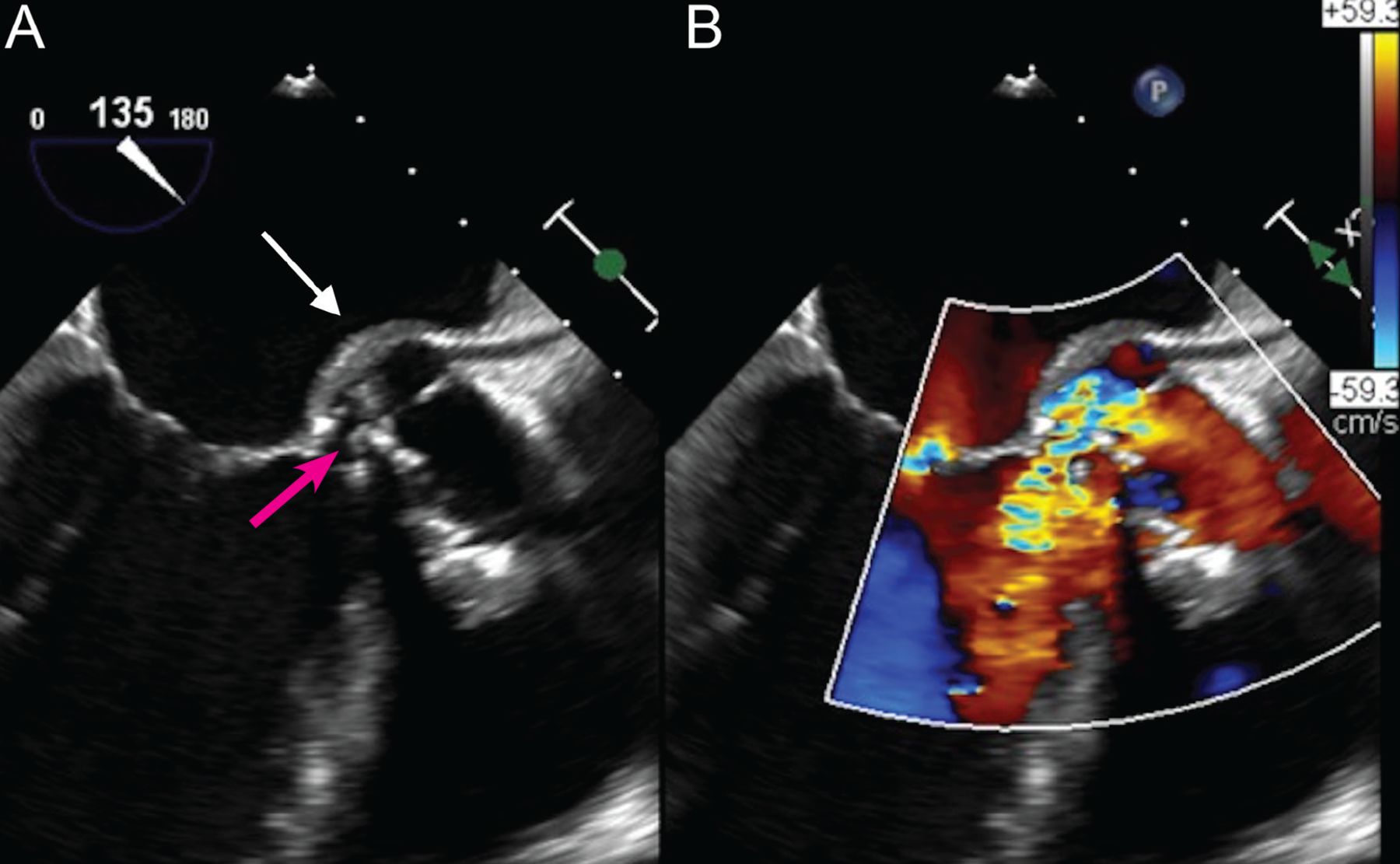
    (A) Transesophageal echocardiography, mid-esophageal long-axis view, demonstrates a prominent aortic root abscess cavity (white arrow) posteriorly in a patient with a prosthetic aortic valve. Also note partial dehiscence of the aortic bioprosthesis (red arrow). (B) Color Doppler analysis demonstrates significant aortic regurgitation between the aortic bioprosthesis and the left ventricular outflow tract through the prominent abscess cavity.

  • Figure 2
    • Download figure
    • Open in new tab
    • Download powerpoint
    Figure 2

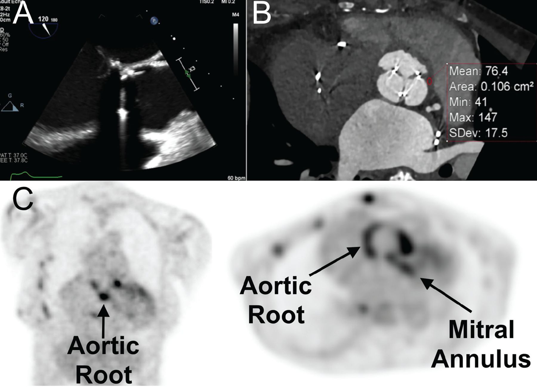
    Utility of adjuvant advanced cardiovascular imaging—multidetector cardiac computed tomography (MDCT) and 18F-fluorodeoxyglucose positron emission tomography/CT (FDG-PET/CT)—in the diagnosis of infective endocarditis. A 68-year-old woman with Staphylococcus aureus bacteremia in the setting of a bioprosthetic aortic valve developed fever and acute right lower limb pain. Initial transesophageal echocardiography (mid-esophageal long-axis view) showed no obvious vegetation associated with the bioprosthesis (A). Due to ongoing clinical suspicion for prosthetic aortic valve endocarditis, MDCT was performed (B) and, at the level of the aortic root, showed abnormal thickening with elevated Hounsfield units (mean: 76.4 units), highly suspicious for periprosthetic aortic root abscess. FDG-PET/CT (C), demonstrated abnormal increased activity at the aortic root and mitral annulus (arrows).

  • Figure 3
    • Download figure
    • Open in new tab
    • Download powerpoint
    Figure 3

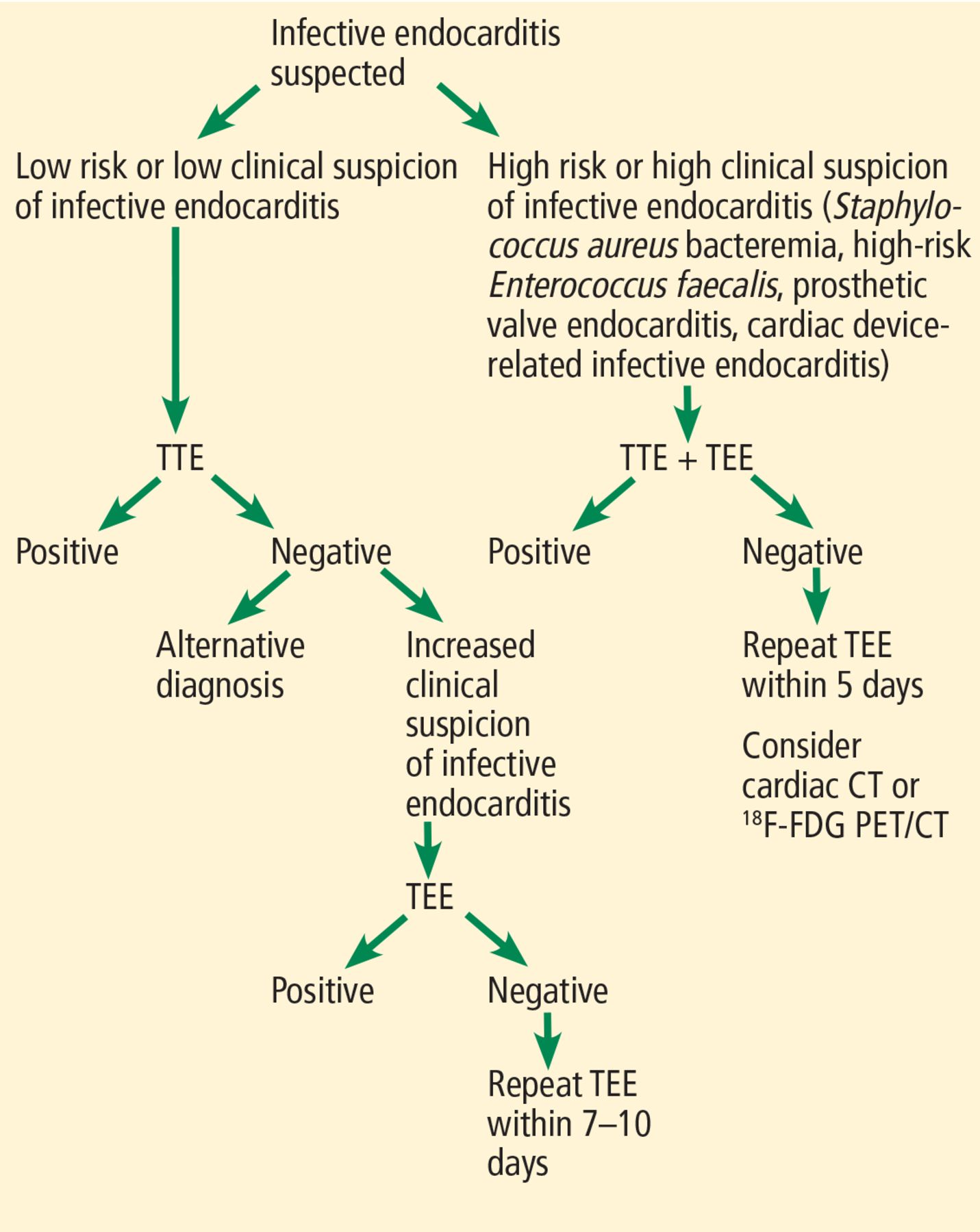
    A proposed diagnostic algorithm for infective endocarditis.

    TEE = transesophageal echocardiography; TTE = transthoracic echocardiography

Tables

  • Figures
    • View popup
    TABLE 1

    Imaging for endocarditis: ESC10 and AHA4 recommendationsa

    Obtain echocardiography as soon as endocarditis is suspected
    (ESC and AHA)
    For initial investigation:
    Transthoracic echocardiography (TTE) should be used (ESC)
    Both TTE and transesophageal echocardiography (TEE) should be used (AHA)
    Perform TEE:
    If TTE is not diagnostic in patients with known or suspected infective endocarditis (ESC and AHA)
    If complications are suspected (eg, new murmur, embolism, persisting fever, heart failure, abscess, atrioventricular block) (ESC and AHA)
    If intracardiac device, leads, or prosthetic valves are present (ESC and AHA)
    Repeat TEE if initial TTE is negative but clinical suspicion of infective endocarditis remains high:
    7-10 days later (ESC), or
    3-5 days later (AHA)
    • ↵a All recommendations listed are class 1 (strong).

    • AHA = American Heart Association; ESC = European Society of Cardiology

PreviousNext
Back to top

In this issue

Cleveland Clinic Journal of Medicine: 88 (3)
Cleveland Clinic Journal of Medicine
Vol. 88, Issue 3
1 Mar 2021
  • Table of Contents
  • Table of Contents (PDF)
  • Index by author
  • Complete Issue (PDF)
Print
Download PDF
Article Alerts
Sign In to Email Alerts with your Email Address
Email Article

Thank you for your interest in spreading the word on Cleveland Clinic Journal of Medicine.

NOTE: We only request your email address so that the person you are recommending the page to knows that you wanted them to see it, and that it is not junk mail. We do not capture any email address.

Enter multiple addresses on separate lines or separate them with commas.
Imaging to evaluate suspected infective endocarditis
(Your Name) has sent you a message from Cleveland Clinic Journal of Medicine
(Your Name) thought you would like to see the Cleveland Clinic Journal of Medicine web site.
CAPTCHA
This question is for testing whether or not you are a human visitor and to prevent automated spam submissions.
Citation Tools
Imaging to evaluate suspected infective endocarditis
Ikram-Ul Haq, Iqraa Haq, Brian Griffin, Bo Xu
Cleveland Clinic Journal of Medicine Mar 2021, 88 (3) 163-172; DOI: 10.3949/ccjm.88a.19142

Citation Manager Formats

  • BibTeX
  • Bookends
  • EasyBib
  • EndNote (tagged)
  • EndNote 8 (xml)
  • Medlars
  • Mendeley
  • Papers
  • RefWorks Tagged
  • Ref Manager
  • RIS
  • Zotero
Share
Imaging to evaluate suspected infective endocarditis
Ikram-Ul Haq, Iqraa Haq, Brian Griffin, Bo Xu
Cleveland Clinic Journal of Medicine Mar 2021, 88 (3) 163-172; DOI: 10.3949/ccjm.88a.19142
Twitter logo Facebook logo Mendeley logo
  • Tweet Widget
  • Facebook Like
  • Linkedin Share Button

Jump to section

  • Article
    • ABSTRACT
    • AN OLD PROBLEM IN A NEW DEMOGRAPHIC
    • ECHOCARDIOGRAPHY IS ESSENTIAL
    • USE BOTH TTE AND TEE FOR MANY PATIENTS
    • WHEN SHOULD TTE OR TEE BE REPEATED IF NEGATIVE, BUT BACTEREMIA PERSISTS?
    • NATIVE VS PROSTHETIC VALVE
    • CARDIAC DEVICE-RELATED INFECTIONS
    • CAUSATIVE ORGANISMS
    • ALTERNATIVE IMAGING METHODS
    • OUR ALGORITHM FOR EVALUATING SUSPECTED INFECTIVE ENDOCARDITIS
    • CASE CONCLUDED
    • DISCLOSURES
    • REFERENCES
  • Figures & Data
  • Info & Metrics
  • PDF

Related Articles

  • PubMed
  • Google Scholar

Cited By...

  • No citing articles found.
  • Google Scholar

More in this TOC Section

  • Alpha-gal syndrome: Recognizing and managing a tick-bite–related meat allergy
  • Managing right ventricular failure in the setting of pulmonary embolism
  • Psychedelics, spirituality, and existential distress in patients at the end of life
Show more Review

Similar Articles

Subjects

  • Cardiology
  • Hospital Medicine
  • Imaging
  • Infectious Diseases

Navigate

  • Current Issue
  • Past Issues
  • Supplements
  • Article Type
  • Specialty
  • CME/MOC Articles
  • CME/MOC Calendar
  • Media Kit

Authors & Reviewers

  • Manuscript Submission
  • Authors & Reviewers
  • Subscriptions
  • About CCJM
  • Contact Us
  • Cleveland Clinic Center for Continuing Education
  • Consult QD

Share your suggestions!

Copyright © 2025 The Cleveland Clinic Foundation. All rights reserved. The information provided is for educational purposes only. Use of this website is subject to the website terms of use and privacy policy. 

Powered by HighWire