
Main menu

  • Home
  • Content
    • Current Issue
    • Ahead of Print
    • Past Issues
    • Supplements
    • Article Type
  • Specialty
    • Articles by Specialty
  • CME/MOC
    • Articles
    • Calendar
  • Info For
    • Manuscript Submission
    • Authors & Reviewers
    • Subscriptions
    • About CCJM
    • Contact Us
    • Media Kit
  • Conversations with Leaders
  • Conference Coverage
    • Kidney Week 2024
    • CHEST 2024
    • ACR Convergence 2023
    • Kidney Week 2023
    • ObesityWeek 2023
    • IDWeek 2023
    • CHEST 2023
    • MDS 2023
    • IAS 2023
    • ACP 2023
    • AAN 2023
    • ACC / WCC 2023
    • AAAAI Meeting 2023
    • ACR Convergence 2022
    • Kidney Week 2022
    • AIDS 2022
  • Other Publications
    • www.clevelandclinic.org

User menu

  • Register
  • Log in

Search

  • Advanced search
Cleveland Clinic Journal of Medicine
  • Other Publications
    • www.clevelandclinic.org
  • Register
  • Log in
Cleveland Clinic Journal of Medicine

Advanced Search

  • Home
  • Content
    • Current Issue
    • Ahead of Print
    • Past Issues
    • Supplements
    • Article Type
  • Specialty
    • Articles by Specialty
  • CME/MOC
    • Articles
    • Calendar
  • Info For
    • Manuscript Submission
    • Authors & Reviewers
    • Subscriptions
    • About CCJM
    • Contact Us
    • Media Kit
  • Conversations with Leaders
  • Conference Coverage
    • Kidney Week 2024
    • CHEST 2024
    • ACR Convergence 2023
    • Kidney Week 2023
    • ObesityWeek 2023
    • IDWeek 2023
    • CHEST 2023
    • MDS 2023
    • IAS 2023
    • ACP 2023
    • AAN 2023
    • ACC / WCC 2023
    • AAAAI Meeting 2023
    • ACR Convergence 2022
    • Kidney Week 2022
    • AIDS 2022
The Clinical Picture

Mucormycosis presenting as dental pain

Pankaj Garg, MS, MBBS, Aalam Sohal, MD and Sankalp Mittal, MDS
Cleveland Clinic Journal of Medicine October 2022, 89 (10) 553-555; DOI: https://doi.org/10.3949/ccjm.89a.22001
Pankaj Garg
Department of Surgery, Indus International Hospital, Punjab, India
  • Find this author on Google Scholar
  • Find this author on PubMed
  • Search for this author on this site
  • For correspondence: [email protected]
Aalam Sohal
Department of Internal Medicine, University of California San Francisco Fresno Center for Medical Education and Research, Fresno, CA
  • Find this author on Google Scholar
  • Find this author on PubMed
  • Search for this author on this site
Sankalp Mittal
Department of Oral and maxillofacial surgery, Mittal Dental Clinic, Jaipur, India
  • Find this author on Google Scholar
  • Find this author on PubMed
  • Search for this author on this site
  • Article
  • Figures & Data
  • Info & Metrics
  • PDF
Loading

A 19-year-old indian female was referred to the emergency department by her dentist. She had presented to the dentist with 15 days of constant pain in the left premolar tooth, describing the pain 8 out of 10 and radiating to the left cheek. The dentist performed a root canal procedure, which did not resolve the pain, and 1 week after the procedure she developed mobility in her left upper teeth and sinus discharges on the upper buccal mucosa.

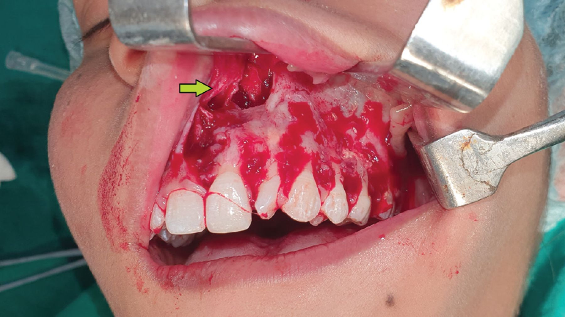
On presentation in the emergency department, the patient said that she had recovered from COVID-19 about 2 weeks earlier and had been treated with dexamethasone. Evaluation in the emergency department showed that she was hemodynamically stable and afebrile. The left side of her face was tender to palpation. On oral examination, mobility of the left upper teeth was noted, with purulent discharge on the upper buccal mucosa (Figure 1).

Figure 1
  • Download figure
  • Open in new tab
  • Download powerpoint
Figure 1

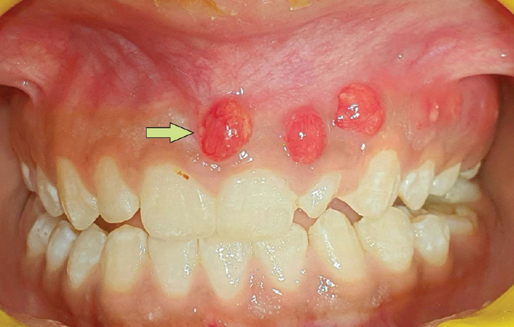
Multiple tracts in the buccal vestibule were noted on the left gum. The arrow indicates the most medial tract.

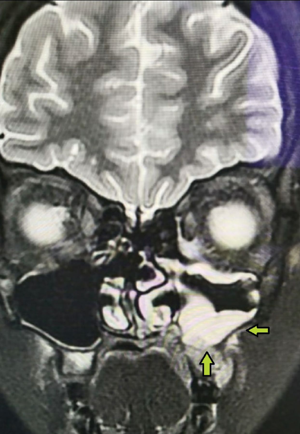
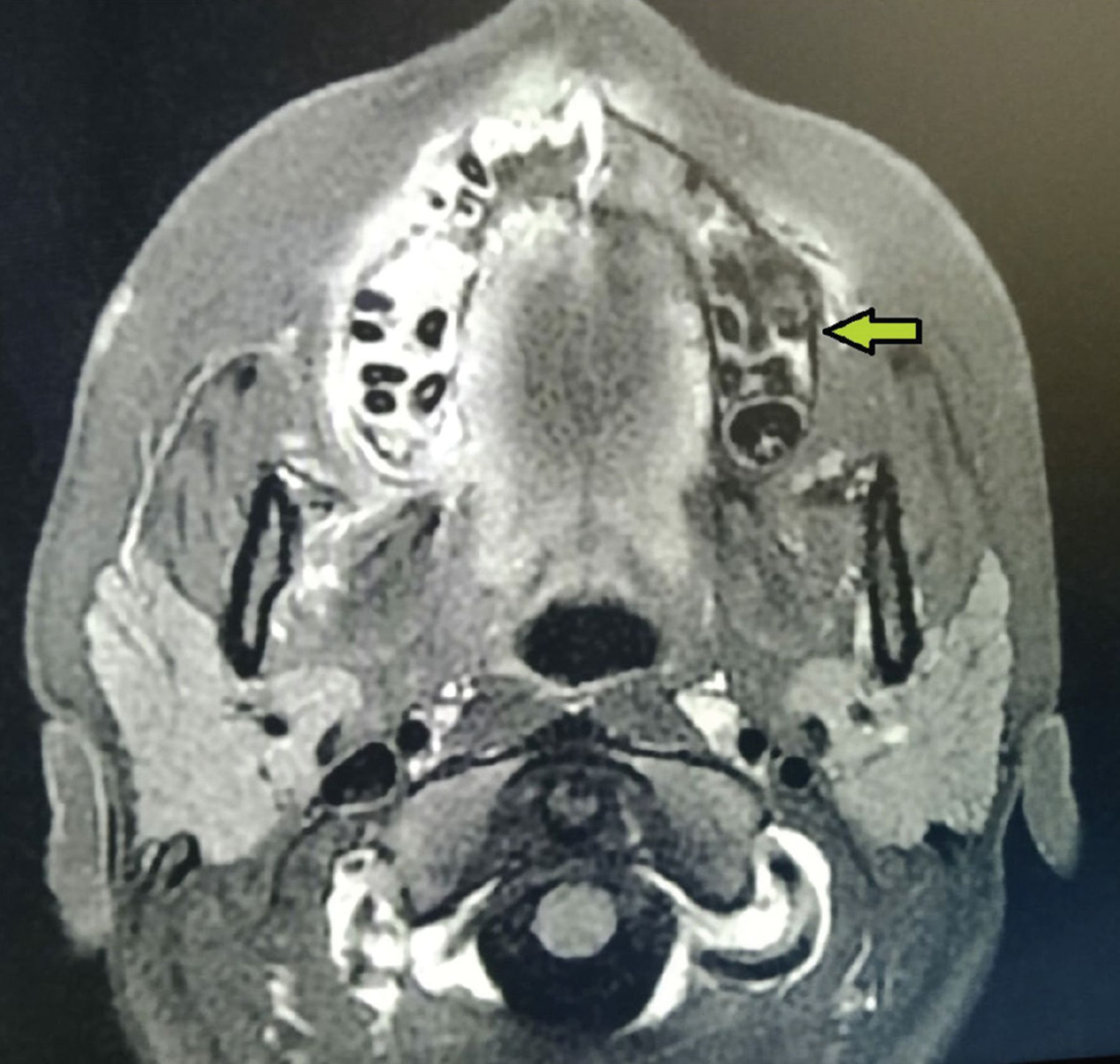
Results of a complete blood cell count and basal metabolic panel were unremarkable. Magnetic resonance imaging with contrast revealed markedly inflamed and hypertrophic mucosa of the left maxillary sinus cavity (Figure 2). The left maxillary bone was noted to be necrotic and nonenhancing (Figure 3). The findings on oral examination and imaging raised concern for mucormycosis, and the patient was referred for emergency surgery.

Figure 2
  • Download figure
  • Open in new tab
  • Download powerpoint
Figure 2

Markedly inflamed and hypertrophic mucosa of the left maxillary sinus cavity were noted on magnetic resonance imaging. The arrows point to the lateral and inferior sides of the left maxillary sinus.

Figure 3
  • Download figure
  • Open in new tab
  • Download powerpoint
Figure 3

Necrotic left maxillary bone noted on magnetic resonance imaging. The arrow indicates the lateral side of the left maxillary sinus.

She underwent thorough debridement and left subtotal maxillectomy. Intraoperatively, the left maxillary sinus cavity could be seen to communicate with the buccal vestibule (Figure 4). Microscopic examination of the excised tissue demonstrated hyphae of irregular width and branching angles of 90 degrees. A diagnosis of maxillary mucormycosis invading into the buccal vestibule was made. The patient was started on amphotericin B, and at 4 weeks after surgery, she had improved completely.

Figure 4
  • Download figure
  • Open in new tab
  • Download powerpoint
Figure 4

Communication between the buccal vestibule and the maxillary sinuses (arrow) was noted on intraoperative evaluation.

MUCORMYCOSIS: SYMPTOMS, DIAGNOSIS, AND TREATMENT

Mucormycosis is an infection caused by filamentous molds that commonly affect immunocompromised patients such as those with cancer or uncontrolled diabetes.1 In 2021, a systematic review found numerous cases of invasive fungal infection and mucormycosis reported in patients with COVID-19.2 This has been attributed to diabetes as a common predisposing comorbidity and to treatment with steroids. Our patient was healthy at baseline. We believe that her treatment with steroids and her immunocompromised state secondary to COVID-19 may have predisposed her to mucormycosis.

Our patient had initial symptoms 25 days after COVID-19 infection, which aligns with the already available literature.3 Several hospitals in India have opened outpatient departments for mucormycosis to monitor COVID-19-recovered patients from 10 days to 6 weeks, when they are most vulnerable to the fungal infection.3

Symptoms

Initial symptoms of mucormycosis are paresthesia and swelling over the face, nasal congestion and discharge, fever, headache, and lethargy, which are similar to symptoms of sinusitis and periorbital cellulitis. However, the appearance of black lesions on the nasal bridge or upper inside of the mouth that are progressively increasing in number or size and associated pulmonary symptoms like cough, chest pain, and dyspnea in a patient with diabetes or immunosuppression should raise a strong suspicion for maxillary mucormycosis. Nevertheless, in rare cases, dental pain can also be a presenting feature of maxillary mucormycosis.4 As this disease is rapidly progressive, early diagnosis is crucial in the prognosis of the patient. Fortunately, our patient’s dentist was able to recognize this alarming sign on the second visit and transfer the patient for an emergency evaluation.

Diagnosis and treatment

Microscopic examination of excised tissue in mucormycosis shows hyphae of irregular width and branching angles of 90 degrees. On histopathology, the invasive disease is characterized by prominent infarcts and angioinvasion. Rapid identification of invasive mucormycosis is necessary to minimize the mortality and morbidity of this disease. Thus, it is worthwhile for clinicians to be able to rapidly identify the characteristics of hyphae of mucormycosis.

It is pertinent to mention that mucormycosis is not contagious. A person can get mucormycosis through contact with fungal spores in the environment. For example, the lung or sinus forms of the infection can occur after someone inhales spores from the air.

Treatment includes immediate hospitalization, surgical debridement and systemic antifungal therapy.1 The liposomal formulation of amphotericin B (AmBisome) is the drug of choice based on efficacy and safety data.

TAKE-HOME MESSAGE

Our patient’s case illustrates the need for early diagnosis and urgent debridement for better outcomes. Mucormycosis should be considered in the differential diagnosis for any patient presenting with dental pain in the setting of recent COVID-19 infection.

DISCLOSURES

The authors report no relevant financial relationships which, in the context of their contributions, could be perceived as a potential conflict of interest.

  • Copyright © 2022 The Cleveland Clinic Foundation. All Rights Reserved.

REFERENCES

  1. ↵
    1. Hernández JL,
    2. Buckley CJ
    . Mucormycosis. In: StatPearls. Treasure Island, FL: StatPearls Publishing; April 30, 2022.
  2. ↵
    1. Singh AK,
    2. Singh R,
    3. Joshi SR,
    4. Misra A
    . Mucormycosis in COVID-19: a systematic review of cases reported worldwide and in India. Diabetes Metab Syndr 2021; 15(4):102146. doi:10.1016/j.dsx.2021.05.019
    OpenUrlCrossRefPubMed
  3. ↵
    1. Nambiar M,
    2. Varma SR,
    3. Damdoum M
    . Post-COVID alliance-mucormycosis, a fatal sequel to the pandemic in India. Saudi J Biol Sci 2021; 28(11):6461–6464. doi:10.1016/j.sjbs.2021.07.004
    OpenUrlCrossRef
  4. ↵
    1. Nicolatou-Galitis O,
    2. Sachanas S,
    3. Drogari-Apiranthitou M, et al
    . Mucormycosis presenting with dental pain and palatal ulcer in a patient with chronic myelomonocytic leukaemia: case report and literature review. JMM Case Reports 2015. Available at: https://www.microbiologyresearch.org/content/journal/jmmcr/10.1099/jmmcr.0.000014. Accessed September 1, 2022.
PreviousNext
Back to top

In this issue

Cleveland Clinic Journal of Medicine: 89 (10)
Cleveland Clinic Journal of Medicine
Vol. 89, Issue 10
1 Oct 2022
  • Table of Contents
  • Table of Contents (PDF)
  • Index by author
  • Complete Issue (PDF)
Print
Download PDF
Article Alerts
Sign In to Email Alerts with your Email Address
Email Article

Thank you for your interest in spreading the word on Cleveland Clinic Journal of Medicine.

NOTE: We only request your email address so that the person you are recommending the page to knows that you wanted them to see it, and that it is not junk mail. We do not capture any email address.

Enter multiple addresses on separate lines or separate them with commas.
Mucormycosis presenting as dental pain
(Your Name) has sent you a message from Cleveland Clinic Journal of Medicine
(Your Name) thought you would like to see the Cleveland Clinic Journal of Medicine web site.
CAPTCHA
This question is for testing whether or not you are a human visitor and to prevent automated spam submissions.
Citation Tools
Mucormycosis presenting as dental pain
Pankaj Garg, Aalam Sohal, Sankalp Mittal
Cleveland Clinic Journal of Medicine Oct 2022, 89 (10) 553-555; DOI: 10.3949/ccjm.89a.22001

Citation Manager Formats

  • BibTeX
  • Bookends
  • EasyBib
  • EndNote (tagged)
  • EndNote 8 (xml)
  • Medlars
  • Mendeley
  • Papers
  • RefWorks Tagged
  • Ref Manager
  • RIS
  • Zotero
Share
Mucormycosis presenting as dental pain
Pankaj Garg, Aalam Sohal, Sankalp Mittal
Cleveland Clinic Journal of Medicine Oct 2022, 89 (10) 553-555; DOI: 10.3949/ccjm.89a.22001
Twitter logo Facebook logo Mendeley logo
  • Tweet Widget
  • Facebook Like
  • Linkedin Share Button

Jump to section

  • Article
    • MUCORMYCOSIS: SYMPTOMS, DIAGNOSIS, AND TREATMENT
    • TAKE-HOME MESSAGE
    • DISCLOSURES
    • REFERENCES
  • Figures & Data
  • Info & Metrics
  • PDF

Related Articles

  • No related articles found.
  • PubMed
  • Google Scholar

Cited By...

  • No citing articles found.
  • Google Scholar

More in this TOC Section

  • Tinea incognito
  • Prolonged venous filling time and dependent rubor in a patient with peripheral artery disease
  • Sarcoidosis with diffuse purplish erythematous plaques on the hands
Show more The Clinical Picture

Similar Articles

Subjects

  • Infectious Diseases
  • Physical Exam

Navigate

  • Current Issue
  • Past Issues
  • Supplements
  • Article Type
  • Specialty
  • CME/MOC Articles
  • CME/MOC Calendar
  • Media Kit

Authors & Reviewers

  • Manuscript Submission
  • Authors & Reviewers
  • Subscriptions
  • About CCJM
  • Contact Us
  • Cleveland Clinic Center for Continuing Education
  • Consult QD

Share your suggestions!

Copyright © 2025 The Cleveland Clinic Foundation. All rights reserved. The information provided is for educational purposes only. Use of this website is subject to the website terms of use and privacy policy. 

Powered by HighWire