
Main menu

  • Home
  • Content
    • Current Issue
    • Ahead of Print
    • Past Issues
    • Supplements
    • Article Type
  • Specialty
    • Articles by Specialty
  • CME/MOC
    • Articles
    • Calendar
  • Info For
    • Manuscript Submission
    • Authors & Reviewers
    • Subscriptions
    • About CCJM
    • Contact Us
    • Media Kit
  • Conversations with Leaders
  • Conference Coverage
    • ACC / WCC 2023
    • AAAAI Meeting 2023
    • ACR Convergence 2022
    • Kidney Week 2022
    • AIDS 2022
    • CHEST 2021
    • IDWeek 2021
    • IAS 2021
    • ADA 2021
    • ATS 2021
    • ACC 2021
    • ACP 2021
    • AAN 2021
  • Other Publications
    • www.clevelandclinic.org

User menu

  • Register
  • Log in

Search

  • Advanced search
Cleveland Clinic Journal of Medicine
  • Other Publications
    • www.clevelandclinic.org
  • Register
  • Log in
Cleveland Clinic Journal of Medicine

Advanced Search

  • Home
  • Content
    • Current Issue
    • Ahead of Print
    • Past Issues
    • Supplements
    • Article Type
  • Specialty
    • Articles by Specialty
  • CME/MOC
    • Articles
    • Calendar
  • Info For
    • Manuscript Submission
    • Authors & Reviewers
    • Subscriptions
    • About CCJM
    • Contact Us
    • Media Kit
  • Conversations with Leaders
  • Conference Coverage
    • ACC / WCC 2023
    • AAAAI Meeting 2023
    • ACR Convergence 2022
    • Kidney Week 2022
    • AIDS 2022
    • CHEST 2021
    • IDWeek 2021
    • IAS 2021
    • ADA 2021
    • ATS 2021
    • ACC 2021
    • ACP 2021
    • AAN 2021
Review

Common skin signs of COVID-19 in adults: An update

Samantha Polly, MD and Anthony P. Fernandez, MD, PhD
Cleveland Clinic Journal of Medicine March 2022, 89 (3) 161-167; DOI: https://doi.org/10.3949/ccjm.89a.21126
Samantha Polly
Complex Medical Dermatology Fellow, Department of Dermatology, Cleveland Clinic, Cleveland, OH
  • Find this author on Google Scholar
  • Find this author on PubMed
  • Search for this author on this site
Anthony P. Fernandez
Assistant Professor of Clinical Medicine, Cleveland Clinic Lerner College of Medicine of Case Western Reserve University, Cleveland, OH; Director of Medical Dermatology; W. D. Steck Chair of Clinical Dermatology, Departments of Dermatology and Pathology, Cleveland Clinic, Cleveland, OH
  • Find this author on Google Scholar
  • Find this author on PubMed
  • Search for this author on this site
  • For correspondence: fernana6@ccf.org
  • Find this author on Cleveland Clinic
  • Article
  • Figures & Data
  • Info & Metrics
  • PDF
Loading

ABSTRACT

Cutaneous findings can be clues to diagnosis and infection severity in viral illnesses, including COVID-19. The authors provide an update on the diagnostic and prognostic value of the 5 most common cutaneous abnormalities associated with COVID-19 in adult patients: morbilliform rash, urticaria, vesicles, pseudo-chilblains, and vaso-occlusive lesions.

KEY POINTS
  • The common cutaneous abnormalities that occur in COVID-19 patients were recognized early in the pandemic, and evidence concerning their pathogenesis and clinical relevance continues to accumulate.

  • Urticarial and vesicular eruptions may precede other COVID-19-associated symptoms and, along with morbilliform rashes, are typically associated with overall high survival rates.

  • The association of pseudo-chilblains with COVID-19 remains controversial, and no definitive evidence linking them to SARS-CoV-2 infection has been reported.

  • The most worrisome manifestations are vaso-occlusive skin lesions, which most often occur in hospitalized patients with COVID-19 and are associated with a poorer prognosis than other skin lesions.

As experience with caring for patients with COVID-19 has accumulated since the onset of the pandemic, so has our understanding of its associated cutaneous manifestations and their clinical implications.

It is beneficial to watch for cutaneous manifestations of COVID-19, both in and out of the hospital. For example, a study of more than 330,000 community-based patients in the United Kingdom1 found that patient-reported skin rash was associated with positive COVID-19 testing and was more predictive than fever. Additionally, an analysis of 296 hospitalized patients with COVID-19 in the United States2 found that mucocutaneous findings were associated with the need for mechanical ventilation, even when adjusted for age, body mass index, and comorbidities.

COVID-19-associated cutaneous abnormalities are often grouped into 5 major categories (Table 1)3:

  • Morbilliform rash (containing macules and papules, resembling measles)

  • Urticaria (itchy red welts)

  • Vesicles (small blisters)

  • Pseudo-chilblains (also known as “COVID toes,” painful inflammation of the digits in response to cold)

  • Vaso-occlusive lesions (due to thrombosis and occlusion of small arteries, with subsequent ischemia).

View this table:
  • View inline
  • View popup
TABLE 1

Major categories of cutaneous eruptions in COVID-19

MORBILLIFORM RASH: THE MOST COMMON SKIN MANIFESTATION

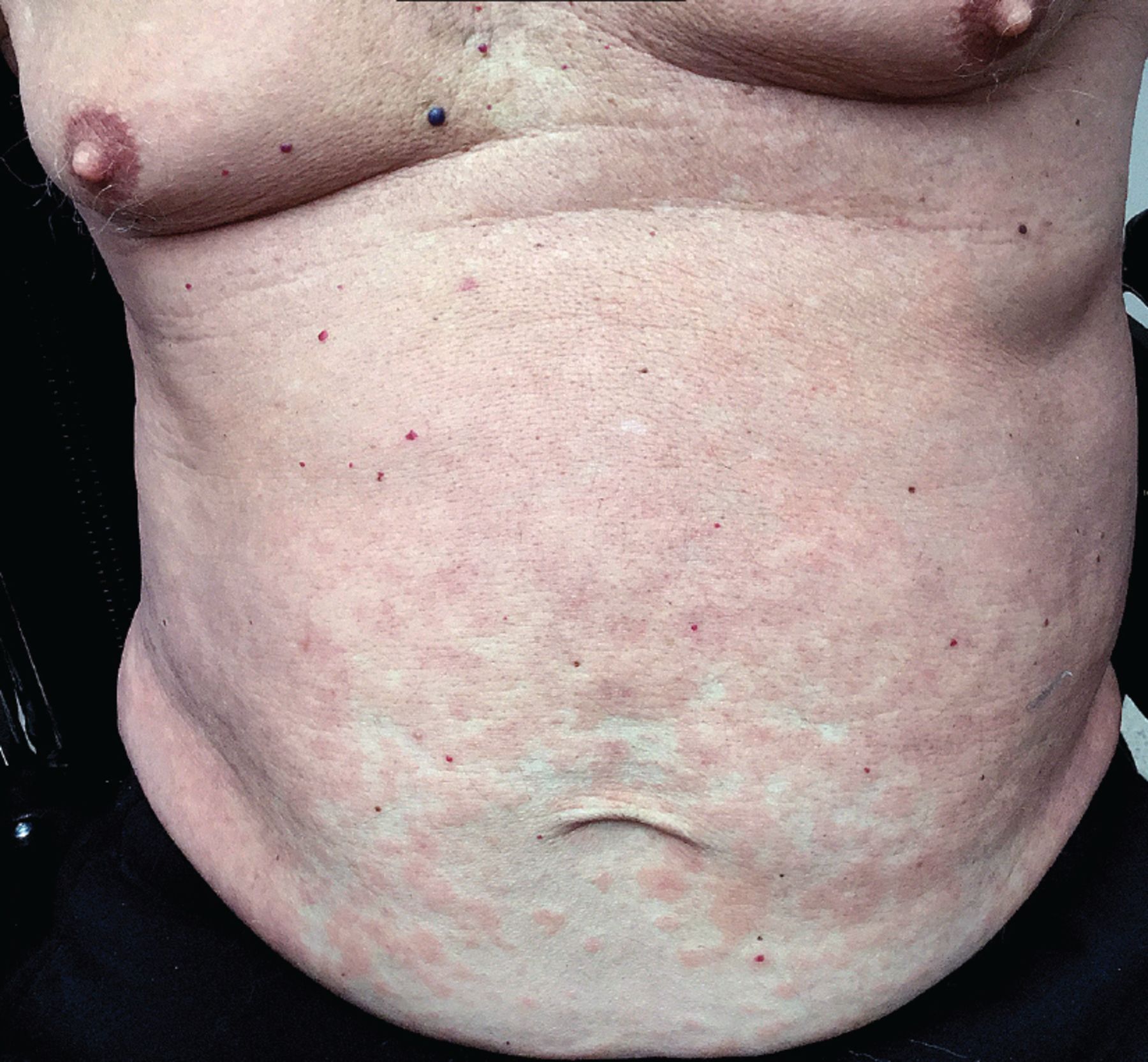
Morbilliform eruptions are common in many viral illnesses and were reported in patients with COVID-19 early in the pandemic.4,5 International registry data indicate that morbilliform eruptions are the most common cutaneous manifestation in patients with laboratory-confirmed COVID-19.6 Typical clinical features include a generalized, symmetric maculopapular rash with pruritus (Figure 1).7

Figure 1
  • Download figure
  • Open in new tab
  • Download powerpoint
Figure 1

Morbilliform rash in COVID-19. A 77-year-old man was hospitalized with COVID-19 and developed bilateral pneumonia and acute hypoxic respiratory failure. Four days after discharge, while continuing to have low-grade fevers, he developed pink papules confluent over the trunk and extremities consistent with a morbilliform eruption.

Patients with COVID-19-associated morbilliform eruptions have an excellent prognosis, with survival rates of 96.9%8 to 97.5%.3

URTICARIA CAN BE THE FIRST SIGN OF COVID-19

Urticaria is also common in COVID-19. The clinical features do not appear to differ from those of idiopathic urticaria and typically consist of generalized pruritic wheals.9,10 On average, urticaria lasts less than 1 week11 and is associated with relatively mild disease and survival rates of 97.8%6 to 98.2%.3

Histologic features also mimic those of idiopathic urticaria and thus limit the value of skin biopsy.9,10 However, urticarial vasculitis has been described in association with COVID-19, suggesting that biopsy should be considered in patients with persistent urticarial plaques with associated purpura.12

Interestingly, in a systematic review of 895 patients with COVID-19,13 105 (12%) had urticarial lesions, and in 17 (16%) of these 105 the urticaria began before the onset of the other COVID-19 symptoms, suggesting that it can be a clue to diagnosis in appropriate clinical settings and can help guide early testing (Figure 2).13 Additionally, an analysis of 200 patients with COVID-19 with cutaneous manifestations14 found a significant association between urticaria and gastrointestinal symptoms, which could assist clinicians in their anticipatory management.

Figure 2
  • Download figure
  • Open in new tab
  • Download powerpoint
Figure 2

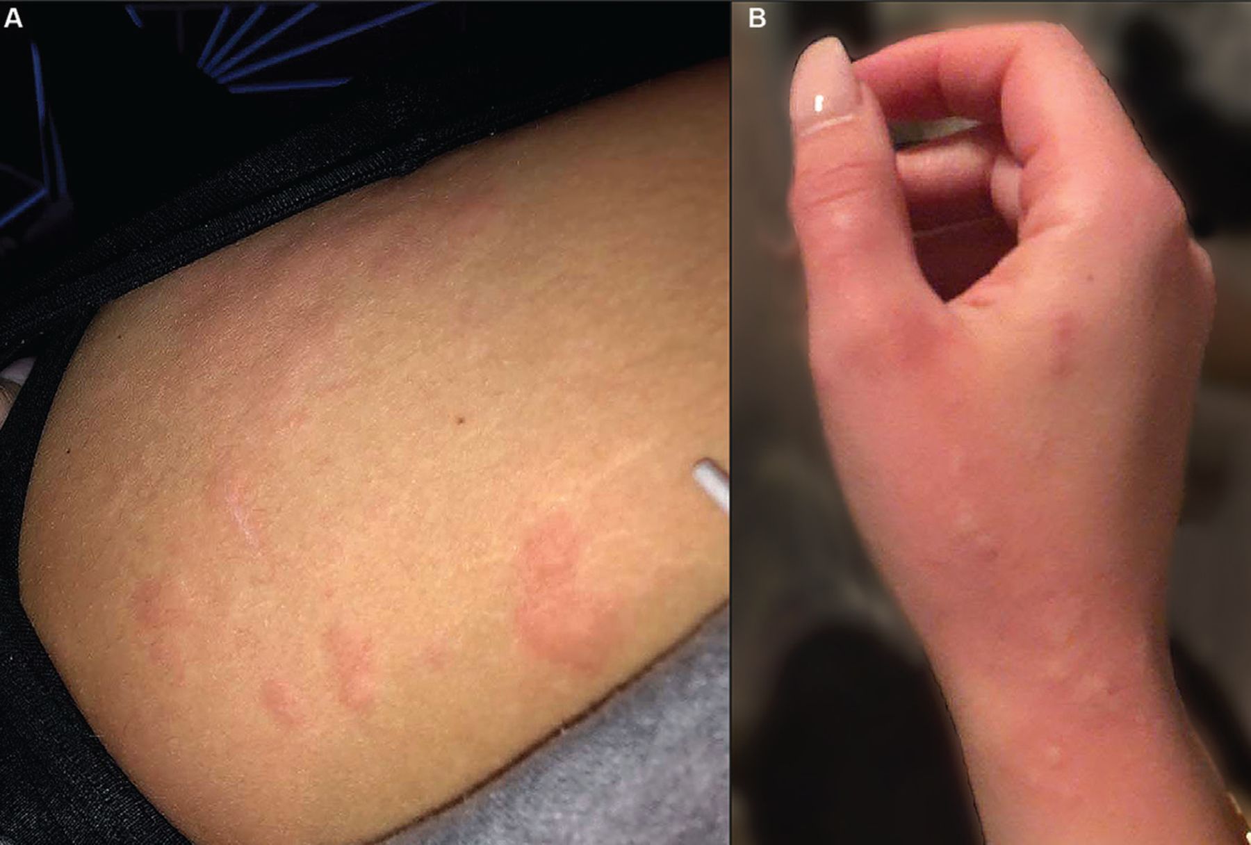
Urticarial lesions preceding COVID-19 diagnosis. A 21-year-old woman with no known previous skin problems developed urticarial lesions in various locations, including the thighs (A) and hands (B) several days before testing positive for COVID-19 as part of a routine screening protocol. She subsequently experienced systemic symptoms including palpitations, cough, fatigue, and loss of taste and smell, but was able to be managed on an outpatient basis with supportive care.

VESICLES CAN ALSO BE THE FIRST SIGN OF COVID-19

Initially described as “varicella-like,”15 vesicular eruptions in COVID-19 have been described in both localized and diffuse distributions. The localized pattern is characterized by monomorphic vesicles in the same stage of evolution that are confined to the trunk (Figure 3). But the diffuse pattern may be more common. A cohort study16 reported that it accounted for 18 (75%) of 24 cases. The diffuse pattern consists of polymorphic papules, vesicles, and pustules that develop simultaneously on the trunk and spread distally, sometimes involving the palms and soles. Lesions tend to resolve after about 8 days without scarring.15

Figure 3
  • Download figure
  • Open in new tab
  • Download powerpoint
Figure 3

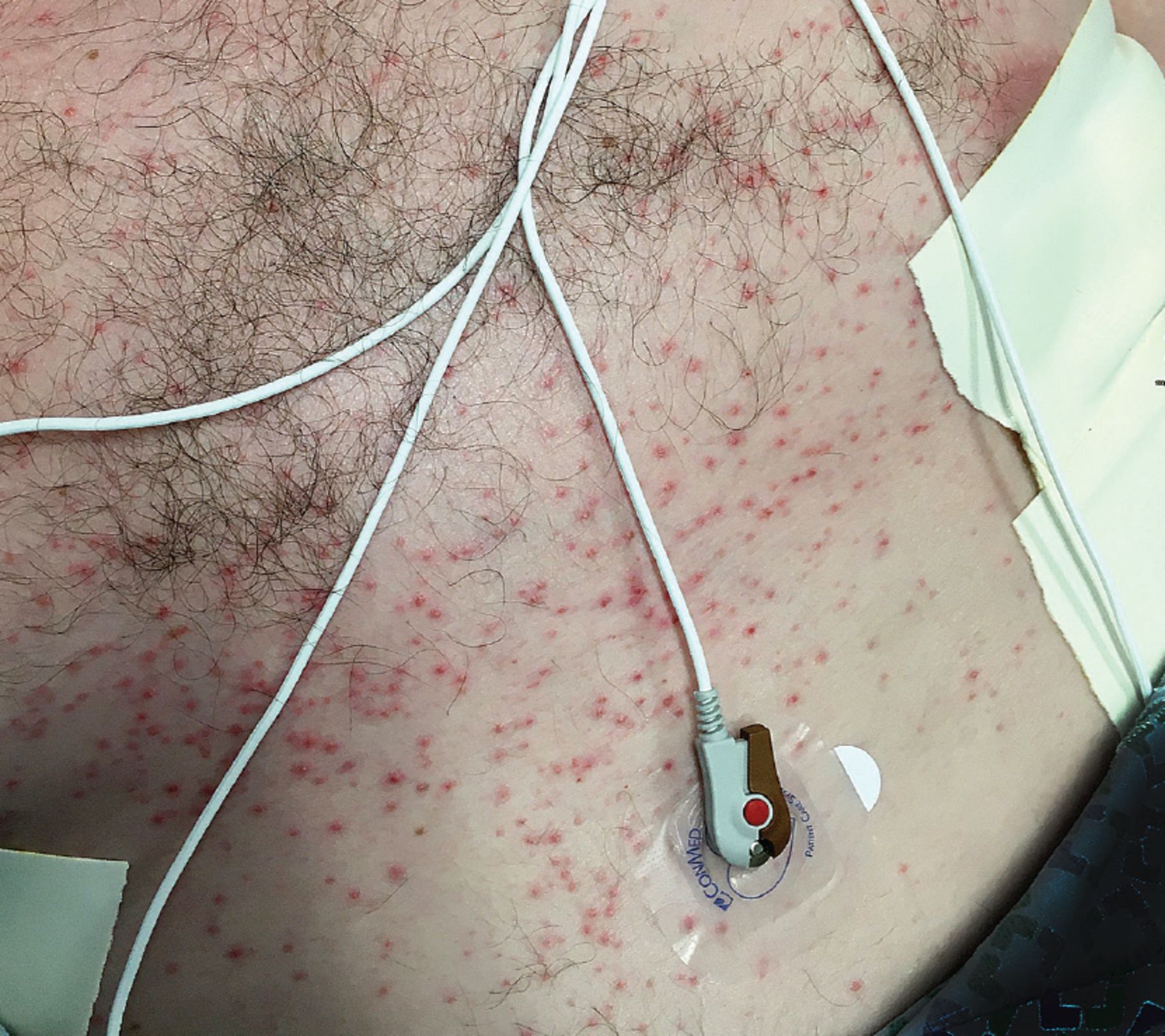
Vesicular eruption in COVID-19. A 52-year-old man developed a vesiculopustular eruption on his trunk during hospitalization for COVID-19 requiring intensive care unit admission and mechanical ventilation for acute respiratory failure due to respiratory distress syndrome. He recovered and was eventually discharged.

Lesional skin biopsies reveal histologic features consistent with viral exanthems, namely vacuolar degeneration of the basal epidermal layer with occasional dyskeratotic keratinocytes and superficial dermal inflammation.15 However, some reports describe prominent keratinocyte acantholysis contributing to formation of intraepidermal vesicles, which is a relatively unusual histologic finding.17 Additionally, while there have been reports of SARS-CoV-2 spike proteins detected with immunohistochemistry in sweat glands and dermal endothelial cells in skin biopsies from COVID-19 patients, 2 studies of COVID-19-associated vesicular rashes detected no SARS-CoV-2 in vesicular fluid by reverse transcriptase polymerase chain reaction testing.16–18

Like urticaria, vesicular eruptions were also commonly noted before other COVID-19 symptoms (in 8.5%3 to 15%13 of cases of COVID-19-associated urticaria) in multiple studies, and therefore may similarly provide an indication for COVID-19 testing and isolation in the appropriate clinical context.3,14,19 Additionally, a systematic review8 reported a possible link between vesicular eruptions and neurologic symptoms including headache, dysgeusia, irritability, and confusion. Like those with morbilliform rash or urticaria, patients with COVID-19 with vesicular eruptions have high survival rates (96.1%3 to 96.6%8).

PSEUDO-CHILBLAINS: LINK TO COVID-19 DEBATED

Although pernio-like acral lesions (Figure 4) were the first cutaneous manifestations to generate significant attention, whether they are truly linked to COVID-19 has been debated.

Figure 4
  • Download figure
  • Open in new tab
  • Download powerpoint
Figure 4

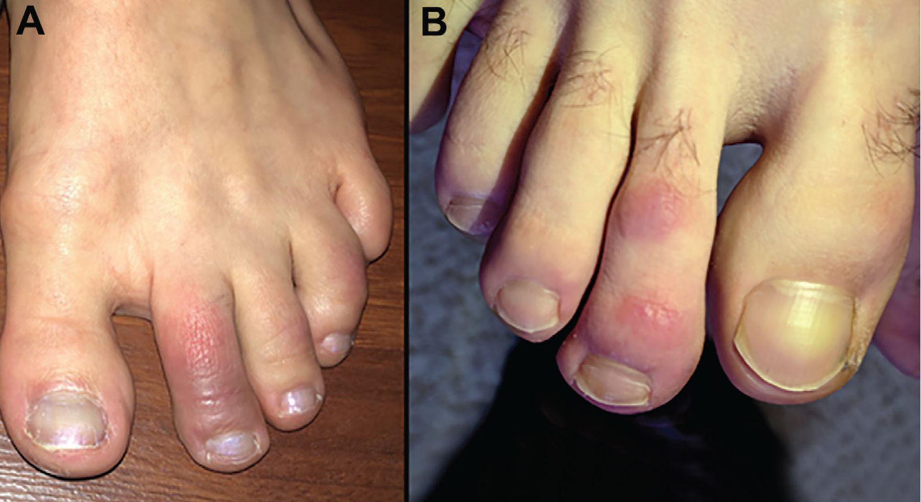
Pseudo-chilblains (“COVID toes”). (A) A 24-year-old woman developed painful erythematous and violaceous macules involving her dorsal toes after testing positive for COVID-19. She had no other symptoms. (B) A young adult man sought care in a telemedicine encounter after developing painful, erythematous papules on his toes. His eventual COVID-19 status is unknown.

Challenging the link are strikingly low rates of positive COVID-19 testing in affected patients, as well as results of several studies20–24 that suggest these lesions are most consistent with typical perniosis, with an increased incidence related to changes in daily routine (eg, quarantining, working from home) during the pandemic rather than infection with SARS-CoV-2. Additionally, a systematic review8 found that pre-existing rheumatologic conditions were more common in patients with presumed COVID-19-related pernio-like lesions, raising the possibility that underlying diagnoses contributed to development of the acral lesions.

However, proponents of the association with COVID-19 point to “outbreaks” of chilblain-like lesions corresponding to COVID-19 waves and propose that an efficient, type I interferon-driven antiviral response could induce pernio-like lesions and suppress both symptoms and confirmatory testing.25–29 Interestingly, information is accumulating about pernio-like lesions in “long-hauler” patients, with a significant association reported between persistent cutaneous and extracutaneous symptoms.30–32

While the debate continues, if these lesions are truly a COVID-19 manifestation, they are fortunately associated with high survival rates (96.4%6 to 98.7%3) and few or no systemic symptoms.33,34

VASO-OCCLUSIVE LESIONS ARE ASSOCIATED WITH HIGHER RISK

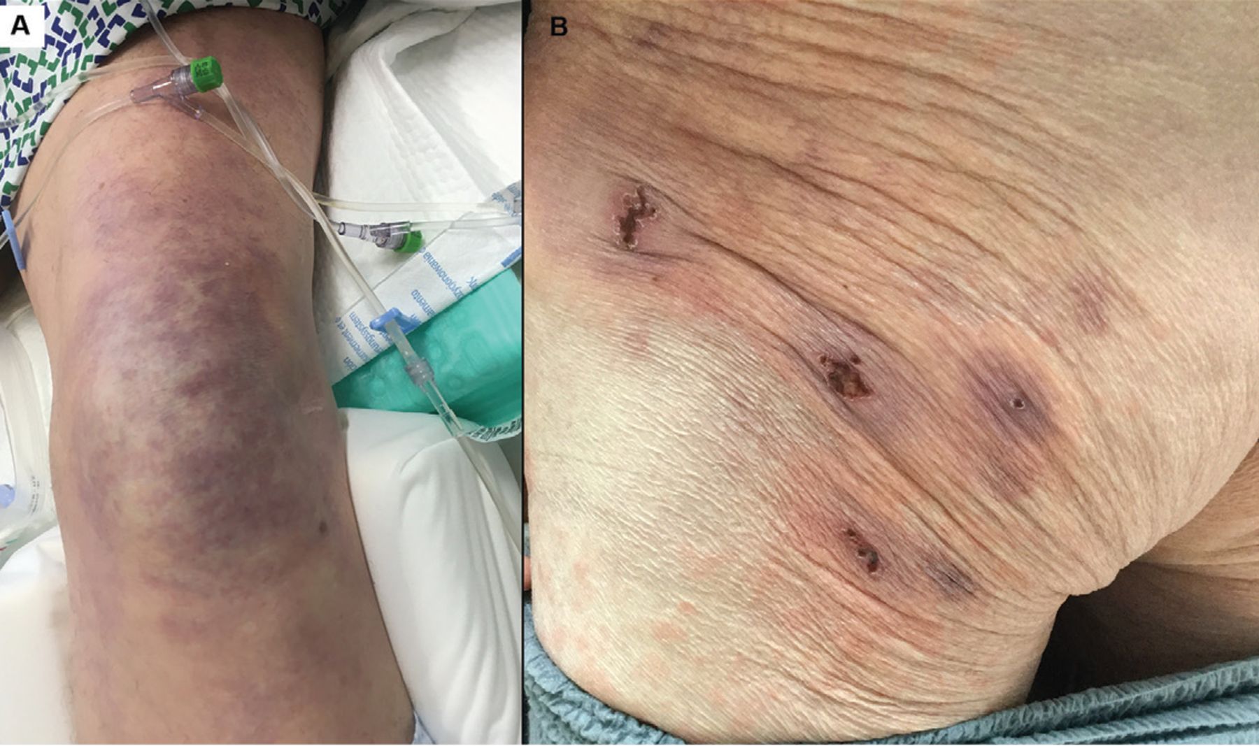
Vaso-occlusive lesions (Figure 5) have been reported in patients with COVID-19 with varied clinical presentations, including fixed livedo racemosa, retiform purpura, and acral ischemia, which may be clinically confused with COVID toes.19 These lesions are most commonly seen in hospitalized patients with moderate to severe COVID-1919 and are associated with higher risks of severe pneumonia and intensive care unit admission and relatively low survival rates (78.9%3 to 81.8%8).

Figure 5
  • Download figure
  • Open in new tab
  • Download powerpoint
Figure 5

Vaso-occlusive lesions in COVID-19. (A) A 62-year-old man with COVID-19 developed an irregular, mottled, purpuric patch on his knee extending onto his thigh during an extended hospitalization complicated by septic shock and acute respiratory failure requiring mechanical ventilation. He died of his illness 3.5 weeks after admission. (B) A 77-year-old man (also described in Figure 1) developed purpuric patches with central hemorrhagic crusts on the left buttock shortly after hospitalization for COVID-19.

Similar patterns of microvascular thrombosis have been found in skin biopsies and pulmonary tissue of COVID-19 patients with vaso-occlusive cutaneous lesions, suggesting that this manifestation could be a marker of systemic microvascular injury.35 Additionally, systemic thrombotic events including deep vein thrombosis and pulmonary embolism have been reported in patients with retiform and necrotic lesions, with rates as high as 64%.6,36 Whether early recognition of these lesions can prompt treatment decisions that decrease systemic thrombotic events or increase overall survival requires further research.

OTHER CUTANEOUS FINDINGS

Other cutaneous findings that have been reported with COVID-194,7,37–39 include oral lesions; reactivation of viral infections; rash resembling symmetrical drug-related intertriginous and flexural exanthema; small-vessel vasculitis; cutaneous hyperesthesia; papulosquamous eruptions; and erythema nodosum-like lesions.

Oral lesions. A study of 666 patients40 reported various oral mucosal findings in 78 (26%) of 304 patients who had mucocutaneous manifestations, and the authors hypothesized that lesions in the mouth may be under reported due to contact precautions and assisted ventilation that limits examination of the oral mucosa.40

Reactivation of herpes simplex virus (HSV) and varicella-zoster virus (VZV) infections has been reported in conjunction with COVID-19 infection. A cross-sectional study of nearly 900 patients with COVID-19 found a significantly higher prevalence of HSV-1 and VZV than in the hospital population, even when adjustments were made for numerous comorbidities.41 Some reports suggest that HSV reactivation may be associated with more severe COVID-19 infection, including acute respiratory distress syndrome and viremia,42,43 but the prognostic implications of treating these reactivations has not yet been robustly investigated.

DISCLOSURES

Dr. Fernandez reports consulting for Abbvie Pharmaceuticals, Boehringer Ingelheim, Bristol-Myers Squibb, Mallinckrodt, Novartis, and UCB; teaching and speaking (non-promotional) for Abbvie Pharmaceuticals, Kyowa Kirin, Mallinckrodt, and Novartis; research/independent contracting for Abbvie Pharmaceuticals, Mallinckrodt, Novartis, and Pfizer; and work as advisor or review panel participant for Abbvie Pharmaceuticals. Dr. Polly reports no relevant financial relationships which, in the context of her contributions, could be perceived as a potential conflict of interest.

Acknowledgment

The authors would like to thank Janine Sot, MBA, for her help and expertise in preparing the figures in this article.

  • Copyright © 2022 The Cleveland Clinic Foundation. All Rights Reserved.

REFERENCES

  1. ↵
    1. Visconti A,
    2. Bataille V,
    3. Rossi N, et al
    . Diagnostic value of cutaneous manifestation of SARS-CoV-2 infection. Br J Dermatol 2021; 184(5):880–887. doi:10.1111/bjd.19807
    OpenUrlCrossRef
  2. ↵
    1. Rekhtman S,
    2. Tannenbaum R,
    3. Strunk A, et al
    . Eruptions and related clinical course among 296 hospitalized adults with confirmed COVID-19. J Am Acad Dermatol 2021; 84(4):946–952. doi:10.1016/j.jaad.2020.12.046
    OpenUrlCrossRef
  3. ↵
    1. Tan SW,
    2. Tam YC,
    3. Oh CC
    . Skin manifestations of COVID-19: a worldwide review. JAAD Int 2021; 2:119–133. doi:10.1016/j.jdin.2020.12.003
    OpenUrlCrossRefPubMed
  4. ↵
    1. Galván Casas C,
    2. Català A,
    3. Carretero Hernández G, et al
    . Classification of the cutaneous manifestations of COVID-19: a rapid prospective nationwide consensus study in Spain with 375 cases. Br J Dermatol 2020; 183(1):71–77. doi:10.1111/bjd.19163
    OpenUrlCrossRefPubMed
  5. ↵
    1. Sachdeva M,
    2. Gianotti R,
    3. Shah M, et al
    . Cutaneous manifestations of COVID-19: Report of three cases and a review of literature. J Dermatol Sci 2020; 98(2):75–81. doi:10.1016/j.jdermsci.2020.04.011
    OpenUrlCrossRef
  6. ↵
    1. Freeman EE,
    2. McMahon DE,
    3. Lipoff JB, et al
    . The spectrum of COVID-19-associated dermatologic manifestations: an international registry of 716 patients from 31 countries. J Am Acad Dermatol 2020; 83(4):1118–1129. doi:10.1016/j.jaad.2020.06.1016
    OpenUrlCrossRef
  7. ↵
    1. Genovese G,
    2. Moltrasio C,
    3. Berti E,
    4. Marzano AV
    . Skin manifestations associated with COVID-19: current knowledge and future perspectives. Dermatology 2021; 237(1):1–12. doi:10.1159/000512932
    OpenUrlCrossRefPubMed
  8. ↵
    1. Jamshidi P,
    2. Hajikhani B,
    3. Mirsaeidi M,
    4. Vahidnezhad H,
    5. Dadashi M,
    6. Nasiri MJ
    . Skin manifestations in COVID-19 patients: are they indicators for disease severity? A systematic review. Front Med (Lausanne) 2021. Published 2021 Feb 16. doi:10.3389/fmed.2021.634208
    OpenUrlCrossRef
  9. ↵
    1. Rongioletti F,
    2. Ferreli C,
    3. Sena P,
    4. Caputo V,
    5. Atzori L
    . Clinicopathologic correlations of COVID-19-related cutaneous manifestations with special emphasis on histopathologic patterns. Clin Dermatol 2021; 39(1):149–162. doi:10.1016/j.clindermatol.2020.12.004
    OpenUrlCrossRef
  10. ↵
    1. Almeida G,
    2. Arruda S,
    3. Marques E,
    4. Michalany N,
    5. Sadick N
    . Presentation and management of cutaneous manifestations of COVID-19. J Drugs Dermatol 2021; 20(1):76–83. doi:10.36849/JDD.5676
    OpenUrlCrossRef
  11. ↵
    1. Algaadi SA
    . Urticaria and COVID-19: a review. Dermatol Ther 2020; 33(6):e14290. doi:10.1111/dth.14290
    OpenUrlCrossRefPubMed
  12. ↵
    1. de Perosanz-Lobo D,
    2. Fernandez-Nieto D,
    3. Burgos-Blasco P, et al
    . Urticarial vasculitis in COVID-19 infection: a vasculopathy-related symptom? J Eur Acad Dermatol Venereol 2020; 34(10):e566–e568. doi:10.1111/jdv.16713
    OpenUrlCrossRef
  13. ↵
    1. Gisondi P,
    2. Di Leo S,
    3. Bellinato F,
    4. Cazzaniga S,
    5. Piaserico S,
    6. Naldi L
    . Time of onset of selected skin lesions associated with COVID-19: a systematic review. Dermatol Ther (Heidelb) 2021; 11(3):695–705. doi:10.1007/s13555-021-00526-8
    OpenUrlCrossRef
  14. ↵
    1. Marzano AV,
    2. Genovese G,
    3. Moltrasio C, et al
    . The clinical spectrum of COVID-19-associated cutaneous manifestations: an Italian multicenter study of 200 adult patients. J Am Acad Dermatol 2021; 84(5):1356–1363. doi:10.1016/j.jaad.2021.01.023
    OpenUrlCrossRef
  15. ↵
    1. Marzano AV,
    2. Genovese G,
    3. Fabbrocini G, et al
    . Varicella-like exanthem as a specific COVID-19-associated skin manifestation: multicenter case series of 22 patients. J Am Acad Dermatol 2020; 83(1):280–285. doi:10.1016/j.jaad.2020.04.044
    OpenUrlCrossRefPubMed
  16. ↵
    1. Fernandez-Nieto D,
    2. Ortega-Quijano D,
    3. Jimenez-Cauhe J, et al
    . Clinical and histological characterization of vesicular COVID-19 rashes: a prospective study in a tertiary care hospital. Clin Exp Dermatol 2020; 45(7):872–875. doi:10.1111/ced.14277
    OpenUrlCrossRef
  17. ↵
    1. Mahé A,
    2. Birckel E,
    3. Merklen C, et al
    . Histology of skin lesions establishes that the vesicular rash associated with COVID-19 is not ‘varicella-like’. J Eur Acad Dermatol Venereol 2020; 34(10):e559–e561. doi:10.1111/jdv.16706
    OpenUrlCrossRef
  18. ↵
    1. Liu J,
    2. Li Y,
    3. Liu L, et al
    . Infection of human sweat glands by SARS-CoV-2. Cell Discov 2020; 6(1):84. doi:10.1038/s41421-020-00229-y
    OpenUrlCrossRef
  19. ↵
    1. Galván Casas C,
    2. Català A,
    3. Carretero Hernández G, et al
    . Classification of the cutaneous manifestations of COVID-19: a rapid prospective nationwide consensus study in Spain with 375 cases. Br J Dermatol 2020; 183(1):71–77. doi:10.1111/bjd.19163
    OpenUrlCrossRefPubMed
  20. ↵
    1. McCleskey PE,
    2. Zimmerman B,
    3. Lieberman A, et al
    . Epidemiologic analysis of chilblains cohorts before and during the COVID-19 pandemic. JAMA Dermatol 2021; 157(8):947–953. doi:10.1001/jamadermatol.2021.2120
    OpenUrlCrossRef
    1. Herman A,
    2. Peeters C,
    3. Verroken A, et al
    . Evaluation of chilblains as a manifestation of the COVID-19 pandemic. JAMA Dermatol 2020; 156(9):998–1003. doi:10.1001/jamadermatol.2020.2368
    OpenUrlCrossRefPubMed
    1. Baeck M,
    2. Peeters C,
    3. Herman A
    . Chilblains and COVID-19: further evidence against a causal association. J Eur Acad Dermatol Venereol 2021; 35(1):e2–e3. doi:10.1111/jdv.16901
    OpenUrlCrossRef
    1. Baeck M,
    2. Herman A
    . COVID toes: where do we stand with the current evidence? Int J Infect Dis 2021; 102:53–55. doi:10.1016/j.ijid.2020.10.021
    OpenUrlCrossRef
  21. ↵
    1. Le Cleach L,
    2. Dousset L,
    3. Assier H, et al
    . Most chilblains observed during the COVID-19 outbreak occur in patients who are negative for COVID-19 on polymerase chain reaction and serology testing. Br J Dermatol 2020; 183(5):866–874. doi:10.1111/bjd.19377
    OpenUrlCrossRef
  22. ↵
    1. Giraud-Kerleroux L,
    2. Mongereau M,
    3. Cassius C, et al
    . Detection of a second outbreak of chilblain-like lesions during COVID-19 pandemic through teledermatology. J Eur Acad Dermatol Venereol 2021; 35(9):e556–e558. doi:10.1111/jdv.17378
    OpenUrlCrossRef
    1. Hubiche T,
    2. Le Duff F,
    3. Fontas E,
    4. Rapp J,
    5. Chiaverini C,
    6. Passeron T
    . Relapse of chilblain-like lesions during the second wave of the COVID-19 pandemic: a cohort follow-up. Br J Dermatol 2021; 185(4):858–859. doi:10.1111/bjd.20584
    OpenUrlCrossRef
    1. Hubiche T,
    2. Cardot-Leccia N,
    3. Le Duff F, et al
    . Clinical, laboratory, and interferon-alpha response characteristics of patients with chilblain-like lesions during the COVID-19 pandemic. JAMA Dermatol 2021; 157(2):202–206. doi:10.1001/jamadermatol.2020.4324
    OpenUrlCrossRef
    1. Cappel MA,
    2. Cappel JA,
    3. Wetter DA
    . Pernio (chilblains), SARS-CoV-2, and COVID toes unified through cutaneous and systemic mechanisms. Mayo Clin Proc 2021; 96(4):989–1005. doi:10.1016/j.mayocp.2021.01.009
    OpenUrlCrossRef
  23. ↵
    1. Arkin LM,
    2. Moon JJ,
    3. Tran JM, et al
    . From your nose to your toes: a review of severe acute respiratory syndrome coronavirus 2 pandemic–associated pernio. J Invest Dermatol 2021; 141(12):2791–2796. doi:10.1016/j.jid.2021.05.024
    OpenUrlCrossRef
  24. ↵
    1. McMahon DE,
    2. Gallman AE,
    3. Hruza GJ, et al
    . Long COVID in the skin: a registry analysis of COVID-19 dermatological duration. Lancet Infect Dis 2021; 21(3):313–314. doi:10.1016/S1473-3099(20)30986-5
    OpenUrlCrossRef
    1. Mehta P,
    2. Bunker CB,
    3. Ciurtin C, et al
    . Chilblain-like acral lesions in long COVID-19: management and implications for understanding microangiopathy. Lancet Infect Dis 2021; 21(7):912. doi:10.1016/S1473-3099(21)00133-X
    OpenUrlCrossRef
  25. ↵
    1. Moghadam P,
    2. Frumholtz L,
    3. Jaume L, et al
    . Frequency of relapse and persistent cutaneous symptoms after a first episode of chilblain-like lesion during the COVID-19 pandemic. J Eur Acad Dermatol Venereol 2021; 35(9):e566–e568. doi:10.1111/jdv.17393
    OpenUrlCrossRef
  26. ↵
    1. Piccolo V,
    2. Neri I,
    3. Filippeschi C, et al
    . Chilblain-like lesions during COVID-19 epidemic: a preliminary study on 63 patients. J Eur Acad Dermatol Venereol 2020; 34(7):e291– 293. doi:10.1111/jdv.16526
    OpenUrlCrossRef
  27. ↵
    1. de Masson A,
    2. Bouaziz JD,
    3. Sulimovic L, et al
    . Chilblains is a common cutaneous finding during the COVID-19 pandemic: A retrospective nationwide study from France. J Am Acad Dermatol 2020; 83(2):667–670. doi:10.1016/j.jaad.2020.04.161
    OpenUrlCrossRefPubMed
  28. ↵
    1. Magro C,
    2. Mulvey JJ,
    3. Berlin D, et al
    . Complement associated microvascular injury and thrombosis in the pathogenesis of severe COVID-19 infection: a report of five cases. Transl Res 2020; 220:1–13. doi:10.1016/j.trsl.2020.04.007
    OpenUrlCrossRefPubMed
  29. ↵
    1. Droesch C,
    2. Do MH,
    3. DeSancho M,
    4. Lee EJ,
    5. Magro C,
    6. Harp J
    . Livedoid and purpuric skin eruptions associated with coagulopathy in severe COVID-19. JAMA Dermatol 2020; 156(9):1–3. doi:10.1001/jamadermatol.2020.2800
    OpenUrlCrossRef
  30. ↵
    1. Mahé A,
    2. Birckel E,
    3. Krieger S,
    4. Merklen C,
    5. Bottlaender L
    . A distinctive skin rash associated with coronavirus disease 2019? J Eur Acad Dermatol Venereol 2020; 34(6): e246–e247. doi:10.1111/jdv.16471
    OpenUrlCrossRef
    1. Krajewski PK,
    2. Maj J,
    3. Szepietowski JC
    . Cutaneous hyperaesthesia in SARS-CoV-2 infection: rare but not unique clinical manifestation. Acta Derm Venereol 2021; 101(1):adv00366. Published 2021 Jan 5. doi:10.2340/00015555-3729
    OpenUrlCrossRef
  31. ↵
    1. Kuriyama Y,
    2. Shimizu A,
    3. Oka H, et al
    . Erythema nodosumlike eruption in coronavirus disease 2019: a case report and literature review of Asian countries. J Dermatol 2021; 48(10):1588–1592. doi:10.1111/1346-8138.16071
    OpenUrlCrossRef
  32. ↵
    1. Nuno-Gonzalez A,
    2. Martin-Carrillo P,
    3. Magaletsky K, et al
    . Prevalence of mucocutaneous manifestations in 666 patients with COVID-19 in a field hospital in Spain: oral and palmoplantar findings. Br J Dermatol 2021; 184(1): 184–185. doi:10.1111/bjd.19564
    OpenUrlCrossRef
  33. ↵
    1. Katz J,
    2. Yue S,
    3. Xue W
    . Herpes simplex and herpes zoster viruses in COVID-19 patients. Ir J Med Sci 2021; 1–5. doi:10.1007/s11845-021-02714-z
    OpenUrlCrossRef
  34. ↵
    1. Le Balc’h P,
    2. Pinceaux K,
    3. Pronier C,
    4. Seguin P,
    5. Tadié JM,
    6. Reizine F
    . Herpes simplex virus and cytomegalovirus reactivations among severe COVID-19 patients. Crit Care 2020; 24(1):530. doi:10.1186/s13054-020-03252-3
    OpenUrlCrossRefPubMed
  35. ↵
    1. Xu R,
    2. Zhou Y,
    3. Cai L, et al
    . Co-reactivation of the human herpesvirus alpha subfamily (herpes simplex virus-1 and varicella zoster virus) in a critically ill patient with COVID-19. Br J Dermatol 2020; 183(6):1145–1147. doi:10.1111/bjd.19484
    OpenUrlCrossRef
PreviousNext
Back to top

In this issue

Cleveland Clinic Journal of Medicine: 89 (3)
Cleveland Clinic Journal of Medicine
Vol. 89, Issue 3
1 Mar 2022
  • Table of Contents
  • Table of Contents (PDF)
  • Index by author
  • Complete Issue (PDF)
Print
Download PDF
Article Alerts
Sign In to Email Alerts with your Email Address
Email Article

Thank you for your interest in spreading the word on Cleveland Clinic Journal of Medicine.

NOTE: We only request your email address so that the person you are recommending the page to knows that you wanted them to see it, and that it is not junk mail. We do not capture any email address.

Enter multiple addresses on separate lines or separate them with commas.
Common skin signs of COVID-19 in adults: An update
(Your Name) has sent you a message from Cleveland Clinic Journal of Medicine
(Your Name) thought you would like to see the Cleveland Clinic Journal of Medicine web site.
CAPTCHA
This question is for testing whether or not you are a human visitor and to prevent automated spam submissions.
Citation Tools
Common skin signs of COVID-19 in adults: An update
Samantha Polly, Anthony P. Fernandez
Cleveland Clinic Journal of Medicine Mar 2022, 89 (3) 161-167; DOI: 10.3949/ccjm.89a.21126

Citation Manager Formats

  • BibTeX
  • Bookends
  • EasyBib
  • EndNote (tagged)
  • EndNote 8 (xml)
  • Medlars
  • Mendeley
  • Papers
  • RefWorks Tagged
  • Ref Manager
  • RIS
  • Zotero
Share
Common skin signs of COVID-19 in adults: An update
Samantha Polly, Anthony P. Fernandez
Cleveland Clinic Journal of Medicine Mar 2022, 89 (3) 161-167; DOI: 10.3949/ccjm.89a.21126
Reddit logo Twitter logo Facebook logo Mendeley logo
  • Tweet Widget
  • Facebook Like
  • Linkedin Share Button

Jump to section

  • Article
    • ABSTRACT
    • MORBILLIFORM RASH: THE MOST COMMON SKIN MANIFESTATION
    • URTICARIA CAN BE THE FIRST SIGN OF COVID-19
    • VESICLES CAN ALSO BE THE FIRST SIGN OF COVID-19
    • PSEUDO-CHILBLAINS: LINK TO COVID-19 DEBATED
    • VASO-OCCLUSIVE LESIONS ARE ASSOCIATED WITH HIGHER RISK
    • OTHER CUTANEOUS FINDINGS
    • DISCLOSURES
    • Acknowledgment
    • REFERENCES
  • Figures & Data
  • Info & Metrics
  • PDF

Related Articles

  • No related articles found.
  • PubMed
  • Google Scholar

Cited By...

  • No citing articles found.
  • Google Scholar

More in this TOC Section

  • Smallpox and monkeypox: Looking back and looking ahead
  • To repeat or not to repeat? Measuring bone mineral density during anti-resorptive therapy or a drug holiday
  • Colovesical fistula in men with chronic urinary tract infection: A diagnostic challenge
Show more Review

Similar Articles

Subjects

  • Infectious Diseases
  • Pulmonology
  • Dermatology
  • Covid-19

Navigate

  • Current Issue
  • Past Issues
  • Supplements
  • Article Type
  • Specialty
  • CME/MOC Articles
  • CME/MOC Calendar
  • Media Kit

Authors & Reviewers

  • Manuscript Submission
  • Authors & Reviewers
  • Subscriptions
  • About CCJM
  • Contact Us
  • Cleveland Clinic Center for Continuing Education
  • Consult QD

Share your suggestions!

Copyright © 2023 The Cleveland Clinic Foundation. All rights reserved. The information provided is for educational purposes only. Use of this website is subject to the website terms of use and privacy policy. 

Powered by HighWire