


ABSTRACT
Vitamin D supplementation is common in the United States, with about one-fifth of the adult population taking a daily supplement in one form or another. Although the detrimental effects of insufficient sun exposure in childhood was established centuries ago, the beneficial effects of vitamin D sufficiency have only recently been established, given the myriad investigations associating vitamin D deficiency with numerous chronic diseases. But it is far less clear precisely how to replete low 25-hydroxyvitamin D (25[OH]D) levels, how long treatment should be continued, if there are potential hazards in doing so, and how to assess and counsel patients regarding the use of vitamin D. This article provides a brief historical review, examines how to assess and counsel patients on the use of vitamin D, presents scenarios that clinicians are likely to encounter, and reviews the literature on recommendations for vitamin D supplementation.
Typical vitamin D replacement requires at least 2,000 IU/ day, with some authors recommending 5,000 IU/day.
The richest food sources of vitamin D, consumed in manageable portions, provide only a small percentage of the recommended daily intake of 800 IU.
Several mechanisms contribute to the ability of vitamin D3 to attain and maintain goal serum concentrations of 25(OH)D more efficiently than vitamin D2, including that vitamin D2 has a lower affinity for D binding protein and D 25-hydroxylase converts D to 25(OH)D3 substantially faster.
Vitamin D supplementaton is ubiquitous in the United States, and 20% of all adults take a dietary supplement containing vitamin D. Supplement use is highest in the very young and in people age 60 and older.1 Observations of the detrimental effects of inadequate sun exposure date back centuries. In 1650, scientists noted that children who lived in polluted and crowded cities in Northern Europe developed debilitating skeletal abnormalities, including bowed legs.2 In the 1890s, epidemiologic studies in Great Britain noted the higher incidence of significant skeletal abnormalities in children in industrialized cities compared with those who lived in rural areas of the British highlands.3 In the United States, it took until the 1920s to achieve wide acceptance that routinely exposing children to sunshine could prevent debilitating skeletal abnormalities.4
During the 18th and 19th centuries, cod-liver oil was commonly used to prevent and treat skeletal abnormalities in children.5 The antirachitic factor of cod-liver oil was later isolated and became known as vitamin D. Investigations early in the 20th century led to the vitamin D fortification of milk and infant formulas that became common practice by the 1930s. As a result, rickets, once the most common disease in children, was eradicated in the United States 100 years ago.
VITAMIN D DEFICIENCY AND CURRENT RECOMMENDATIONS
Eradication of rickets was a giant step forward in skeletal health of youngsters. However, fortifying foods and beverages with enough vitamin D to prevent rickets but avoid hypercalcemia did not eliminate vitamin D deficiency.
Vitamin D deficiency is common in the United States and around the globe. The most common cause of deficiency is insufficient intake (oral or dermal). In a study using National Health and Nutrition Examination Survey (NHANES) data from 2011 to 2014, almost 20% of the US population had serum 25-hydroxyvitamin D (25[OH]D) values categorized as “at risk for inadequacy” (defined as 30 to 49 nmol/L or 12 to 19 ng/mL), and 5% were categorized as “at risk for deficiency” (< 30 nmol/L or 12 ng/mL).6 These reference ranges may be lower than what most clinicians consider to be deficient. For example, numerous studies have found a recommended threshold of 50 nmol/L (20 ng/mL) for bone health to be insufficient for fall or fracture risk reduction.7
Immunologic effects
Vitamin D supplementation to prevent and treat immune-related diseases including COVID-19 was reviewed by Charoenngam et al.8 In an extensive examination of the immunologic effects of vitamin D supplements, the authors described the immunomodulatory hormonal effects of vitamin D, noted significant biologic effects on the innate and adaptive immune systems, cited the immunomodulatory and antiviral effects of the active form of vitamin D (1,25 dihydroxyvitamin D), and suggested that vitamin D supplementation might reduce the risk and severity of COVID-19 infection. They concluded that although the optimal level of vitamin D remains unclear, maintaining a serum 25(OH)D level of 100 to 150 nmol/L (40 to 60 ng/mL) is recommended.8
As reported at an American Academy of Dermatology conference in 2005,9 repeated exposure to ultraviolet (UV) light activates both the innate and adaptive arms of the immune system, and UV light from solar radiation has dose-dependent effects on cells, with cellular and DNA damage that can cause immunosuppression.9
Guidelines for replacement
In 2011, the Endocrine Society issued clinical practice guidelines that defined vitamin D deficiency as less than 30 ng/mL and recommended minimum replacement dosages (Table 1).10
Minimum requirements for vitamin D as defined by the Endocrine Society guidelines
The guidelines identify minimum requirements to maximize bone health and muscle function. However, achieving blood levels above 30 ng/mL (which is considered below normal or low-normal in most laboratory reference ranges) may require more than 2,000 IU daily. Patients with obesity may require several times that dose to attain and maintain a normal level. The Endocrine Society guidelines, in addressing the issue of assay variability, note that in the clinical setting, achieving a level of 40 ng/mL will not result in toxicity but will ensure that an individual’s true value is greater than 30 ng/mL.10
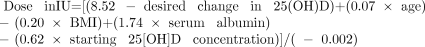
Investigators have considered whether a predictive equation could help clinicians select the correct replacement dose of vitamin D for their patients. Singh et al11 addressed this question with a retrospective observational study. After reviewing the response to vitamin D supplementation in more than 1,300 ambulatory and nursing home patients and employing multiple regression analyses, they published a series of equations that predict the dose of vitamin D needed to achieve a given change in the serum concentration of 25(OH)D in these patient populations. Their equation for calculating the dose in international units (IU) that incorporates body mass index (BMI) was as follows11:
Singh et al speculated that lack of sun exposure explained the need for higher doses of vitamin D in nursing home patients, since their analyses concluded that increased age alone was not a negative factor in response to vitamin D treatment.11 Their analyses did not address the duration of treatment, but Singh et al acknowledged that many patients require long-term maintenance therapy. They further observed that 5,000 IU per day is usually needed to correct deficiency, and a typical maintenance dose should be at least 2,000 IU daily.11
HOW TO REPLACE VITAMIN D
Vitamin D dietary supplements are widely available, and in 2020, the industry’s estimated market value exceeded $1.1 billion, projected to reach close to $1.6 billion by 2025.12 The annual growth rate is more than 7% due to people paying more attention to their nutrition and their health in general.
The popularity of vitamin D supplements has been fueled at least in part by campaigns educating the public about the risks of skin cancer due to excess sun exposure, the association of vitamin D deficiency with many chronic diseases, and the association of vitamin D levels with optimal immune function.
Vitamin D supplements are available by prescription, over-the-counter, and online. In 2021, the cost per 100 tablets of 2,000 IU vitamin D3 was around $0.05 per tablet, while 100 capsules of 50,000 IU vitamin D2 or D3 started at $0.25 per capsule.
Is it possible to get sufficient vitamin D exclusively from diet?
Despite fortification of commonly consumed products such as milk, food sources of vitamin D are few, and even the richest sources consumed in manageable portions provide only a small percentage of the recommended daily intake (Table 2).1
Vitamin D content of selected foods
CLINICAL SCENARIO 1: VITAMIN D2 OR D3?
An otherwise healthy 30-year-old woman with a BMI of 37 kg/m2 was referred for vitamin D deficiency “unresponsive to D repletion.” Her initial 25(OH)D level was 14 ng/mL. After taking vitamin D2 at a dose of 50,000 IU once weekly with her morning coffee for 4 weeks, her 25(OH)D level remained at 21 ng/mL, still below the normal range.
The clinical challenges with this patient are to consider whether vitamin D2 (ergocalciferol) or D3 (cholecalciferol) makes a difference, and whether taking it on an empty stomach is optimal for absorption.
Several recent articles have addressed the question of whether D2 and D3 supplements are equivalent in raising serum 25(OH)D.13–15 Houghton and Vieth13 questioned assumptions about their equivalency and proposed several mechanisms that may contribute to the ability of vitamin D3 to maintain higher serum concentrations over time, including the following:
Supplementation with vitamin D2 produces serum 25(OH)D2, but its lower affinity for D binding protein results in a shorter half-life than that of 25(OH)D3
Mitochondrial vitamin D 25-hydroxylase converts vitamin D3 to 25(OH)D3 five times faster than it converts vitamin D2 to 25(OH)D2.
In a systematic review and meta-analysis, Tripkovic et al14 concluded that supplementation with vitamin D3 had a significant and positive effect in the raising of serum 25(OH)D concentrations compared with the effect of D2 (P = .001). In a study that explored the relative potency of vitamin D2 and vitamin D3, Armas et al15 found the 2 forms to be equivalent in absorption. Further, they both produced similar increases in serum 25(OH)D in the first 72 hours, but the 25(OH)D level continued to rise in the D3-treated patients, peaking at day 14. Their calculated area under the curve at 28 days indicated that D3 was 9.5 times more potent than D2.
Any difference in how it is taken?
Does it matter if the supplement is taken on an empty stomach vs with a meal? In a small study, Mulligan and Licata16 found that taking either vitamin D2 or D3 with the largest meal of the day increased the average serum 25(OH)D level by 50.2% (± 13.4%).
Similarly, a systematic review by Silva and Furlanetto17 included randomized controlled trials examining the response to a single dose of vitamin D taken with a fat-free meal vs meals that contained 15 g or more of fat. Mean serum 25(OH)D concentrations were higher in those who took the supplement with a meal that included at least 15 g of fat.17
Recommended treatment for this patient
This 30-year-old female patient, deemed unresponsive to vitamin D repletion, was treated with vitamin D3 50,000 IU weekly for 8 weeks taken with dinner. Her 25(OH)D level rose to 42.8 ng/mL.
CLINICAL SCENARIO 2: PHOTOTHERAPY TO COUNTER MALABSORPTION?
A 38-year-old man with a history of fistulizing Crohn disease had undergone multiple small-bowel resections and had become dependent on parenteral nutrition. His 25(OH)D level was 12 ng/mL despite taking vitamin D2 50,000 IU daily. In an effort to overcome his malabsorption issue, he would bite into the gel cap to release the contents before swallowing the supplement.
Dual x-ray absorptiometry was notable for an extraordinarily low hip Z-score of −3.4, his long bones were painful to palpation, and his parathyroid hormone level was significantly elevated at 248 pg/mL (reference range 15–65 pg/mL). Osteomalacia is not uncommon in this patient population, but treating the vitamin D deficiency can be very challenging.
In addition to Crohn disease, other conditions can interfere with vitamin D absorption, including a history of malabsorptive-type bariatric surgery, celiac disease, cystic fibrosis, steatorrhea, short bowel disease, inflammatory bowel disease, and severe cholestasis.17 A vitamin D challenge test is one way to confirm the absorptive capability for vitamin D supplementation in these patients.
When vitamin D is taken orally, it is incorporated into the chylomicron fraction, and about 80% of the dose is absorbed into the lymphatics. The blood level of 25(OH)D will peak about 12 hours after a single dose of 50,000 IU. Knowing this about oral absorption of vitamin D allows for provocative testing in patients with suspected malabsorption of the vitamin.
To test for malabsorption, a blood sample is drawn immediately before administering a 50,000-IU oral dose of vitamin D. The blood draw is repeated in 12 to 24 hours. If no increase in 25(OH)D is noted, the patient has “complete” malabsorption of vitamin D.18 Incidentally, if this is the finding, then the patient may need testing for deficiencies of other fat-soluble vitamins such as vitamin A.
In addition to supplementation, vitamin D synthesis can take place when the skin is exposed to UV-B light. The therapeutic benefits of phototherapy are recognized for a wide variety of skin conditions, and with careful skin-typing and carefully metered exposure to UV-B light, phototherapy can also achieve normal 25(OH)D levels.9,19
This 38-year-old patient was referred to dermatology for phototherapy. UV-B light was administered 3 days per week under the close supervision of an experienced dermatologist, and his 25(OH)D level rose to 48 ng/mL within a few weeks.
Sunbathing and tanning booths: A word of caution
Sunbathing and tanning booths are not therapies for vitamin D deficiency. Sunshine is composed of approximately 95% UV-A and 5% UV-B, but only UV-B is required for vitamin D synthesis. UV-A is the predominant or sole light source used in tanning beds, and the dose of UV-A in tanning beds can be up to 12 times that provided by the sun.19
Skin cancers comprise one-half of all cancers, and UV-A and UV-B are both implicated. UV-A is thought to damage skin and increase the risk of melanoma by causing oxidative stress-induced DNA damage. UV-B damage is more direct, with photoproducts that are implicated in skin carcinogenesis. Skin type and age are factors in the response to UV exposure, but in general, exposing 5% of the body surface twice weekly for 20 minutes during the summer months may be equivalent to 430 IU of vitamin D per day, with a plateau being reached after 20 minutes.9
CLINICAL SCENARIO 3: VITAMIN D DEFICIENCY WITH HYPERCALCEMIA
A 78-year-old otherwise healthy woman with primary hyperparathyroidism also has vitamin D deficiency, with a 25(OH)D level of 15 ng/mL in the presence of an elevated serum calcium level of 11.4 mg/dL (reference range 8.5–10.2 mg/dL), high parathyroid hormone of 128 pg/mL (reference range 15–65 pg/mL), low phosphorus of 1.7 mg/dL (reference range 3.0–4.5 mg/dL), and high 24-hour urine calcium of 472 mg (reference range 100–300/day).
In a meta-analysis of 10 studies that included 340 patients with primary hyperparathyroidism, Shah et al20 assessed the effect of 25(OH)D replacement in patients with coexistent vitamin D deficiency. The studies included the use of vitamin D2 and D3 supplements, and the time span of administration ranged from 1 to 12 months. Interestingly, this study noted a nonsignificant but modest decline in serum calcium after vitamin D replacement. Only 2.2% developed more severe hypercalcemia (> 12 mg/dL) that responded to stopping the supplement or to reducing the dose. The authors concluded that vitamin D replacement in patients with primary hyperparathyroidism does not worsen hypercalcemia.
This patient was placed on 5,000 IU of vitamin D3 daily, taken with her largest meal, and was maintained on that dose following parathyroid surgery. At her 3-month postoperative visit, the 25(OH)D level was normal at 52 ng/mL, and her parathyroid hormone and serum calcium levels were also normal.
A word of caution: Vitamin D toxicity
Vitamin D toxicity can result from overcorrection of vitamin D deficiency. Case reports have implicated manufacturing errors, overdosing by patients or prescribers, or a combination of these factors.21 Perhaps no report is more poignant than the report by Zhou et al22 of an 80-year-old man who presented with signs and symptoms consistent with vitamin D toxicity including confusion, dysarthria, and ataxic gait, and was found to have a serum calcium of 14.4 mg/dL in the presence of a parathyroid hormone level of 11 pg/mL and 25(OH)D of 365 ng/mL. He had been prescribed a weekly 50,000-IU vitamin D tablet, but at some point, he began to take it daily with his other medications. All of his symptoms resolved after a brief hospital stay, during which the vitamin D supplement was stopped and the hypercalcemia was addressed.
CLOSING THOUGHTS
Vitamin D deficiency is relatively common. The detrimental effects of vitamin D deficiency have been well documented, dating to the 1600s, but only during the early 1900s did we discover and implement palatable fortification of milk and other foods that led to the eradication of rickets in children. However, fortification of milk alone failed to eliminate vitamin D deficiency.
Fortunately, vitamin D supplements are easily prescribed, inexpensive, and available over the counter. It is important for clinicians to be attentive to the likelihood of vitamin D deficiency, especially in patients with certain diseases and conditions; to advise patients on the best ways to attain and maintain an adequate 25(OH)D level; to counsel patients taking supplements on avoiding oversupplementation; to advise against inappropriate reliance on sun exposure and tanning beds for vitamin D supplementation; and to recognize symptomatic vitamin D toxicity.
DISCLOSURES
The author reports no relevant financial relationships which, in the context of her contributions, could be perceived as a potential conflict of interest.
- Copyright © 2022 The Cleveland Clinic Foundation. All Rights Reserved.
REFERENCES
In this issue


Jump to section
Related Articles
Cited By...
- No citing articles found.