
Main menu

  • Home
  • Content
    • Current Issue
    • Ahead of Print
    • Past Issues
    • Supplements
    • Article Type
  • Specialty
    • Articles by Specialty
  • CME/MOC
    • Articles
    • Calendar
  • Info For
    • Manuscript Submission
    • Authors & Reviewers
    • Subscriptions
    • About CCJM
    • Contact Us
    • Media Kit
  • Conversations with Leaders
  • Conference Coverage
    • Kidney Week 2024
    • CHEST 2024
    • ACR Convergence 2023
    • Kidney Week 2023
    • ObesityWeek 2023
    • IDWeek 2023
    • CHEST 2023
    • MDS 2023
    • IAS 2023
    • ACP 2023
    • AAN 2023
    • ACC / WCC 2023
    • AAAAI Meeting 2023
    • ACR Convergence 2022
    • Kidney Week 2022
    • AIDS 2022
  • Other Publications
    • www.clevelandclinic.org

User menu

  • Register
  • Log in

Search

  • Advanced search
Cleveland Clinic Journal of Medicine
  • Other Publications
    • www.clevelandclinic.org
  • Register
  • Log in
Cleveland Clinic Journal of Medicine

Advanced Search

  • Home
  • Content
    • Current Issue
    • Ahead of Print
    • Past Issues
    • Supplements
    • Article Type
  • Specialty
    • Articles by Specialty
  • CME/MOC
    • Articles
    • Calendar
  • Info For
    • Manuscript Submission
    • Authors & Reviewers
    • Subscriptions
    • About CCJM
    • Contact Us
    • Media Kit
  • Conversations with Leaders
  • Conference Coverage
    • Kidney Week 2024
    • CHEST 2024
    • ACR Convergence 2023
    • Kidney Week 2023
    • ObesityWeek 2023
    • IDWeek 2023
    • CHEST 2023
    • MDS 2023
    • IAS 2023
    • ACP 2023
    • AAN 2023
    • ACC / WCC 2023
    • AAAAI Meeting 2023
    • ACR Convergence 2022
    • Kidney Week 2022
    • AIDS 2022
The Clinical Picture

Evolving distal signs

Zackary D. Goff, MD, Sandra E. Zaeh, MD, MS, Michael K. Poku, MD, John N. Aucott, MD and Allan C. Gelber, MD
Cleveland Clinic Journal of Medicine June 2021, 88 (6) 310-312; DOI: https://doi.org/10.3949/ccjm.88a.20126
Zackary D. Goff
Thayer Firm, Osler Medical Service, Johns Hopkins Hospital; Department of Medicine, Johns Hopkins University School of Medicine, Baltimore, MD
  • Find this author on Google Scholar
  • Find this author on PubMed
  • Search for this author on this site
Sandra E. Zaeh
Thayer Firm, Osler Medical Service, Johns Hopkins Hospital; Postdoctoral Fellow, Division of Pulmonary and Critical Care Medicine, Department of Medicine, Johns Hopkins School of Medicine, Baltimore, MD
  • Find this author on Google Scholar
  • Find this author on PubMed
  • Search for this author on this site
Michael K. Poku
Thayer Firm, Osler Medical Service, Johns Hopkins Hospital; Department of Medicine, Johns Hopkins University School of Medicine, Baltimore, MD
  • Find this author on Google Scholar
  • Find this author on PubMed
  • Search for this author on this site
John N. Aucott
Thayer Firm, Osler Medical Service, Johns Hopkins Hospital; Associate Professor of Medicine, Director of Lyme Disease Research Center, Department of Medicine, Johns Hopkins University School of Medicine, Baltimore, MD
  • Find this author on Google Scholar
  • Find this author on PubMed
  • Search for this author on this site
Allan C. Gelber
Thayer Firm, Osler Medical Service, Johns Hopkins Hospital; Professor of Medicine, Department of Medicine, Johns Hopkins University School of Medicine, Baltimore, MD
  • Find this author on Google Scholar
  • Find this author on PubMed
  • Search for this author on this site
  • For correspondence: [email protected]
  • Article
  • Figures & Data
  • Info & Metrics
  • PDF
Loading

A 40-year-old man who hunted large and small game and had frequent exposure to ticks was admitted to the hospital with persistent fevers, peaking at 102.6°F (39.2°C), and a transient episode of monocular vision loss.

About 9 months earlier, he developed night sweats, fatigue, and arthralgias, and over the next several months he noted ongoing fatigue, intermittent night sweats, and a 10-lb weight loss. He also had an episode of transient loss of vision in his left eye. When he sought medical care about 4 months earlier, tests for Borrelia burgdorferi immunoglobulin M (IgM) and Ehrlichia chaffeensis IgG were positive; he was started on doxycycline and experienced modest improvement.

See related editorial, page 316

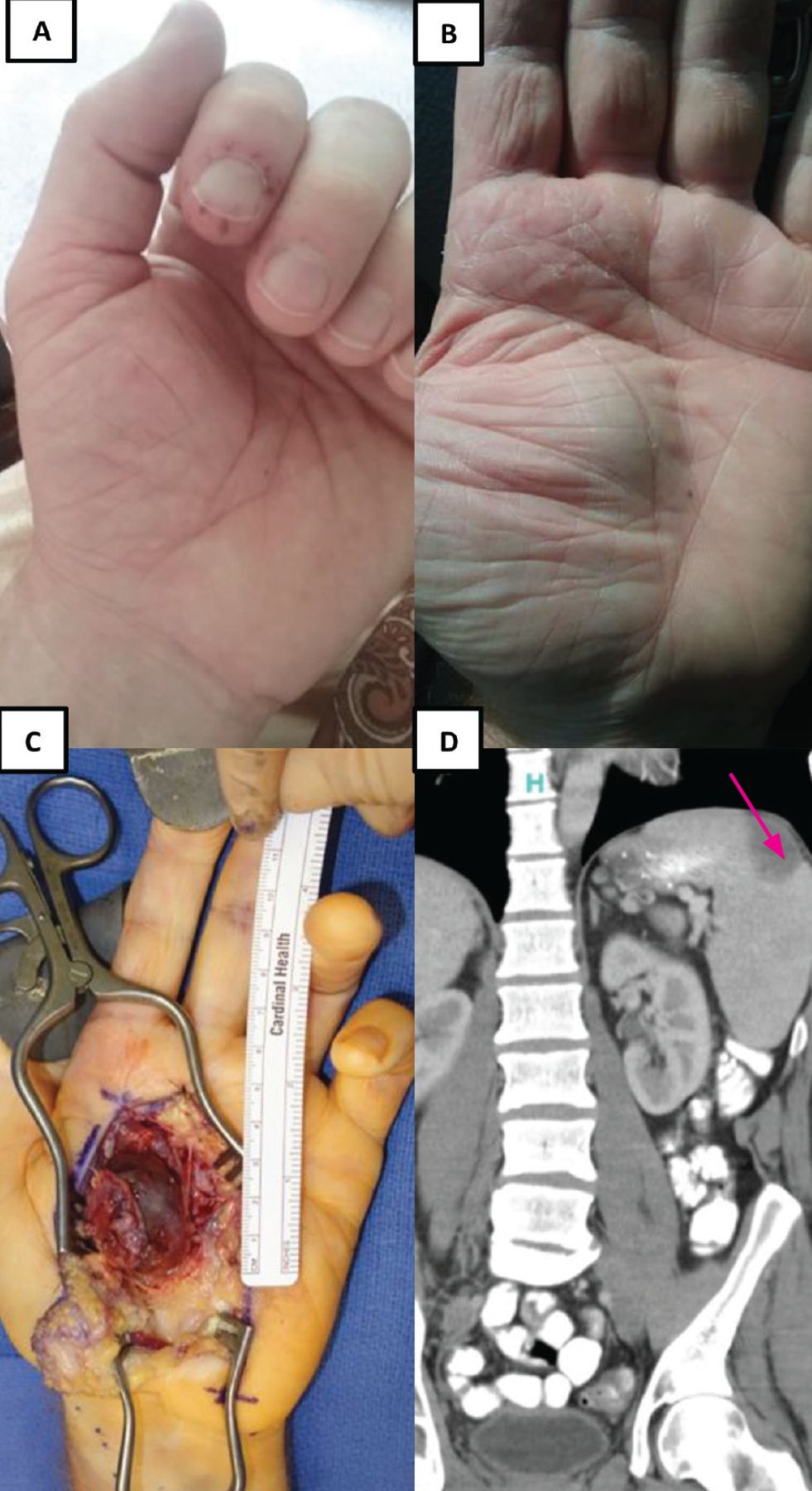
However, during the next month, he developed pain in his left index finger and right second toe, with periungual and digital petechiae (Figure 1A), and then developed a painful pulsatile palmar mass (Figure 1B). Magnetic resonance angiography at another medical facility revealed a superficial palmar arch aneurysm that was subsequently excised (Figure 1C). He also developed new left scapular pain, and computed tomography showed evidence of a wedge-shaped splenic infarct (Figure 1D).

Figure 1
  • Download figure
  • Open in new tab
  • Download powerpoint
Figure 1

Findings before presentation: (A) painful periungual petechial rash on the left index finger; (B) pulsatile palmar mass; (C) intraoperative image of the aneurysm arising from the superficial palmar arch; and (D) abdominal computed tomography showing a wedge-shaped splenic infarct (arrow).

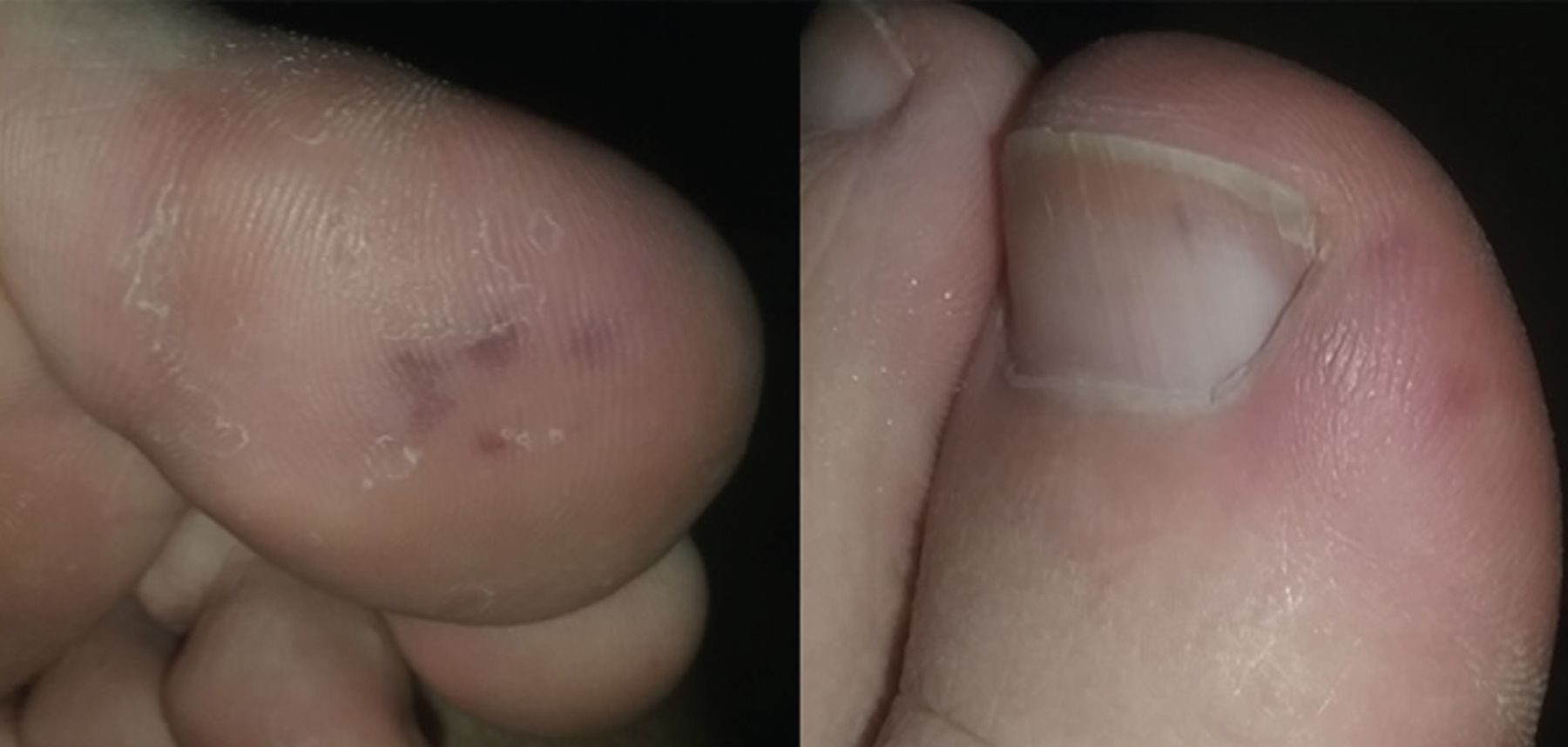
On admission to our hospital, he did not appear ill. Cardiac examination revealed a grade III/IV blowing diastolic murmur at the left lower sternal border. There were painful grouped petechiae and splinter hemorrhages affecting the left great toe (Figure 2). Results of laboratory testing were as follows:

  • White blood cell count 6.48 × 10⁹/L (reference range 4.5–11.0)

  • Platelet count 280 × 109/L (150–350)

  • Hemoglobin level 11.0 g/dL (13.9–16.3)

  • Hematocrit 35% (41–53)

  • Erythrocyte sedimentation rate 33 mm/h (1–15)

  • C-reactive protein 5.6 mg/dL (< 0.5)

  • Rheumatoid factor 649 IU/mL (0–35).

Figure 2
  • Download figure
  • Open in new tab
  • Download powerpoint
Figure 2

Painful petechial rash affecting the left great toe, compatible with Osler nodes, and splinter hemorrhage of the toenail.

B burgdorferi antibody, Babesia microti IgG and IgM, and E chaffeensis polymerase chain reaction testing were negative.

Transthoracic echocardiography detected a vegetation on the left coronary cusp of the aortic valve. Blood cultures on the first and second hospital days were positive for Streptococcus mutans.

The patient’s evolving distal signs, aortic regurgitation murmur, results of echocardiography, and microbiologic data were confirmatory of subacute bacterial endocarditis, and an extended course of antimicrobial therapy was implemented.

EMBOLIC MANIFESTATIONS OF ENDOCARDITIS

Early in this patient’s course, the repeated exposure to ticks and the positive serology for Lyme disease and ehrlichiosis prompted treatment with doxycycline. However, the development of distal embolic phenomena, transient visual impairment, splenic infarct, and pulsatile palmar mass compelled reconsideration of the underlying infectious process.

Janeway lesions (painless hemorrhagic or erythematous lesions on the palms and soles developing as septic emboli) and Osler nodes (painful petechial eruptions on the finger pads and toes), understood to arise from immune complex deposition, are classic features of endocarditis.1,2 They are helpful clues to uncover the diagnosis. This patient’s sudden visual disturbances, albeit transient, and shoulder pain were instructive additive symptoms arising as immunologic and vascular occlusive insults to the retina and spleen, respectively.1,3

Elevated rheumatoid factor is present in one-third of patients with confirmed endocarditis, arising from sustained inflammation, thereby inducing polyclonal hypergammaglobulinemia.3,4 The painful palmar swelling arose from a mycotic aneurysm of the superficial palmar artery. Surgical pathology identified a dilated arterial wall with granulation tissue, acute and chronic inflammation, and prominent luminal thrombus. Interestingly, mycotic aneurysms may occur in 10% to 20% of patients in large endocarditis series, and were described by Sir William Osler as “mycotic endarteritis” in 1885.5 They may arise in the cerebral, splenic, coronary, pulmonary, and mesenteric arteries and in the abdominal aorta.1,2

THE PATIENT’S MANAGEMENT

In our patient, the evolving distal signs, along with the lack of specificity and potential for false-positive results of serologic bloodwork for Lyme disease and ehrlichiosis, necessitated a reformulation of the underlying disease process and the establishment of the diagnosis of bacterial endocarditis.6

On completion of a 4-week course of intravenous penicillin G, our patient achieved complete resolution of symptoms.

DISCLOSURES

The authors report no relevant financial relationships which, in the context of their contributions, could be perceived as a potential conflict of interest.

  • Copyright © 2021 The Cleveland Clinic Foundation. All Rights Reserved.

REFERENCES

  1. ↵
    1. Hoen B,
    2. Duval X
    . Clinical practice. Infective endocarditis. N Engl J Med 2013; 368(15):1425–1433. doi:10.1056/NEJMcp1206782
    OpenUrlCrossRefPubMed
  2. ↵
    1. Servy A,
    2. Valeyrie-Allanore L,
    3. Alla F, et al
    . Prognostic value of skin manifestations of infective endocarditis. JAMA Dermatol 2014; 150(5):494–500. doi:10.1001/jamadermatol.2013.8727
    OpenUrlCrossRefPubMed
  3. ↵
    1. Durack DT,
    2. Lukes AS,
    3. Bright DK
    . New criteria for diagnosis of infective endocarditis: utilization of specific echocardiographic findings. Duke Endocarditis Service. Am J Med 1994; 96(3):200–209. doi:10.1016/0002-9343(94)90143-0
    OpenUrlCrossRefPubMed
  4. ↵
    1. Gouriet F,
    2. Bothelo-Nevers E,
    3. Coulibaly B,
    4. Raoult D,
    5. Casalta JP
    . Evaluation of sedimentation rate, rheumatoid factor, C-reactive protein, and tumor necrosis factor for the diagnosis of infective endocarditis. Clin Vaccine Immunol 2006; 13(2):301. doi:10.1128/CVI.13.2.301.2006
    OpenUrlFREE Full Text
  5. ↵
    1. Osler W
    . The gulstonian lectures, on malignant endocarditis. Br Med J 1885; 1(1262):467–470. doi:10.1136/bmj.1.1262.467
    OpenUrlFREE Full Text
  6. ↵
    1. Kaell AT,
    2. Volkman DJ,
    3. Gorevic PD,
    4. Dattwyler RJ
    . Positive Lyme serology in subacute bacterial endocarditis. A study of four patients. JAMA 1990; 264(22):2916–2918. pmid:2232087
    OpenUrlCrossRefPubMed
PreviousNext
Back to top

In this issue

Cleveland Clinic Journal of Medicine: 88 (6)
Cleveland Clinic Journal of Medicine
Vol. 88, Issue 6
1 Jun 2021
  • Table of Contents
  • Table of Contents (PDF)
  • Index by author
  • Complete Issue (PDF)
Print
Download PDF
Article Alerts
Sign In to Email Alerts with your Email Address
Email Article

Thank you for your interest in spreading the word on Cleveland Clinic Journal of Medicine.

NOTE: We only request your email address so that the person you are recommending the page to knows that you wanted them to see it, and that it is not junk mail. We do not capture any email address.

Enter multiple addresses on separate lines or separate them with commas.
Evolving distal signs
(Your Name) has sent you a message from Cleveland Clinic Journal of Medicine
(Your Name) thought you would like to see the Cleveland Clinic Journal of Medicine web site.
CAPTCHA
This question is for testing whether or not you are a human visitor and to prevent automated spam submissions.
Citation Tools
Evolving distal signs
Zackary D. Goff, Sandra E. Zaeh, Michael K. Poku, John N. Aucott, Allan C. Gelber
Cleveland Clinic Journal of Medicine Jun 2021, 88 (6) 310-312; DOI: 10.3949/ccjm.88a.20126

Citation Manager Formats

  • BibTeX
  • Bookends
  • EasyBib
  • EndNote (tagged)
  • EndNote 8 (xml)
  • Medlars
  • Mendeley
  • Papers
  • RefWorks Tagged
  • Ref Manager
  • RIS
  • Zotero
Share
Evolving distal signs
Zackary D. Goff, Sandra E. Zaeh, Michael K. Poku, John N. Aucott, Allan C. Gelber
Cleveland Clinic Journal of Medicine Jun 2021, 88 (6) 310-312; DOI: 10.3949/ccjm.88a.20126
Twitter logo Facebook logo Mendeley logo
  • Tweet Widget
  • Facebook Like
  • Linkedin Share Button

Jump to section

  • Article
    • EMBOLIC MANIFESTATIONS OF ENDOCARDITIS
    • THE PATIENT’S MANAGEMENT
    • DISCLOSURES
    • REFERENCES
  • Figures & Data
  • Info & Metrics
  • PDF

Related Articles

  • No related articles found.
  • PubMed
  • Google Scholar

Cited By...

  • No citing articles found.
  • Google Scholar

More in this TOC Section

  • Tinea incognito
  • Prolonged venous filling time and dependent rubor in a patient with peripheral artery disease
  • Sarcoidosis with diffuse purplish erythematous plaques on the hands
Show more The Clinical Picture

Similar Articles

Subjects

  • Cardiology
  • Drug Therapy
  • Emergency Medicine
  • Hospital Medicine
  • Infectious Diseases
  • Rheumatology

Navigate

  • Current Issue
  • Past Issues
  • Supplements
  • Article Type
  • Specialty
  • CME/MOC Articles
  • CME/MOC Calendar
  • Media Kit

Authors & Reviewers

  • Manuscript Submission
  • Authors & Reviewers
  • Subscriptions
  • About CCJM
  • Contact Us
  • Cleveland Clinic Center for Continuing Education
  • Consult QD

Share your suggestions!

Copyright © 2025 The Cleveland Clinic Foundation. All rights reserved. The information provided is for educational purposes only. Use of this website is subject to the website terms of use and privacy policy. 

Powered by HighWire