
Main menu

  • Home
  • Content
    • Current Issue
    • Ahead of Print
    • Past Issues
    • Supplements
    • Article Type
  • Specialty
    • Articles by Specialty
  • CME/MOC
    • Articles
    • Calendar
  • Info For
    • Manuscript Submission
    • Authors & Reviewers
    • Subscriptions
    • About CCJM
    • Contact Us
    • Media Kit
  • Conversations with Leaders
  • Conference Coverage
    • Kidney Week 2024
    • CHEST 2024
    • ACR Convergence 2023
    • Kidney Week 2023
    • ObesityWeek 2023
    • IDWeek 2023
    • CHEST 2023
    • MDS 2023
    • IAS 2023
    • ACP 2023
    • AAN 2023
    • ACC / WCC 2023
    • AAAAI Meeting 2023
    • ACR Convergence 2022
    • Kidney Week 2022
    • AIDS 2022
  • Other Publications
    • www.clevelandclinic.org

User menu

  • Register
  • Log in

Search

  • Advanced search
Cleveland Clinic Journal of Medicine
  • Other Publications
    • www.clevelandclinic.org
  • Register
  • Log in
Cleveland Clinic Journal of Medicine

Advanced Search

  • Home
  • Content
    • Current Issue
    • Ahead of Print
    • Past Issues
    • Supplements
    • Article Type
  • Specialty
    • Articles by Specialty
  • CME/MOC
    • Articles
    • Calendar
  • Info For
    • Manuscript Submission
    • Authors & Reviewers
    • Subscriptions
    • About CCJM
    • Contact Us
    • Media Kit
  • Conversations with Leaders
  • Conference Coverage
    • Kidney Week 2024
    • CHEST 2024
    • ACR Convergence 2023
    • Kidney Week 2023
    • ObesityWeek 2023
    • IDWeek 2023
    • CHEST 2023
    • MDS 2023
    • IAS 2023
    • ACP 2023
    • AAN 2023
    • ACC / WCC 2023
    • AAAAI Meeting 2023
    • ACR Convergence 2022
    • Kidney Week 2022
    • AIDS 2022
Review

Endoscopic ultrasonography: An inside view

C. Roberto Simons-Linares, MD, Praneet Wander, MD, John Vargo, MD and Prabhleen Chahal, MD
Cleveland Clinic Journal of Medicine March 2020, 87 (3) 175-183; DOI: https://doi.org/10.3949/ccjm.87a.19003
C. Roberto Simons-Linares
Department of Gastroenterology, Hepatology, and Nutrition, Cleveland Clinic
  • Find this author on Google Scholar
  • Find this author on PubMed
  • Search for this author on this site
Praneet Wander
Department of Gastroenterology, Hofstra Northwell School of Medicine, Hempstead, NY
  • Find this author on Google Scholar
  • Find this author on PubMed
  • Search for this author on this site
John Vargo
Medical Director of Advanced Endoscopy, Department of Gastroenterology, Hepatology, and Nutrition, Cleveland Clinic
Associate Professor, Cleveland Clinic Lerner College of Medicine of Case Western Reserve University, Cleveland, OH
  • Find this author on Google Scholar
  • Find this author on PubMed
  • Search for this author on this site
Prabhleen Chahal
Department of Gastroenterology, Hepatology, and Nutrition, Cleveland Clinic
Clinical Assistant Professor, Cleveland Clinic Lerner College of Medicine of Case Western Reserve University, Cleveland, OH
  • Find this author on Google Scholar
  • Find this author on PubMed
  • Search for this author on this site
  • For correspondence: [email protected]
  • Article
  • Figures & Data
  • Info & Metrics
  • PDF
Loading

ABSTRACT

Endoscopic ultrasonography (EUS) has been used since the mid-1980s. Initially a diagnostic tool, it has since evolved into a therapeutic, minimally invasive surgical tool with significant impact on the diagnosis and management of a range of benign and malignant conditions. The authors review current indications, safety, and efficacy of EUS for diseases of the upper and lower gastrointestinal tract, posterior mediastinum, pancreas, bile duct, gallbladder, retroperitoneum, liver, adrenal glands, and kidneys.

KEY POINTS
  • EUS is commonly used in the diagnosis and management of biliary and pancreatic diseases, including guidance of biliary drainage in bile duct or pancreatic duct obstruction.

  • EUS-guided fine-needle aspiration aids staging of malignancies of the upper and lower digestive tract.

  • EUS is now increasingly used to drain abdominal fluid collections of nonpancreatic etiology.

Endoscopic ultrasonography (EUS) has evolved from a diagnostic tool to a therapeutic procedure for a wide range of conditions of the upper and lower gastrointestinal tract, hepatobiliary system, pancreas, adrenal glands, kidneys, retroperitoneum, and lymph nodes around the gastrointestinal tract. It can help differentiate benign from malignant disease and in many cases is an alternative to surgery.

This review of current and evolving diagnostic and therapeutic indications for EUS will help physicians identify patients who may benefit from this procedure.

EUS VS OTHER IMAGING TESTS

EUS offers advantages over other imaging tests. It does not involve radiation as in computed tomography (CT) or positron emission tomography, and it is not subject to the contraindications of magnetic resonance imaging (MRI) such as metal implants or claustrophobia. EUS offers high-resolution real-time imaging and can be combined with Doppler to evaluate vasculature and perform diagnostic procedures, angiotherapy, fine-needle aspiration biopsy, and core biopsy for tissue diagnosis. In addition, EUS allows therapeutic interventions.1

PROCEDURAL REQUIREMENTS FOR THE PATIENT

EUS is an outpatient procedure that usually takes 30 to 60 minutes and can be done under moderate sedation or general anesthesia.

Periprocedural considerations

According to guidelines of the American Society for Gastrointestinal Endoscopy (ASGE),2 patients fast for 8 hours before the procedure.

For patients taking antithrombotics, guidelines recommend a platelet level greater than 50 × 109/L and an international normalized ratio below 1.5.

The endoscope

EUS is performed with a flexible wide endoscope with a small ultrasound probe and camera at the tip; other equipment in the scope depend on the indication and can include a fine-aspiration needle, a core biopsy needle, a celiac plexus blockade and neurolysis needle, and a metal or plastic stent. EUS can be performed using either a radial (360°) or a linear (approximately 120°) view. A narrow angle of view is required to allow endoscopic fine-needle aspiration.

Expanding indications

The indications for EUS are rapidly expanding. Uses identified by the American Cancer Society and the ASGE now include evaluation and staging of upper gastrointestinal malignancies, mediastinal adenopathy, pancreatic lesions and cancers, submucosal tumors, rectal cancer, and lung cancer staging.3 In particular, EUS has changed the approach to diagnosis and management of biliary and pancreatic diseases.

PANCREATIC DISORDERS

Pancreatic cancer

Pancreatic cancer has a poor prognosis since it is usually diagnosed at an advanced stage. According to the American Cancer Society, the 1-year relative survival rate is 20%, and the 5-year rate is 8% for all stages of pancreatic cancer. Current studies show that EUS has a sensitivity of 90% to 95% for detecting malignant pancreatic tumors measuring 2 cm to 3 cm, which is far superior to other imaging modalities.4 EUS is considered complementary to CT or MRI for diagnosis and staging of pancreatic adenocarcinoma. EUS is also used to rule out pancreatic cancer if results of CT or MRI are ambiguous.

EUS in combination with fine-needle aspiration biopsy improves diagnostic accuracy for pancreatic masses and helps in histologic confirmation.5 Immediate cytologic evaluation or rapid on-site cytologic evaluation helps improve the yield.

The sensitivity of EUS declines in the setting of severe underlying chronic pancreatitis or severe acute pancreatitis. The challenges of detecting pancreatic malignancy in the setting of chronic pancreatitis are being addressed with the advent of EUS elastography, which evaluates tissue stiffness and helps with characterization of the lesion.6

Acute pancreatitis

Gallstones are the most common cause of acute pancreatitis. EUS has higher sensitivity than ultrasonography, CT, and magnetic resonance cholangiopancreatography in detecting common bile duct stones and sludge in patients with acute pancreatitis or recurrent pancreatitis.7

The ASGE recommends EUS for the assessment of choledocholithiasis in patients at intermediate risk. If EUS confirms bile duct stones, therapeutic endoscopic retrograde cholangiopancreatography (ERCP) can be performed with the patient under the same sedation; and if a stone is not present, an additional diagnostic procedure can be avoided.2

EUS also helps determine other causes of acute pancreatitis such as pancreas divisum, small pancreatic tumors undetected on CT or MRI, autoimmune pancreatitis, and chronic pancreatitis, and it can be an important investigative test in patients with idiopathic pancreatitis.8

Pancreatic fluid collections

The revised Atlanta classification categorizes pancreatic fluid collections according to 4 types9:

  • Acute peripancreatic fluid collection, occurring in interstitial edematous pancreatitis

  • Pancreatic pseudocyst, occurring as a delayed complication (> 4 weeks) of interstitial edematous pancreatitis

  • Acute necrotic collection, occurring in necrotizing pancreatitis

  • Walled-off necrosis, which has a radiologically identifiable capsule (> 4 weeks).

Surgical and percutaneous approaches have traditionally been used to drain symptomatic pancreatic and peripancreatic fluid collections.10 However, disadvantages of these procedures include higher cost, longer hospital stay, possibly higher morbidity risk, and discomfort of external catheters requiring multiple exchanges.

In the past few years, EUS-guided transgastric or transduodenal drainage has been used more frequently with comparable success and lower morbidity and costs than surgical and percutaneous drainage. The procedure is feasible in more than 90% of patients, is minimally invasive, and results in shorter hospital stays than surgical drainage, and compared with CT or MRI, it more accurately differentiates pseudocyst from cystic neoplasms.11

Complications of endoscopic drainage of pancreatic fluid collections are minimal and include bleeding, perforation, and infection, all with rates of less than 5%.12

Chronic pancreatitis

Chronic pancreatitis is a complex disease with an ill-defined epidemiology but significant rates of morbidity and mortality. Smoking and alcohol consumption are main risk factors. Chronic abdominal pain is the most common presentation.

Diagnosis can be challenging in early, mild, or moderate disease. Studies13–16 have shown that the diagnostic accuracy of EUS is comparable to that of ERCP and pancreatic function testing. In addition, technologic advances in EUS such as contrast-enhanced harmonics and elastography offer improved diagnostic accuracy in patients with chronic pancreatitis.17,18 As a result, EUS is being increasingly used as a frontline test for this indication.

Pancreatic cystic neoplasms

The challenge in management of pancreatic cystic neoplasms lies in the timely and accurate diagnosis of premalignant mucinous cysts.

Differentiation of premalignant mucinous cysts from benign nonmucinous cysts by EUS with fine-needle fluid aspiration has become a valuable tool, providing high-quality imaging of the cyst and samples for fluid analysis, leading to increased diagnostic accuracy.

Periprocedural antibiotics are administered to minimize the risk of infection from fine-needle aspiration. Cyst fluid samples are sent to the laboratory for cytologic study and for analysis for carcinoembryonic antigen, glucose, amylase, and mucin.19,20 Premalignant mucinous cysts are managed according to the international consensus guidelines.21,22

GALLBLADDER AND BILIARY DISORDERS

Choledocholithiasis

Common bile duct stones can be present in 20% of patients with cholelithiasis. Noninvasive imaging with abdominal ultrasonography or CT has a diagnostic accuracy of only 50% for these stones. Magnetic resonance cholangiopancreatography is the most accurate noninvasive imaging test (diagnostic accuracy from 81% to 99%).23–26

ERCP and intraoperative cholangiography are accurate but invasive and can cause complications. Same-session EUS and ERCP (if stones are confirmed on EUS) for common bile duct stones are usually performed in patients with intermediate probability of stones according to the ASGE criteria or in patients with contraindications to magnetic resonance cholangiopancreatography.

Obstructive jaundice

Obstructive jaundice can result from benign and malignant diseases. Common benign causes are choledocholithiasis, postcholecystectomy bile duct injury, liver transplant, portal cavernoma cholangiopathy, primary sclerosing cholangitis, acquired immunodeficiency syndrome cholangiopathy, chronic pancreatitis, and immunoglobulin G4 cholangiopathy. Malignant causes include cholangiocarcinoma, gallbladder carcinoma, pancreatic malignancies, ampullary carcinoma, metastasis, metastatic lymph nodal compression on bile duct, and hepatocellular carcinoma. The proximity of the stomach and duodenum to the extrahepatic biliary system makes EUS useful for imaging the biliary anatomy. EUS-guided fine-needle aspiration from the bile duct wall and surrounding lymph nodes enables histologic diagnosis.

ERCP is the standard procedure for biliary drainage in benign or malignant biliary obstruction. However, it is not feasible in surgically altered anatomy as in Roux-en-Y anastomosis and duodenal bulb infiltration by tumor. In these cases, EUS-guided biliary drainage is being used with high success rates.27,28 EUS-guided cholangiopancreatography has been done in patients in whom ERCP could not be performed. This procedure requires a high level of technical expertise and can cause complications such as perforation and bile leak.

Cholecystitis

Cholecystectomy is the optimal treatment for acute calculous or acalculous cholecystitis. Patients deemed high-risk surgical candidates cannot undergo the procedure and traditionally undergo percutaneous placement of a cholecystostomy tube by interventional radiology for decompression of the gallbladder. EUS can be used to perform transgastric or transduodenal gallbladder drainage with a covered metal stent. Case series have shown outcomes comparable to those of percutaneous tube placement.29,30

GASTROINTESTINAL TRACT DISEASE

Upper tract conditions

EUS can be used to identify benign tumors of the upper gastrointestinal tract, includ ing submucosal esophagogastric tumors.31 EUS-guided fine-needle aspiration and biopsy can aid the cytohistologic diagnosis of esophagogastric solid subepithelial lesions.32

EUS is routinely used for diagnosis and staging of malignant esophageal tumors. The procedure may not be technically feasible if high-grade malignant esophageal stricture precludes passage of echoendoscope.

EUS-guided evaluation and biopsy are particularly useful to diagnose linitis plastica, a gastric cancer characterized by marked thickening of deeper layers of gastric wall. It is difficult to diagnose with superficial mucosal biopsies rendered with esophagogastroduodenoscopy.

Mediastinal adenopathy and non-small-cell lung cancer

Nearly 26% of lung cancer patients present with mediastinal lymph node involvement.33 CT, MRI, and positron emission tomography are the common modalities for diagnosing mediastinal lymph node enlargement. However, lack of tissue sampling results only in a presumptive diagnosis. Posterior and inferior mediastinal lesions are especially suitable for EUS-guided fine-needle aspiration and biopsy for histologic diagnosis, thus avoiding an invasive surgical intervention like mediastinoscopy. However, EUS is unable to visualize anterior upper mediastinal nodes.

Mediastinal staging in lung cancer is an area in which EUS has made most significant impact.34 It also allows for evaluation of distant metastases involving adrenal glands.

Submucosal lesions

EUS provides a detailed image of gastrointestinal wall layers and therefore has become the principal tool for assessment of submucosal masses in this area.

Gastrointestinal stromal tumors are the most common mesenchymal tumors, and the stomach is the most common site. EUS-guided fine-needle aspiration is considered the procedure of choice for preoperative diagnosis of these tumors, although it may provide inadequate material in one-third of cases. However, with the advent of newer biopsy needles, the histologic yield has improved significantly.

In addition to the diagnosis of gastrointestinal stromal tumors, EUS can distinguish between other benign lesions (leiomyoma, pancreatic rest, indolent neuroendocrine tumor, granular cell tumor, schwannoma, duplication cyst) and malignant lesions (primary or metastatic). EUS elastography is a promising technique that may also improve the distinction of benign from malignant lesions.

Retroperitoneal masses

For evaluation of idiopathic abdominal masses, EUS-guided fine-needle aspiration and biopsy provide a minimally invasive technique to obtain tissue samples for safe and accurate diagnosis, thus avoiding the need for exploratory laparotomy; it also helps guide subsequent therapy.35 These procedures are useful for evaluation of periintestinal and periesophageal lymph nodes and are more accurate than CT.36

Apart from providing adequate diagnostic tissue in lymphoproliferative tumors, EUS is highly useful in nonpancreatic retroperitoneal masses such as adrenal metastasis, leiomyosarcoma, paraganglioma, and lymphangioma. EUS-associated seeding of the needle tract has been reported but is rare.

Lower tract disease

Accurate preoperative staging of rectal cancer leads to targeted treatment strategies, increased cure rates, and reduction in short-term and long-term treatment failure. EUS and MRI have comparable accuracy in TNM staging of colorectal cancer, but MRI is perhaps more advantageous than EUS for nodal staging since it images the entire mesorectum.

CELIAC PLEXUS BLOCK AND NEUROLYSIS

Chronic abdominal pain is common and often disabling in patients with intra-abdominal malignancies and chronic pancreatitis. Pain relief from first-line medications including opiates may be inadequate or fraught with intolerances and adverse effects.37

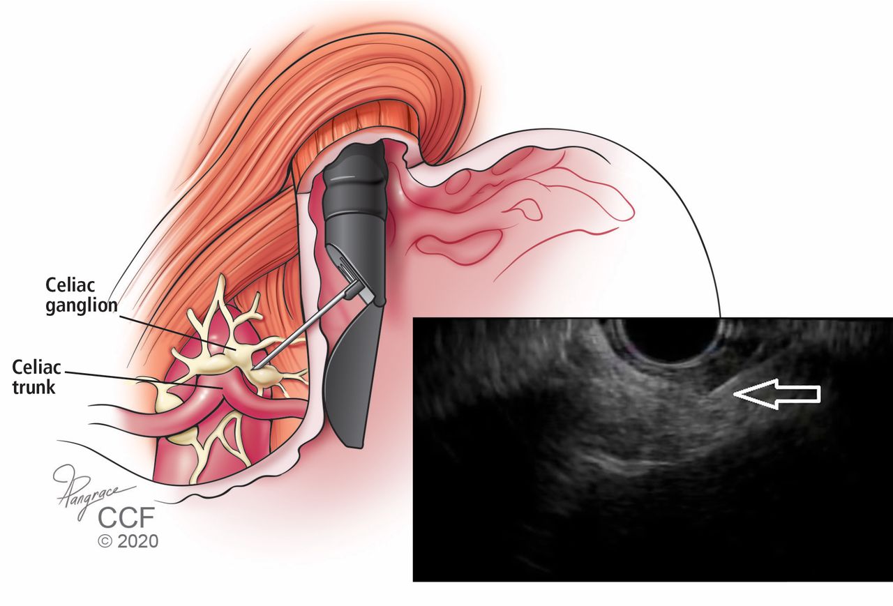
Celiac plexus block with CT-guided percutaneous and EUS-guided neurolysis has been shown to be safe and technically easy to perform (Figure 1). In a Cochrane review of 6 randomized controlled trials with a total of 358 pancreatic cancer patients,38 the authors found that celiac plexus block with either approach caused fewer adverse effects than opioid analgesia. More study is needed to confirm a statistically significant improvement in pain scores.

Figure 1
  • Download figure
  • Open in new tab
  • Download powerpoint
Figure 1

Control of chronic abdominal pain with first-line medications in patients with chronic pancreatitis or intra-abdominal malignancy may be inadequate or fraught with adverse effects. One alternative is endoscopic ultrasonography-guided fine-needle (EUS) celiac plexus block, performed under echoendoscopic guidance with passage of an injection needle. This procedure has been shown to be safe, is technically easy to perform, and is a safe alternative to percutaneous block guided by computed tomography. The arrow shows the EUS-guided placement of the needle.

RECENT DEVELOPMENTS, NEWER INDICATIONS

EUS-guided fine-needle injection

EUS-guided fine-needle injection is being evaluated for intratumoral chemotherapy in pancreatic and esophageal cancers. It has been used for tissue ablation by injection of ethanol in nonmalignant pancreatic cystic tumors and in radiofrequency ablation and brachytherapy in unresectable pancreatic cancers. Portal injection of chemotherapy has been shown to be safe and feasible. It may prove useful in the management of primary liver malignancies.

EUS-guided botulinum toxin injection for achalasia and for management of obesity has been done in few cases. Botox administration in the stomach promotes early satiety and weight loss through inhibition of acetylcholine-mediated peristalsis, ultimately delaying gastric emptying.39

EUS-guided gastrojejunostomy

Gastric outlet obstruction can result from malignancies involving stomach, duodenum, gallbladder, or pancreas. EUS-guided gastrojejunostomy has been reported to be a safe, durable, and successful treatment option.40–42 Compared with enteral (duodenal) stenting, which can be complicated by tumor ingrowth resulting in stent occlusion, EUS-guided gastrojejunostomy has been shown to provide longer-lasting symptom relief since it is performed remotely from the tumor location. It also offers shorter recovery time and is more cost-effective than surgical gastrojejunostomy.43,44

Contrast-enhanced harmonic EUS

Contrast-enhanced harmonic EUS has been developed to detect a microvascular pattern of lesions, which can help differentiate benign from malignant disease. An intravascular contrast agent is injected that contains microbubbles. On exposure to ultrasound, these microbubbles oscillate, and the transducer can make out the appearance of the peripheral microvasculature.45

Molecular marker analysis

DNA analysis of specimens obtained by EUS-guided fine-needle aspiration can aid diagnosis. Microdissection-based genotyping is now available at some centers. This has specific value in distinction of pancreatic cysts seen on CT. DNA quantification can help distinguish benign cysts from malignant ones.

EUS-directed transgastric ERCP in patients with gastric bypass

Duodenal and ampullary access with ERCP is difficult in patients who have undergone Roux-en-Y gastric bypass. Classically, enteroscopy-assisted ERCP is associated with a low success rate. Surgically assisted ERCP (laparoscopy or laparotomy) has a good success rate but is more invasive, has higher complication rates and longer length of stay in the hospital, with associated costs.

EUS-directed transgastric ERCP involves accessing the excluded stomach from the gastric pouch or Roux limb by creation of a fistula with placement of a removable lumen-apposing metal stent. Conventional ERCP can then be performed through the stent. A multicenter study comparing the EUS-directed procedure and laparoscopic ERCP found that the 2 procedures had similar success rates and adverse events.46 The EUS-directed procedure has the benefit of being an outpatient minimally invasive procedure with significantly shorter procedure time and cost.46,47

Drainage of other nonpancreatic abdominopelvic fluid collections

EUS is safe and effective in accessing and draining nonpancreatic abdominopelvic fluid collections from the stomach, duodenum, and colon. Multiple case series have reported safe and successful drainage of subphrenic abscesses, hepatic abscesses, bilomas, and pelvic and retroperitoneal abscesses. In addition, EUS has been increasingly utilized to drain postsurgical fluid collections, which can form anywhere in the abdominal cavity and can become symptomatic or infected. There is mounting recent evidence of successful EUS-guided drainage of such collections after common surgical procedures including Whipple surgery and Roux-en-Y gastric bypass.48

EUS-guided angiotherapy

EUS-guided angiotherapy is a relatively novel application that allows control of variceal bleeding by injecting coils and cyanoacrylate glue directly into the varices and confirming the thrombosis in real time with Doppler. It is purported that the combination of coil and glue might decrease the risk of glue embolization. However, further study is needed to establish the safety and superiority of this technique.34,49,50

DRAWBACKS AND COMPLICATIONS

Adverse events associated with sedation and standard endoscopic procedures are also applicable to EUS. However, since the echoendoscope has a larger diameter with a stiffer tip than the standard endoscope, it can be difficult to maneuver around the cricopharyngeus and duodenal bulb. It is also more time-consuming than standard routine endoscopy if fine-needle aspiration or intervention is planned. Despite this, complications, as reviewed below, are infrequent.

Perforation

Perforation is reported in the cervical esophagus and less often in the duodenum. The risk is usually similar to that with routine endoscopy (0.03%). A prospective study of 4,894 patients undergoing upper EUS found a cervical esophageal perforation rate of 0.06% (3 patients, with a curved linear array endoscope).51 A recent systematic review reported a perforation rate of 0.02% with EUS.52 The risks may be higher in patients with strictures, malignancy, older age, or history of difficult intubation, and with a less experienced operator.

Infection

The risk of bacteremia after EUS-guided fine-needle aspiration is lower than with diagnostic endoscopy, and prophylactic antibiotics are not recommended for aspiration of solid masses and lymph nodes.53,54 Some experts recommend prophylactic antibiotics as well as 48 hours of antibiotics after fine-needle aspiration of the perirectal space.55

EUS-guided aspiration of cystic lesions may carry an increased risk of febrile episodes and possibly sepsis. Therefore, prophylactic antibiotics followed by a short postprocedure course has been recommended.56

There have been isolated reports of streptococcal sepsis, mediastinitis, retroperitoneal abscess, perirectal abscess, and cholangitis.57

Pancreatitis

EUS-guided fine-needle aspiration of pancreatic lesions involves direct passage of the needle through pancreatic tissue. Reported rates of pancreatitis associated with this procedure range from 0% to 2%.58 A recent meta-analysis of 51 studies found a rate of of 0.44%.59

Hemorrhage

A recent meta-analysis of related adverse events reported a bleeding rate of 0.13%.59

Avoiding complications

Careful patient selection, familiarity with the equipment, and planning of the procedure are keys to avoiding complications. Early recognition of complications and prompt intervention can reduce morbidity and mortality risk.

  • Copyright © 2020 The Cleveland Clinic Foundation. All Rights Reserved.

REFERENCES

  1. ↵
    1. Williams DB,
    2. Sahai AV,
    3. Aabakken L, et al
    . Endoscopic ultrasound guided fine needle aspiration biopsy: a large single centre experience. Gut 1999; 44(5):720–726. doi:10.1136/gut.44.5.720
    OpenUrlAbstract/FREE Full Text
  2. ↵
    1. ASGE Standards of Practice Committee;
    2. Maple JT,
    3. Ben-Menachem T,
    4. Anderson MA, et al
    . The role of endoscopy in the evaluation of suspected choledocholithiasis. Gastrointest Endosc 2010; 71(1):1–9. doi:10.1016/j.gie.2009.09.041
    OpenUrlCrossRefPubMed
  3. ↵
    1. Mekky MA,
    2. Yamao K,
    3. Sawaki A, et al
    . Diagnostic utility of EUS-guided FNA in patients with gastric submucosal tumors. Gastrointest Endosc 2010; 71(6):913–919. doi:10.1016/j.gie.2009.11.044
    OpenUrlCrossRefPubMed
  4. ↵
    1. Akahoshi K,
    2. Chijiiwa Y,
    3. Nakano I, et al
    . Diagnosis and staging of pancreatic cancer by endoscopic ultrasound. Br J Radiol 1998; 71(845):492–496. doi:10.1259/bjr.71.845.9691893
    OpenUrlAbstract
  5. ↵
    1. Gonzalo-Marin J,
    2. Vila JJ,
    3. Perez-Miranda M.
    Role of endoscopic ultrasound in the diagnosis of pancreatic cancer. World J Gastrointest Oncol 2014; 6(9):360–368. doi:10.4251/wjgo.v6.i9.360
    OpenUrlCrossRef
  6. ↵
    1. Müller MF,
    2. Meyenberger C,
    3. Bertschinger P,
    4. Schaer R,
    5. Marincek B.
    Pancreatic tumors: evaluation with endoscopic US, CT, and MR imaging. Radiology 1994; 190(3):745–751. doi:10.1148/radiology.190.3.8115622
    OpenUrlCrossRefPubMed
  7. ↵
    1. Ridtitid W,
    2. Schmidt SE,
    3. Al-Haddad MA, et al
    . Performance characteristics of EUS for locoregional evaluation of ampullary lesions. Gastrointest Endosc 2015; 81(2):380–388. doi:10.1016/j.gie.2014.08.005
    OpenUrlCrossRef
  8. ↵
    1. Bhattacharya A,
    2. Cruise M,
    3. Chahal P.
    Endoscopic ultrasound guided 22 gauge core needle biopsy for the diagnosis of autoimmune pancreatitis. Pancreatology 2018; 18(2):168–169. doi:10.1016/j.pan.2018.01.003
    OpenUrlCrossRef
  9. ↵
    1. Banks PA,
    2. Bollen TL,
    3. Dervenis C, et al; Acute Pancreatitis Classification Working Group
    . Classification of acute pancreatitis—2012: revision of the Atlanta classification and definitions by international consensus. Gut 2013; 62(1):102–111. doi:10.1136/gutjnl-2012-302779
    OpenUrlAbstract/FREE Full Text
  10. ↵
    1. Park DH,
    2. Lee SS,
    3. Moon SH, et al
    . Endoscopic ultrasound-guided versus conventional transmural drainage for pancreatic pseudocysts: a prospective randomized trial. Endoscopy 2009; 41(10):842–848. doi:10.1055/s-0029-1215133
    OpenUrlCrossRefPubMed
  11. ↵
    1. Lu X,
    2. Zhang S,
    3. Ma C,
    4. Peng C,
    5. Lv Y,
    6. Zou X
    . The diagnostic value of EUS in pancreatic cystic neoplasms compared with CT and MRI. Endosc Ultrasound 2015; 4(4):324–329. doi:10.4103/2303-9027.170425
    OpenUrlCrossRef
  12. ↵
    1. Varadarajulu S,
    2. Christein JD,
    3. Tamhane A,
    4. Drelichman ER,
    5. Wilcox CM.
    Prospective randomized trial comparing EUS and EGD for transmural drainage of pancreatic pseudocysts (with videos). Gastrointest Endosc 2008; 68(6):1102–1111. doi:10.1016/j.gie.2008.04.028
    OpenUrlCrossRefPubMed
  13. ↵
    1. Chong AK,
    2. Hawes R,
    3. Hoffman B, et al
    . Diagnostic performance of endoscopic ultrasound for chronic pancreatitis: a comparison with histopathology. Gastrointest Endosc 2007; 65(6):808–814. doi:10.1016/j.gie.2006.09.026
    OpenUrlCrossRefPubMed
    1. Schmitz-Moormann P,
    2. Himmelmann GW,
    3. Brandes JW, et al
    . Comparative radiological and morphological study of human pancreas. Pancreatitis like changes in post mortem ductograms and their morphological pattern. Possible implication for ERCP. Gut 1985; 26(4):406–414. doi:10.1136/gut.26.4.406
    OpenUrlAbstract/FREE Full Text
    1. Malfertheiner P,
    2. Buchler M,
    3. Stanescu A,
    4. Ditschuneit H
    . Exocrine pancreatic function in correlation to ductal and parenchymal morphology in chronic pancreatitis. Hepatogastroenterology 1986; 33(3):110–114. pmid:3744281
    OpenUrlPubMed
  14. ↵
    1. Heij HA,
    2. Obertop H,
    3. van Blankenstein M, et al
    . Comparison of endoscopic retrograde pancreatography with functional and histologic changes in chronic pancreatitis. Acta Radiol 1987; 28(3):289–293. pmid:2958034
    OpenUrlCrossRefPubMed
  15. ↵
    1. Buxbaum J,
    2. Ko C,
    3. Varghese N, et al
    . Qualitative and quantitative contrast-enhanced endoscopic ultrasound improves evaluation of focal pancreatic lesions. Clin Gastroenterol Hepatol 2019:S1542–3565(19)30976–0. doi:10.1016/j.cgh.2019.08.054 [Epub ahead of print]
    OpenUrlCrossRef
  16. ↵
    1. Harmsen F-JR,
    2. Domagk D,
    3. Dietrich CF,
    4. Hocke M
    . Discriminating chronic pancreatitis from pancreatic cancer: contrast-enhanced EUS and multidetector computed tomography in direct comparison. Endosc Ultrasound 2018; 7(6):395–403. doi:10.4103/eus.eus_24_18
    OpenUrlCrossRef
  17. ↵
    1. Scheiman JM,
    2. Hwang JH,
    3. Moayyedi P.
    American Gastroenterological Association technical review on the diagnosis and management of asymptomatic neoplastic pancreatic cysts. Gastroenterology 2015; 148(4):824–848.e22. doi:10.1053/j.gastro.2015.01.014
    OpenUrlCrossRefPubMed
  18. ↵
    1. Gress F,
    2. Gottlieb K,
    3. Cummings O,
    4. Sherman S,
    5. Lehman G
    . Endoscopic ultrasound characteristics of mucinous cystic neoplasms of the pancreas. Am J Gastroenterol 2000; 95(4):961–965. doi:10.1111/j.1572-0241.2000.01976.x
    OpenUrlCrossRefPubMed
  19. ↵
    1. Tanaka M,
    2. Chari S,
    3. Adsay V, et al; International Association of Pancreatology
    . International consensus guidelines for management of intraductal papillary mucinous neoplasms and mucinous cystic neoplasms of the pancreas. Pancreatology 2006; 6(1–2):17–32. doi:10.1159/000090023
    OpenUrlCrossRefPubMed
  20. ↵
    1. Tanaka M,
    2. Fernández-Del Castillo C,
    3. Kamisawa T3, et al
    . Revisions of international consensus Fukuoka guidelines for the management of IPMN of the pancreas. Pancreatology 2017; 17(5):738–753. doi:10.1016/j.pan.2017.07.007
    OpenUrlCrossRefPubMed
  21. ↵
    1. Reinhold C,
    2. Taourel P,
    3. Bret PM, et al
    . Choledocholithiasis: evaluation of MR cholangiography for diagnosis. Radiology 1998; 209(2):435–442. doi:10.1148/radiology.209.2.9807570
    OpenUrlCrossRefPubMed
    1. Guibaud L,
    2. Bret PM,
    3. Reinhold C,
    4. Atri M,
    5. Barkun AN
    . Diagnosis of choledocholithiasis: value of MR cholangiography. AJR Am J Roentgenol 1994; 163(4):847–850. doi:10.2214/ajr.163.4.8092021
    OpenUrlCrossRefPubMed
    1. Regan F,
    2. Fradin J,
    3. Khazan R,
    4. Bohlman M,
    5. Magnuson T.
    Choledocholithiasis: evaluation with MR cholangiography. AJR Am J Roentgenol 1996; 167(6):1441–1445. doi:10.2214/ajr.167.6.8956574
    OpenUrlCrossRefPubMed
  22. ↵
    1. Becker CD,
    2. Grossholz M,
    3. Becker M,
    4. Mentha G,
    5. de Peyer R,
    6. Terrier F
    . Choledocholithiasis and bile duct stenosis: diagnostic accuracy of MR cholangiopancreatography. Radiology 1997; 205(2):523–530. doi:10.1148/radiology.205.2.9356639
    OpenUrlCrossRefPubMed
  23. ↵
    1. Hathorn KE,
    2. Bazarbashi AN,
    3. Sack JS, et al
    . EUS-guided biliary drainage is equivalent to ERCP for primary treatment of malignant distal biliary obstruction: a systematic review and meta-analysis. Endosc Int Open 2019; 7(11):E1432–E1441. doi:10.1055/a-0990-9488
    OpenUrlCrossRef
  24. ↵
    1. Mohan BP,
    2. Shakhatreh M,
    3. Garg R,
    4. Ponnada S,
    5. Navaneethan U,
    6. Adler DG
    . Efficacy and safety of endoscopic ultrasound-guided choledochoduodenostomy: a systematic review and meta-analysis. J Clin Gastroenterol 2019; 53(4):243–250. doi:10.1097/MCG.0000000000001167
    OpenUrlCrossRef
  25. ↵
    1. Baron TH,
    2. Topazian MD
    . Endoscopic transduodenal drainage of the gallbladder: implications for endoluminal treatment of gallbladder disease. Gastrointest Endosc 2007; 65(4):735–737. doi:10.1016/j.gie.2006.07.041
    OpenUrlCrossRefPubMed
  26. ↵
    1. Choi JH,
    2. Kim HW,
    3. Lee JC, et al
    . Percutaneous transhepatic versus EUS-guided gallbladder drainage for malignant cystic duct obstruction. Gastrointest Endosc 2017; 85(2):357–364. doi:10.1016/j.gie.2016.07.067
    OpenUrlCrossRef
  27. ↵
    1. Debol SM,
    2. Stanley MW,
    3. Mallery S,
    4. Sawinski E,
    5. Bardales RH.
    Glomus tumor of the stomach: cytologic diagnosis by endoscopic ultrasound-guided fine-needle aspiration. Diagn Cytopathol 2003; 28(6):316–321. doi:10.1002/dc.10294
    OpenUrlCrossRefPubMed
  28. ↵
    1. Akahoshi K,
    2. Oya M,
    3. Koga T, et al
    . Clinical usefulness of endoscopic ultrasound-guided fine needle aspiration for gastric subepithelial lesions smaller than 2 cm. J Gastrointestin Liver Dis 2014; 23(4):405–412. doi:10.15403/jgld.2014.1121.234.eug
    OpenUrlCrossRef
  29. ↵
    1. Silvestri GA,
    2. Gonzales AV,
    3. Jantz MA, et al
    . Methods of staging non-small cell lung cancer: diagnosis and management of lung cancer, 3rd ed: American College of Chest Physicians Evidence-Based Clinical Practice Guidelines. Chest 2013; 143(5 suppl):e211S–e250S. doi:10.1378/chest.12-2355
    OpenUrlCrossRefPubMed
  30. ↵
    1. Redondo-Cerezo E,
    2. Martínez-Cara JG,
    3. Esquivias J, et al
    . Endoscopic ultrasonography-fine needle aspiration versus PET-CT in undiagnosed mediastinal and upper abdominal lymphadenopathy: a comparative clinical study. Eur J Gastroenterol Hepatol 2015; 27(4):455–459. doi:10.1097/MEG.0000000000000302
    OpenUrlCrossRef
  31. ↵
    1. Catalano MF,
    2. Sial S,
    3. Chak A, et al
    . EUS-guided fine needle aspiration of idiopathic abdominal masses. Gastrointest Endosc 2002; 55(7):854–858. doi:10.1067/mge.2002.122956
    OpenUrlCrossRefPubMed
  32. ↵
    1. Findlay JM,
    2. Bradley KM,
    3. Maile EJ, et al
    . Pragmatic staging of oesophageal cancer using decision theory involving selective endoscopic ultrasonography, PET and laparoscopy. Br J Surg 2015; 102(12):1488–1499. doi:10.1002/bjs.9905
    OpenUrlCrossRefPubMed
  33. ↵
    1. Wyse JM,
    2. Carone M,
    3. Paquin SC,
    4. Usatii M,
    5. Sahai AV
    . Randomized, double-blind, controlled trial of early endoscopic ultrasound-guided celiac plexus neurolysis to prevent pain progression in patients with newly diagnosed, painful, inoperable pancreatic cancer. J Clin Oncol 2011; 29(26):3541–3546. doi:10.1200/JCO.2010.32.2750
    OpenUrlAbstract/FREE Full Text
  34. ↵
    1. Arcidiacono PG,
    2. Calori G,
    3. Carrara S,
    4. McNicol ED,
    5. Testoni PA
    . Celiac plexus block for pancreatic cancer pain in adults. Cochrane Data Base Syst Rev 2011; 3:CD007519. doi:10.1002/14651858.CD007519.pub2
    OpenUrlCrossRef
  35. ↵
    1. de Moura EGH,
    2. Ribeiro IB,
    3. Frazão MSV, et al
    . EUS-guided intragastric injection of botulinum toxin A in the preoperative treatment of super-obese patients: a randomized clinical trial. Obes Surg 2019; 29(1):32–39. doi:10.1007/s11695-018-3470-y
    OpenUrlCrossRef
  36. ↵
    1. Khashab MA,
    2. Baron TH,
    3. Binmoeller KF,
    4. Itoi T
    . EUS-guided gastroenterostomy: a new promising technique in evolution. Gastrointest Endosc 2015; 81(5):1234–1236. doi:10.1016/j.gie.2014.12.053
    OpenUrlCrossRefPubMed
    1. Ikeuchi N,
    2. Itoi T,
    3. Tsuchiya T,
    4. Nagakawa Y,
    5. Tsuchida A.
    One-step EUS-guided gastrojejunostomy with use of lumen-apposing metal stent for afferent loop syndrome treatment. Gastrointest Endosc 2015; 82(1):166. doi:10.1016/j.gie.2015.01.010
    OpenUrlCrossRef
  37. ↵
    1. Itoi T,
    2. Ishii K,
    3. Tanaka R,
    4. Umeda J,
    5. Tonozuka R.
    Current status and perspective of endoscopic ultrasonography-guided gastrojejunostomy: endoscopic ultrasonography-guided double-balloon-occluded gastrojejunostomy (with videos). J Hepatobiliary Pancreat Sci 2015; 22(1):3–11. doi:10.1002/jhbp.148
    OpenUrlCrossRefPubMed
  38. ↵
    1. Urrehman A,
    2. Khor CJ,
    3. Ka CY,
    4. Asokkumar R,
    5. Tan D
    . Su1449 EUS-guided gastrojejunostomy using a lumen apposing metal stent in patients with symptomatic gastric outlet obstruction. Gastrointestinal Endoscopy 2018; 87(6):AB359. doi: 10.1016/j.gie.2018.04.1779
    OpenUrlCrossRef
  39. ↵
    1. Perez-Miranda M,
    2. Tyberg A,
    3. Poletto D, et al
    . EUS-guided gastrojejunostomy versus laparoscopic gastrojejunostomy: an international collaborative study. J Clin Gastroenterol 2017; 51(10):896–899. doi:10.1097/MCG.0000000000000887
    OpenUrlCrossRef
  40. ↵
    1. Iglesias-Garcia J,
    2. Lindkvist B,
    3. Cruz-Soares JB,
    4. Larino-Noia J,
    5. Dominguez-Munoz E.
    SA1208 Does contrast enhancement play a role as an adjunct to endoscopic ultrasound for the diagnosis of chronic pancreatitis? A pilot study. Gastroenterology 2012; 142(5, suppl 1): S243–S244.
    OpenUrl
  41. ↵
    1. Kedia P,
    2. Sharaiha RZ,
    3. Kumta NA,
    4. Kahaleh M
    . Internal EUS-directed transgastric ERCP (EDGE): game over. Gastroenterology 2014; 147(3):566–568. doi:10.1053/j.gastro.2014.05.045
    OpenUrlCrossRef
  42. ↵
    1. Kedia P,
    2. Tarnasky PR,
    3. Nieto J, et al
    . EUS-directed transgastric ERCP (EDGE) versus laparoscopy-assisted ERCP (LA-ERCP) for Roux-en-Y gastric bypass (RYGB) anatomy: a multicenter early comparative experience of clinical outcomes. J Clin Gastroenterol 2019; 53(4):304–308. doi:10.1097/MCG.0000000000001037
    OpenUrlCrossRefPubMed
  43. ↵
    1. Simons-Linares CR,
    2. Rodriguez J,
    3. Chahal P
    . EUS-guided drainage of post-operative subphrenic fluid collection through gastric pouch with a lumen-apposing metal stent in a patient with Roux-en-Y gastric bypass. Obes Surg 2018; 28(10):3301–3303. doi:10.1007/s11695-018-3422-6
    OpenUrlCrossRef
  44. ↵
    1. Mouri R,
    2. Yoshida S,
    3. Tanaka S,
    4. Oka S,
    5. Yoshihara M,
    6. Chayama K
    . Usefulness of endoscopic ultrasonography in determining the depth of invasion and indication for endoscopic treatment of early gastric cancer. J Clin Gastroenterol 2009; 43(4):318–322. doi:10.1097/MCG.0b013e3181775966
    OpenUrlCrossRefPubMed
  45. ↵
    1. Fujii-Lau LL,
    2. Levy MJ
    . EUS-guided angiotherapy: a sticky situation. Gastrointest Endosc 2016; 83(6):1173–1175. doi:10.1016/j.gie.2015.12.015
    OpenUrlCrossRef
  46. ↵
    1. Eloubeidi MA,
    2. Tamhane A,
    3. Lopes TL,
    4. Morgan DE,
    5. Cerfolio RJ
    . Cervical esophageal perforations at the time of endoscopic ultrasound: a prospective evaluation of frequency, outcomes, and patient management. Am J Gastroenterol 2009; 104(1):53–56. doi:10.1038/ajg.2008.21
    OpenUrlCrossRefPubMed
  47. ↵
    1. Wang KX,
    2. Ben QW,
    3. Jin ZD, et al
    . Assessment of morbidity and mortality associated with EUS-guided FNA: a systematic review. Gastrointest Endosc 2011; 73(2):283–290. doi:10.1016/j.gie.2010.10.045
    OpenUrlCrossRefPubMed
  48. ↵
    1. Touchefeu Y,
    2. Le Rhun M,
    3. Coron E, et al
    . Endoscopic ultrasound-guided fine-needle aspiration for the diagnosis of solid pancreatic masses: the impact on patient-management strategy. Aliment Pharmacol Ther 2009; 30(10):1070–1077. doi:10.1111/j.1365-2036.2009.04138.x
    OpenUrlCrossRefPubMed
  49. ↵
    1. Wilson JL,
    2. Kalade A,
    3. Prasad S, et al
    . Diagnosis of solid pancreatic masses by endoscopic ultrasound-guided fine-needle aspiration. Intern Med J 2009; 39(1):32–37. doi:10.1111/j.1445-5994.2008.01633.x
    OpenUrlCrossRefPubMed
  50. ↵
    1. Hirota WK,
    2. Petersen K,
    3. Baron TH, et al; Standards of Practice Committee of the American Society for Gastrointestinal Endoscopy
    . Guidelines for antibiotic prophylaxis for GI endoscopy. Gastrointest Endosc 2003; 58(4):475–482. doi:10.1067/s0016-5107(03)01883–2
    OpenUrlCrossRefPubMed
  51. ↵
    1. Ryan AG,
    2. Zamvar V,
    3. Roberts SA.
    Iatrogenic candidal infection of a mediastinal foregut cyst following endoscopic ultrasound-guided fine-needle aspiration. Endoscopy 2002; 34(10):838–839. doi:10.1055/s-2002-34262
    OpenUrlCrossRefPubMed
  52. ↵
    1. Gress F,
    2. Schmitt C,
    3. Sherman S,
    4. Ciaccia D,
    5. Ikenberry S,
    6. Lehman G
    . Endoscopic ultrasound-guided celiac plexus block for managing abdominal pain associated with chronic pancreatitis: a prospective single center experience. Am J Gastroenterol 2001; 96(2):409–416. doi:10.1111/j.1572-0241.2001.03551.x
    OpenUrlCrossRefPubMed
  53. ↵
    1. Gress F,
    2. Michael H,
    3. Gelrud D, et al
    . EUS-guided fine-needle aspiration of the pancreas: evaluation of pancreatitis as a complication. Gastrointest Endosc 2002; 56(6):864–867. doi:10.1067/mge.2002.129602
    OpenUrlCrossRefPubMed
  54. ↵
    1. Wang KX,
    2. Ben QW,
    3. Jin ZD, et al
    . Assessment of morbidity and mortality associated with EUS-guided FNA: a systematic review. Gastrointest Endosc 2011; 73(2):283–290. doi:10.1016/j.gie.2010.10.045
    OpenUrlCrossRefPubMed
PreviousNext
Back to top

In this issue

Cleveland Clinic Journal of Medicine: 87 (3)
Cleveland Clinic Journal of Medicine
Vol. 87, Issue 3
1 Mar 2020
  • Table of Contents
  • Table of Contents (PDF)
  • Index by author
  • Complete Issue (PDF)
Print
Download PDF
Article Alerts
Sign In to Email Alerts with your Email Address
Email Article

Thank you for your interest in spreading the word on Cleveland Clinic Journal of Medicine.

NOTE: We only request your email address so that the person you are recommending the page to knows that you wanted them to see it, and that it is not junk mail. We do not capture any email address.

Enter multiple addresses on separate lines or separate them with commas.
Endoscopic ultrasonography: An inside view
(Your Name) has sent you a message from Cleveland Clinic Journal of Medicine
(Your Name) thought you would like to see the Cleveland Clinic Journal of Medicine web site.
CAPTCHA
This question is for testing whether or not you are a human visitor and to prevent automated spam submissions.
Citation Tools
Endoscopic ultrasonography: An inside view
C. Roberto Simons-Linares, Praneet Wander, John Vargo, Prabhleen Chahal
Cleveland Clinic Journal of Medicine Mar 2020, 87 (3) 175-183; DOI: 10.3949/ccjm.87a.19003

Citation Manager Formats

  • BibTeX
  • Bookends
  • EasyBib
  • EndNote (tagged)
  • EndNote 8 (xml)
  • Medlars
  • Mendeley
  • Papers
  • RefWorks Tagged
  • Ref Manager
  • RIS
  • Zotero
Share
Endoscopic ultrasonography: An inside view
C. Roberto Simons-Linares, Praneet Wander, John Vargo, Prabhleen Chahal
Cleveland Clinic Journal of Medicine Mar 2020, 87 (3) 175-183; DOI: 10.3949/ccjm.87a.19003
Twitter logo Facebook logo Mendeley logo
  • Tweet Widget
  • Facebook Like
  • Linkedin Share Button

Jump to section

  • Article
    • ABSTRACT
    • EUS VS OTHER IMAGING TESTS
    • PROCEDURAL REQUIREMENTS FOR THE PATIENT
    • PANCREATIC DISORDERS
    • GALLBLADDER AND BILIARY DISORDERS
    • GASTROINTESTINAL TRACT DISEASE
    • CELIAC PLEXUS BLOCK AND NEUROLYSIS
    • RECENT DEVELOPMENTS, NEWER INDICATIONS
    • DRAWBACKS AND COMPLICATIONS
    • REFERENCES
  • Figures & Data
  • Info & Metrics
  • PDF

Related Articles

  • No related articles found.
  • PubMed
  • Google Scholar

Cited By...

  • No citing articles found.
  • Google Scholar

More in this TOC Section

  • Alpha-gal syndrome: Recognizing and managing a tick-bite–related meat allergy
  • Managing right ventricular failure in the setting of pulmonary embolism
  • Psychedelics, spirituality, and existential distress in patients at the end of life
Show more Review

Similar Articles

Subjects

  • Gastroenterology
  • Imaging
  • Obesity
  • Oncology
  • Pain

Navigate

  • Current Issue
  • Past Issues
  • Supplements
  • Article Type
  • Specialty
  • CME/MOC Articles
  • CME/MOC Calendar
  • Media Kit

Authors & Reviewers

  • Manuscript Submission
  • Authors & Reviewers
  • Subscriptions
  • About CCJM
  • Contact Us
  • Cleveland Clinic Center for Continuing Education
  • Consult QD

Share your suggestions!

Copyright © 2025 The Cleveland Clinic Foundation. All rights reserved. The information provided is for educational purposes only. Use of this website is subject to the website terms of use and privacy policy. 

Powered by HighWire