
Main menu

  • Home
  • Content
    • Current Issue
    • Ahead of Print
    • Past Issues
    • Supplements
    • Article Type
  • Specialty
    • Articles by Specialty
  • CME/MOC
    • Articles
    • Calendar
  • Info For
    • Manuscript Submission
    • Authors & Reviewers
    • Subscriptions
    • About CCJM
    • Contact Us
    • Media Kit
  • Conversations with Leaders
  • Conference Coverage
    • Kidney Week 2024
    • CHEST 2024
    • ACR Convergence 2023
    • Kidney Week 2023
    • ObesityWeek 2023
    • IDWeek 2023
    • CHEST 2023
    • MDS 2023
    • IAS 2023
    • ACP 2023
    • AAN 2023
    • ACC / WCC 2023
    • AAAAI Meeting 2023
    • ACR Convergence 2022
    • Kidney Week 2022
    • AIDS 2022
  • Other Publications
    • www.clevelandclinic.org

User menu

  • Register
  • Log in

Search

  • Advanced search
Cleveland Clinic Journal of Medicine
  • Other Publications
    • www.clevelandclinic.org
  • Register
  • Log in
Cleveland Clinic Journal of Medicine

Advanced Search

  • Home
  • Content
    • Current Issue
    • Ahead of Print
    • Past Issues
    • Supplements
    • Article Type
  • Specialty
    • Articles by Specialty
  • CME/MOC
    • Articles
    • Calendar
  • Info For
    • Manuscript Submission
    • Authors & Reviewers
    • Subscriptions
    • About CCJM
    • Contact Us
    • Media Kit
  • Conversations with Leaders
  • Conference Coverage
    • Kidney Week 2024
    • CHEST 2024
    • ACR Convergence 2023
    • Kidney Week 2023
    • ObesityWeek 2023
    • IDWeek 2023
    • CHEST 2023
    • MDS 2023
    • IAS 2023
    • ACP 2023
    • AAN 2023
    • ACC / WCC 2023
    • AAAAI Meeting 2023
    • ACR Convergence 2022
    • Kidney Week 2022
    • AIDS 2022
Review

Gastric intestinal metaplasia and gastric cancer prevention: Watchful waiting

Shrouq Khazaaleh, MD, Mohammad Alomari, MD, Mamoon Ur Rashid, MD, Daniel Castaneda, MD and Fernando J. Castro, MD
Cleveland Clinic Journal of Medicine January 2024, 91 (1) 33-39; DOI: https://doi.org/10.3949/ccjm.91a.23015
Shrouq Khazaaleh
Department of Internal Medicine, Cleveland Clinic Fairview Hospital, Cleveland, OH
  • Find this author on Google Scholar
  • Find this author on PubMed
  • Search for this author on this site
Mohammad Alomari
  • Find this author on Google Scholar
  • Find this author on PubMed
  • Search for this author on this site
  • For correspondence: [email protected]
Mamoon Ur Rashid
Department of Gastroenterology and Hepatology, Cleveland Clinic Florida, Weston, FL
  • Find this author on Google Scholar
  • Find this author on PubMed
  • Search for this author on this site
Daniel Castaneda
Department of Gastroenterology and Hepatology, Cone Health Annie Penn Hospital, Reidsville, NC
  • Find this author on Google Scholar
  • Find this author on PubMed
  • Search for this author on this site
Fernando J. Castro
  • Find this author on Google Scholar
  • Find this author on PubMed
  • Search for this author on this site
  • Find this author on Cleveland Clinic
  • Article
  • Figures & Data
  • Info & Metrics
  • PDF
Loading

ABSTRACT

Gastric intestinal metaplasia (GIM), a common histologic finding, is associated with increased risk of gastric cancer, and GIM associated with Helicobacter pylori infection is classified as an environmental metaplastic atrophic gastritis. Patients may be asymptomatic or present with various dyspeptic symptoms. Autoimmune metaplastic atrophic gastritis is a less common but important cause of chronic gastritis. The Correa cascade describes the evolution of precancerous mucosal changes that lead to development of GIM, with differentiation of 2 histologic types of GIM (complete and incomplete) and the consequences of each type. The risk of progression to malignancy is higher with incomplete GIM. It is also higher for those who immigrate from regions with a high incidence of H pylori infection to areas where the incidence is low. Guidelines regarding endoscopic management of GIM vary by geographic region.

KEY POINTS
  • Factors in the complex chain of events leading to malignant transformation include genetic predisposition, the anatomic extension of the metaplasia, and histologic differentiation.

  • Environmental risk factor control such as H pylori eradication, smoking cessation, and moderation in alcohol intake may halt the progression of atrophic gastritis to GIM.

  • Careful risk stratification is key: Patients at high risk should undergo endoscopic surveillance.

Gastric intestinal metaplasia (gim), a common histologic finding in clinical practice, is the differentiation of gastric epithelium into a form that resembles intestinal epithelium (Figure 1). It often represents a repair process in response to gastric injury such as from peptic ulcer or gastritis, and therefore most cases have no clinical significance. But if the gastric injury continues without treatment, GIM may be a warning sign of progression to gastric cancer, thus warranting further assessment and risk stratification. However, despite the risk of progression, malignancy develops in only a small minority of patients. Recognition of clinical, endoscopic, and histologic features linked with cancer development is critical to identifying high-risk patients who require endoscopic surveillance.

Figure 1
  • Download figure
  • Open in new tab
  • Download powerpoint
Figure 1

Endoscopic appearance of gastric intestinal metaplasia (GIM). (A) White-light endoscopy reveals macroscopic GIM, with an irregular, even surface. The arrow indicates an elongated, groove-type pit pattern. (B) Enhanced narrow-band imaging of the same surface shows multiple pale, elevated patches.

RISK OF CANCER PROGRESSION

Although uncommon, progression of GIM to gastric cancer is well documented. A total of 10 cohort studies, including 2 that were US-based, involving 25,912 patients with GIM, reported a pooled incidence rate of gastric cancer of more than 12 cases per 10,000 person-years.1 Cancer progression is more likely in patients with GIM who develop dysplasia. In a study from the Netherlands, the annual incidence of gastric cancer was 0.25% with GIM, 0.6% with mild-to-moderate dysplasia, and 6% with severe dysplasia at baseline.2

Barrett esophagus has a similar histopathologic background and established cancer-progression risk. The overall risk of progression to esophageal adenocarcinoma in Barrett esophagus is 0.22% per year.3 The presence of low-grade dysplasia increases the annual cancer risk to 0.5% per year, and high-grade dysplasia increases the risk to 5% to 8% per year.4,5 Although the risk of cancer progression with GIM and Barrett esophagus is similar in the United States, endoscopic surveillance only improved patient-important outcomes in Barrett esophagus, likely because of the lower prevalence of GIM compared with Barrett esophagus.1,6

CHRONIC GASTRITIS AND GIM

Regardless of its cause, chronic gastric inflammation may lead to atrophic gastritis characterized by mucosal thinning and replacement of gastric glandular cells by intestinal epithelium (ie, GIM).

Environmental metaplastic atrophic gastritis

Helicobacter pylori infection remains the leading cause of chronic gastritis, with earlier studies suggesting that it is responsible for more than 90% of cases.7

GIM as a result of H pylori infection is classified as an environmental metaplastic atrophic gastritis (EMAG). H pylori is more prevalent than previously thought, based on estimates that 50% of the world population has been infected in their lifetime,8 and the overall prevalence in the United States is 36%.9 If not eradicated, H pylori infection can progress to atrophic gastritis with damage to the gastric glands. Notably, the virulence of specific H pylori strains can play a critical role in infection outcomes. Strains that express the cytotoxin-associated gene CagA or the vacuolating cytotoxin VacA s1m1 genotype are associated with an increased risk of precancerous lesions and progression to adenocarcinoma.10

Chronic use of proton pump inhibitors (PPIs) has not been shown to prevent or modify histologic changes of GIM. In fact, chronic PPI use often results in decreased H pylori densities and proximal migration of the bacteria from the antrum to the body of the stomach, factors that complicate its diagnosis and timely eradication. Unmonitored long-term use of PPIs should be avoided.11

Other possible causes of EMAG include habits such as high salt intake, cigarette smoking, and alcohol use.12

Clinically, patients with EMAG may be asymptomatic or present with dyspeptic symptoms with variable severity. Autoantibodies to parietal cells and intrinsic factor are lacking, and levels of fasting gastrin tend to be low. In addition to evaluation for H pylori and its timely eradication, EMAG patients should be screened for coexisting conditions such as vitamin B12 and iron deficiency and treated appropriately.

Autoimmune metaplastic atrophic gastritis

A less common but important cause of chronic gastritis is autoimmune metaplastic atrophic gastritis (AMAG). Affecting 0.15% of the adult population,13 AMAG primarily involves the gastric body and fundus while sparing the antrum. Most patients are asymptomatic, but some may present with manifestations of vitamin B12 deficiency or iron-deficiency anemia. In contrast to laboratory findings for EMAG, supportive laboratory findings with AMAG include positive antibodies to intrinsic factor (more specific) and parietal cells (more sensitive), fasting hypergastrinemia, and decreased serum pepsinogen I/II ratio. Screening should be considered for concomitant autoimmune conditions such as type 1 diabetes mellitus and autoimmune thyroid disease.14 Table 1 compares the features associated with EMAG and AMAG.15

View this table:
  • View inline
  • View popup
TABLE 1

Autoimmune vs environmental metaplastic atrophic gastritis

CORREA CASCADE: DIFFERENTIATING COMPLETE AND INCOMPLETE GIM

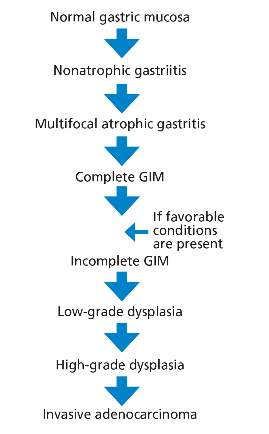
The Correa cascade describes the progression from precancerous histologic changes in the gastric mucosa to the development of GIM and its consequences, including adenocarcinoma (Figure 2). The process begins with development of nonatrophic gastritis and progresses to multifocal atrophic gastritis followed by GIM.

Figure 2
  • Download figure
  • Open in new tab
  • Download powerpoint
Figure 2

The Correa cascade illustrates the progression from precancerous histologic changes in the gastric mucosa to the development of gastric intestinal metaplasia.

GIM can present 2 histologic types:

  • The complete and fully intestinalized pattern has markers for intestinal mucin and absence of gastric mucin.

  • The incomplete gastric and intestinal mixed glands pattern has both gastric mucin and intestinal mucin markers.16

Complete GIM may progress to incomplete GIM if conditions leading to severe inflammation are present (eg, advanced atrophy or hypochlorhydria) before identifiable dysplastic changes.17 Subsequently, the tissue progresses to low-grade dysplasia, followed by high-grade dysplasia, and finally invasive adenocarcinoma.18

Differentiation of the 2 types of GIM is important. Incomplete GIM has been associated with an increased risk of cancer progression, and some experts consider it a mild degree of dysplasia.19,20

RISK FACTORS FOR PROGRESSION TO MALIGNANCY

The risk of developing gastric cancer may be higher in patients with histologic evidence of incomplete and extensive GIM (ie, involvement of the antrum and corpus) than in those with complete and limited GIM.1,21,22 Some studies suggest that the topographic distribution of intestinal metaplasia may affect the risk of cancer progression. In Cassaro et al’s23 cohort study of 135 Colombian patients, a GIM distribution involving the lesser curvature of the stomach from the cardia to the pylorus was associated with higher cancer risk (odds ratio 5.7, 95% confidence interval 1.3–26) compared with “antrum-predominant” or “focal” patterns.23

The incidence of gastric cancer exhibits significant geographic variation worldwide due to potential environmental exposure factors and genetic predisposition. The reported rates are highest in Eastern Asia, Eastern Europe, and South America, and lowest in North America.24 People who immigrate from a region of high incidence to a region of low incidence have an increased risk of gastric cancer.25 Table 2 summarizes risk factors for malignancy.1,26

View this table:
  • View inline
  • View popup
TABLE 2

Risk factors for progression to malignancy in gastric intestinal metaplasia

ENDOSCOPIC MANAGEMENT

The role for endoscopy in GIM is limited to detection and surveillance, as no other methods are currently available for this. Specific recommendations for endoscopy are discussed in the various guidelines below.

NONENDOSCOPIC MANAGEMENT

GIM management should emphasize risk-factor modification, including smoking cessation and moderation in alcohol intake. In patients with H pylori-induced gastritis, early H pylori detection and eradication are crucial to halt progression to gastric cancer. In contrast, the effects of H pylori eradication once GIM occurs are undetermined. GIM changes may be irreversible, and the impact of H pylori eradication on cancer progression once GIM is established may be minimal.27

Observational studies have reported partial GIM reversal and decreased progression to stomach cancer with use of aspirin and nonsteroidal anti-inflammatory drugs such as celecoxib.28 More evidence is needed to support their use.

GLOBAL DIFFERENCES IN GUIDELINE RECOMMENDATIONS

The optimal follow-up of patients with isolated glands of GIM remains controversial, with significant differences in guidelines in the Eastern and Western regions of the world (Table 3).29–32

View this table:
  • View inline
  • View popup
TABLE 3

Variations in society recommendations for the management of gastric intestinal metaplasia

Recommendations for Eastern regions

In countries such as Japan, where the incidence of gastric cancer is high, national screening programs recommend mass endoscopic or radiographic screening of all men and women at age 50 or older.29 For patients found to have GIM without malignancy on initial screening, surveillance endoscopy in 1 to 3 years is recommended if they have GIM with high-risk features such as incomplete GIM, extensive GIM, family history of gastric cancer, smoking, or excessive alcohol use.

Recommendations for Western regions

The British Society of Gastroenterology guidelines33 identify patients with GIM as having increased risk for gastric malignancy and recommend endoscopic surveillance every 3 years if there is extensive GIM (ie, affecting the antrum and corpus), antral GIM with risk factors such as H pylori, or a family history of gastric cancer.33

A 2019 consensus guideline by 4 European organizations agreed with the British Society of Gastroenterology.30 The guideline recommends that patients with GIM who are considered at high risk, including those with histologically proven GIM of the corpus and antrum, undergo endoscopic surveillance every 3 years. The guideline advises consideration of surveillance if GIM is present only in the corpus or antrum but the patient has a family history of gastric cancer, persistent H pylori, incomplete GIM, or autoimmune gastritis.

US recommendations

Two US societies have published guidelines addressing the management of GIM.

The American Gastroenterological Association guidelines31 recommend against routine endoscopic surveillance after GIM is detected in the general population, but if H pylori is detected, treatment is encouraged. Patients with GIM and risk factors associated with progression can be considered for endoscopic surveillance every 3 to 5 years if the patient favors surveillance, which has an unclear impact on mortality risk, vs endoscopic evaluation, which has potential complications.31

The guidelines subcategorized risk factors associated with progression of gastric cancer as follows:

  • Highest risk: incomplete GIM, extensive GIM, or family history of gastric cancer

  • Overall increased risk: certain racial or ethnic minorities immigrating from high-incidence regions.31

The American Society for Gastrointestinal Endoscopy recommendations32 are similar to those of European groups. They advise endoscopic surveillance exclusively for patients with risk factors, but not for the general cohort of patients in whom GIM is detected.

AN ALGORITHMIC APPROACH TO DIAGNOSIS AND MANAGEMENT

Figure 3 suggests an approach to managing patients who have GIM. The updated Sydney protocol includes the collection of 5 nontargeted biopsy specimens: 2 from the antrum (at the lesser and greater curvature), 2 from the corpus (at the lesser and greater curvature), and 1 from the incisura.34 It is recommended that these biopsy specimens be placed in separate jars.

Figure 3
  • Download figure
  • Open in new tab
  • Download powerpoint
Figure 3

An algorithmic approach to the management of gastric intestinal metaplasia (GIM).

Careful inspection should be carried out with high-definition white-light endoscopy rather than standard-definition endoscopy. Adequate air insufflation, use of mucolytic and defoaming agents (for improved visibility), appropriate withdrawal times, and photodocumentation are key for a quality endoscopic examination.35 Additionally, use of narrow-band imaging should be encouraged because it has been shown to improve the detection of GIM.36 It also allows for more targeted biopsies for GIM.

DISCLOSURES

The authors report no relevant financial relationships which, in the context of their contributions, could be perceived as a potential conflict of interest.

  • Copyright © 2024 The Cleveland Clinic Foundation. All Rights Reserved.
View Abstract

REFERENCES

  1. ↵
    1. Gawron AJ,
    2. Shah SC,
    3. Altayar O, et al
    . AGA technical review on gastric intestinal metaplasia: natural history and clinical outcomes. Gastroenterology 2020; 158(3):705–731.e5. doi:10.1053/j.gastro.2019.12.001
    OpenUrlCrossRef
  2. ↵
    1. de Vries AC,
    2. van Grieken NC,
    3. Looman CW, et al
    . Gastric cancer risk in patients with premalignant gastric lesions: a nationwide cohort study in the Netherlands. Gastroenterology 2008; 134(4):945–952. doi:10.1053/j.gastro.2008.01.071
    OpenUrlCrossRefPubMed
  3. ↵
    1. Bhat S,
    2. Coleman HG,
    3. Yousef F, et al
    . Risk of malignant progression in Barrett’s esophagus patients: results from a large population-based study [published correction appears in J Natl Cancer Inst 2013; 105(8):581]. J Natl Cancer Inst 2011; 103(13):1049–1057. doi:10.1093/jnci/djr203
    OpenUrlCrossRefPubMed
  4. ↵
    1. Rastogi A,
    2. Puli S,
    3. El-Serag HB,
    4. Bansal A,
    5. Wani S,
    6. Sharma P
    . Incidence of esophageal adenocarcinoma in patients with Barrett’s esophagus and high-grade dysplasia: a meta-analysis. Gastrointest Endosc 2008; 67(3):394–398. doi:10.1016/j.gie.2007.07.019
    OpenUrlCrossRefPubMed
  5. ↵
    1. Singh S,
    2. Manickam P,
    3. Amin AV, et al
    . Incidence of esophageal adenocarcinoma in Barrett’s esophagus with low-grade dysplasia: a systematic review and meta-analysis. Gastrointest Endosc 2014; 79(6):897–983.e3. doi:10.1016/j.gie.2014.01.009
    OpenUrlCrossRefPubMed
  6. ↵
    1. El-Serag HB,
    2. Naik AD,
    3. Duan Z, et al
    . Surveillance endoscopy is associated with improved outcomes of oesophageal adenocarcinoma detected in patients with Barrett’s oesophagus. Gut 2016; 65(8):1252–1260. doi:10.1136/gutjnl-2014-308865
    OpenUrlAbstract/FREE Full Text
  7. ↵
    1. Sipponen P,
    2. Maaroos HI
    . Chronic gastritis. Scand J Gastroenterol 2015; 50(6):657–667. doi:10.3109/00365521.2015.1019918
    OpenUrlCrossRefPubMed
  8. ↵
    1. Brown LM
    . Helicobacter pylori: epidemiology and routes of transmission. Epidemiol Rev 2000; 22(2):283–297. doi:10.1093/oxfordjournals.epirev.a018040
    OpenUrlCrossRefPubMed
  9. ↵
    1. Smith JG,
    2. Li W,
    3. Rosson RS
    . Prevalence, clinical and endoscopic predictors of Helicobacter pylori infection in an urban population. Conn Med 2009; 73(3):133–137. pmid:19353987
    OpenUrlPubMed
  10. ↵
    1. Murata-Kamiya N
    . Pathophysiological functions of the CagA oncoprotein during infection by Helicobacter pylori. Microbes Infect 2011; 13(10):799–807. doi:10.1016/j.micinf.2011.03.011
    OpenUrlCrossRefPubMed
  11. ↵
    1. Nasser SC,
    2. Slim M,
    3. Nassif JG,
    4. Nasser SM
    . Influence of proton pump inhibitors on gastritis diagnosis and pathologic gastric changes. World J Gastroenterol 2015; 21(15):4599–4606. doi:10.3748/wjg.v21.i15.4599
    OpenUrlCrossRef
  12. ↵
    1. Correa P,
    2. Piazuelo MB,
    3. Wilson KT
    . Pathology of gastric intestinal metaplasia: clinical implications. Am J Gastroenterol 2010; 105(3):493–498. doi:10.1038/ajg.2009.728
    OpenUrlCrossRefPubMed
  13. ↵
    1. Hayter SM,
    2. Cook MC
    . Updated assessment of the prevalence, spectrum and case definition of autoimmune disease. Autoimmun Rev 2012; 11(10):754–765. doi:10.1016/j.autrev.2012.02.001
    OpenUrlCrossRefPubMed
  14. ↵
    1. Park JY,
    2. Lam-Himlin D,
    3. Vemulapalli R
    . Review of autoimmune met-aplastic atrophic gastritis. Gastrointest Endosc 2013; 77(2):284–292. doi:10.1016/j.gie.2012.09.033
    OpenUrlCrossRefPubMed
  15. ↵
    1. Minalyan A,
    2. Benhammou JN,
    3. Artashesyan A,
    4. Lewis MS,
    5. Pisegna JR
    . Autoimmune atrophic gastritis: current perspectives. Clin Exp Gastroenterol 2017; 10:19–27. doi:10.2147/CEG.S109123
    OpenUrlCrossRef
  16. ↵
    1. Reis CA,
    2. David L,
    3. Correa P, et al
    . Intestinal metaplasia of human stomach displays distinct patterns of mucin (MUC1, MUC2, MU-C5AC, and MUC6) expression. Cancer Res 1999; 59(5):1003–1007. pmid:10070955
    OpenUrlAbstract/FREE Full Text
  17. ↵
    1. Sugano K,
    2. Moss SF,
    3. Kuipers EJ
    . Gastric intestinal metaplasia: real culprit or innocent bystander as a precancerous condition for gastric cancer? Gastroenterology 2023; S0016-5085(23)04911-9. doi:10.1053/j.gastro.2023.08.028
    OpenUrlCrossRef
  18. ↵
    1. Correa P,
    2. Haenszel W,
    3. Cuello C,
    4. Tannenbaum S,
    5. Archer M
    . A model for gastric cancer epidemiology. Lancet 1975; 2(7924):58–60. doi:10.1016/s0140-6736(75)90498-5
    OpenUrlCrossRefPubMed
  19. ↵
    1. Tava F,
    2. Luinetti O,
    3. Ghigna MR, et al
    . Type or extension of intestinal metaplasia and immature/atypical “indefinite-for-dysplasia” lesions as predictors of gastric neoplasia. Hum Pathol 2006; 37(11):1489–1497. doi:10.1016/j.humpath.2006.05.012
    OpenUrlCrossRefPubMed
  20. ↵
    1. Tosi P,
    2. Filipe MI,
    3. Luzi P, et al
    . Gastric intestinal metaplasia type III cases are classified as low-grade dysplasia on the basis of morphometry. J Pathol 1993; 169(1):73–78. doi:10.1002/path.1711690112
    OpenUrlCrossRefPubMed
  21. ↵
    1. Silva S,
    2. Filipe MI,
    3. Pinho A
    . Variants of intestinal metaplasia in the evolution of chronic atrophic gastritis and gastric ulcer. A follow-up study. Gut 1990; 31(10):1097–1104. doi:10.1136/gut.31.10.1097
    OpenUrlAbstract/FREE Full Text
  22. ↵
    1. González CA,
    2. Sanz-Anquela JM,
    3. Companioni O, et al
    . Incomplete type of intestinal metaplasia has the highest risk to progress to gastric cancer: results of the Spanish follow-up multicenter study. J Gastroenterol Hepatol 2016; 31(5):953–958. doi:10.1111/jgh.13249
    OpenUrlCrossRefPubMed
  23. ↵
    1. Cassaro M,
    2. Rugge M,
    3. Gutierrez O,
    4. Leandro G,
    5. Graham DY,
    6. Genta RM
    . Topographic patterns of intestinal metaplasia and gastric cancer. Am J Gastroenterol 2000; 95(6):1431–1438. doi:10.1111/j.1572-0241.2000.02074.x
    OpenUrlCrossRefPubMed
  24. ↵
    1. Jemal A,
    2. Jemal A,
    3. Center MM,
    4. Center MM,
    5. Ward E,
    6. Ward E
    . Global cancer statistics [published correction appears in CA Cancer J Clin 2011; 61(2):134]. CA Cancer J Clin 2011; 61(2):69–90. doi:10.3322/caac.20107
    OpenUrlCrossRefPubMed
  25. ↵
    1. Pabla BS,
    2. Shah SC,
    3. Corral JE,
    4. Morgan DR
    . Increased incidence and mortality of gastric cancer in immigrant populations from high to low regions of incidence: a systematic review and meta-analysis. Clin Gastroenterol Hepatol 2020; 18(2):347–359.e5. doi:10.1016/j.cgh.2019.05.032
    OpenUrlCrossRef
  26. ↵
    1. Altayar O,
    2. Davitkov P,
    3. Shah SC, et al
    . AGA technical review on gastric intestinal metaplasia-epidemiology and risk factors. Gastroenterology 2020; 158(3):732–744.e16. doi:10.1053/j.gastro.2019.12.002
    OpenUrlCrossRef
  27. ↵
    1. Chen HN,
    2. Wang Z,
    3. Li X,
    4. Zhou ZG
    . Helicobacter pylori eradication cannot reduce the risk of gastric cancer in patients with intestinal metaplasia and dysplasia: evidence from a meta-analysis. Gastric Cancer 2016; 19(1):166–175. doi:10.1007/s10120-015-0462-7
    OpenUrlCrossRefPubMed
  28. ↵
    1. Tian W,
    2. Zhao Y,
    3. Liu S,
    4. Li X
    . Meta-analysis on the relationship between nonsteroidal anti-inflammatory drug use and gastric cancer. Eur J Cancer Prev 2010; 19(4):288–298. doi:10.1097/CEJ.0b013e328339648c
    OpenUrlCrossRefPubMed
  29. ↵
    1. Hamashima C
    . Cancer screening guidelines and policy making: 15 years of experience in cancer screening guideline development in Japan. Jpn J Clin Oncol 2018; 48(3):278–286. doi:10.1093/jjco/hyx190
    OpenUrlCrossRef
  30. ↵
    1. Pimentel-Nunes P,
    2. Libânio D,
    3. Marcos-Pinto R, et al
    . Management of epithelial precancerous conditions and lesions in the stomach (MAPS II): European Society of Gastrointestinal Endoscopy (ESGE), European Helicobacter and Microbiota Study Group (EHMSG), European Society of Pathology (ESP), and Sociedade Portuguesa de Endoscopia Digestiva (SPED) guideline update 2019. Endoscopy 2019; 51(4):365–388. doi:10.1055/a-0859-1883
    OpenUrlCrossRefPubMed
  31. ↵
    1. Gupta S,
    2. Li D,
    3. El Serag HB, et al
    . AGA clinical practice guidelines on management of gastric intestinal metaplasia. Gastroenterology 2020; 158(3):693–702. doi:10.1053/j.gastro.2019.12.003
    OpenUrlCrossRef
  32. ↵
    1. ASGE Standards of Practice Committee,
    2. Evans JA,
    3. Chandrasekhara V, et al
    . The role of endoscopy in the management of premalignant and malignant conditions of the stomach. Gastrointest Endosc 2015; 82(1):1–8. doi:10.1016/j.gie.2015.03.1967
    OpenUrlCrossRefPubMed
  33. ↵
    1. Banks M,
    2. Graham D,
    3. Jansen M, et al
    . British Society of Gastroenterology guidelines on the diagnosis and management of patients at risk of gastric adenocarcinoma. Gut 2019; 68(9):1545–1575. doi:10.1136/gutjnl-2018-318126
    OpenUrlAbstract/FREE Full Text
  34. ↵
    1. Shah SC,
    2. Gawron AJ,
    3. Li D
    . Surveillance of gastric intestinal metaplasia. Am J Gastroenterol 2020; 115(5):641–644. doi:10.14309/ajg.0000000000000540
    OpenUrlCrossRef
  35. ↵
    1. Veitch AM,
    2. Uedo N,
    3. Yao K,
    4. East JE
    . Optimizing early upper gastrointestinal cancer detection at endoscopy. Nat Rev Gastroenterol Hepatol 2015; 12(11):660–667. doi:10.1038/nrgastro.2015.128
    OpenUrlCrossRefPubMed
  36. ↵
    1. Buxbaum JL,
    2. Hormozdi D,
    3. Dinis-Ribeiro M, et al
    . Narrow-band imaging versus white light versus mapping biopsy for gastric intestinal metaplasia: a prospective blinded trial. Gastrointest Endosc 2017; 86(5):857–865. doi:10.1016/j.gie.2017.03.1528
    OpenUrlCrossRefPubMed
PreviousNext
Back to top

In this issue

Cleveland Clinic Journal of Medicine: 91 (1)
Cleveland Clinic Journal of Medicine
Vol. 91, Issue 1
1 Jan 2024
  • Table of Contents
  • Table of Contents (PDF)
  • Index by author
  • Complete Issue (PDF)
Print
Download PDF
Article Alerts
Sign In to Email Alerts with your Email Address
Email Article

Thank you for your interest in spreading the word on Cleveland Clinic Journal of Medicine.

NOTE: We only request your email address so that the person you are recommending the page to knows that you wanted them to see it, and that it is not junk mail. We do not capture any email address.

Enter multiple addresses on separate lines or separate them with commas.
Gastric intestinal metaplasia and gastric cancer prevention: Watchful waiting
(Your Name) has sent you a message from Cleveland Clinic Journal of Medicine
(Your Name) thought you would like to see the Cleveland Clinic Journal of Medicine web site.
CAPTCHA
This question is for testing whether or not you are a human visitor and to prevent automated spam submissions.
Citation Tools
Gastric intestinal metaplasia and gastric cancer prevention: Watchful waiting
Shrouq Khazaaleh, Mohammad Alomari, Mamoon Ur Rashid, Daniel Castaneda, Fernando J. Castro
Cleveland Clinic Journal of Medicine Jan 2024, 91 (1) 33-39; DOI: 10.3949/ccjm.91a.23015

Citation Manager Formats

  • BibTeX
  • Bookends
  • EasyBib
  • EndNote (tagged)
  • EndNote 8 (xml)
  • Medlars
  • Mendeley
  • Papers
  • RefWorks Tagged
  • Ref Manager
  • RIS
  • Zotero
Share
Gastric intestinal metaplasia and gastric cancer prevention: Watchful waiting
Shrouq Khazaaleh, Mohammad Alomari, Mamoon Ur Rashid, Daniel Castaneda, Fernando J. Castro
Cleveland Clinic Journal of Medicine Jan 2024, 91 (1) 33-39; DOI: 10.3949/ccjm.91a.23015
Twitter logo Facebook logo Mendeley logo
  • Tweet Widget
  • Facebook Like
  • Linkedin Share Button

Jump to section

  • Article
    • ABSTRACT
    • RISK OF CANCER PROGRESSION
    • CHRONIC GASTRITIS AND GIM
    • CORREA CASCADE: DIFFERENTIATING COMPLETE AND INCOMPLETE GIM
    • RISK FACTORS FOR PROGRESSION TO MALIGNANCY
    • ENDOSCOPIC MANAGEMENT
    • NONENDOSCOPIC MANAGEMENT
    • GLOBAL DIFFERENCES IN GUIDELINE RECOMMENDATIONS
    • AN ALGORITHMIC APPROACH TO DIAGNOSIS AND MANAGEMENT
    • DISCLOSURES
    • REFERENCES
  • Figures & Data
  • Info & Metrics
  • PDF

Related Articles

  • No related articles found.
  • PubMed
  • Google Scholar

Cited By...

  • No citing articles found.
  • Google Scholar

More in this TOC Section

  • Managing right ventricular failure in the setting of pulmonary embolism
  • Alpha-gal syndrome: Recognizing and managing a tick-bite–related meat allergy
  • Psychedelics, spirituality, and existential distress in patients at the end of life
Show more Review

Similar Articles

Subjects

  • Gastroenterology
  • Oncology
  • Preventive Care

Navigate

  • Current Issue
  • Past Issues
  • Supplements
  • Article Type
  • Specialty
  • CME/MOC Articles
  • CME/MOC Calendar
  • Media Kit

Authors & Reviewers

  • Manuscript Submission
  • Authors & Reviewers
  • Subscriptions
  • About CCJM
  • Contact Us
  • Cleveland Clinic Center for Continuing Education
  • Consult QD

Share your suggestions!

Copyright © 2025 The Cleveland Clinic Foundation. All rights reserved. The information provided is for educational purposes only. Use of this website is subject to the website terms of use and privacy policy. 

Powered by HighWire