
Main menu

  • Home
  • Content
    • Current Issue
    • Ahead of Print
    • Past Issues
    • Supplements
    • Article Type
  • Specialty
    • Articles by Specialty
  • CME/MOC
    • Articles
    • Calendar
  • Info For
    • Manuscript Submission
    • Authors & Reviewers
    • Subscriptions
    • About CCJM
    • Contact Us
    • Media Kit
  • Conversations with Leaders
  • Conference Coverage
    • Kidney Week 2024
    • CHEST 2024
    • ACR Convergence 2023
    • Kidney Week 2023
    • ObesityWeek 2023
    • IDWeek 2023
    • CHEST 2023
    • MDS 2023
    • IAS 2023
    • ACP 2023
    • AAN 2023
    • ACC / WCC 2023
    • AAAAI Meeting 2023
    • ACR Convergence 2022
    • Kidney Week 2022
    • AIDS 2022
  • Other Publications
    • www.clevelandclinic.org

User menu

  • Register
  • Log in

Search

  • Advanced search
Cleveland Clinic Journal of Medicine
  • Other Publications
    • www.clevelandclinic.org
  • Register
  • Log in
Cleveland Clinic Journal of Medicine

Advanced Search

  • Home
  • Content
    • Current Issue
    • Ahead of Print
    • Past Issues
    • Supplements
    • Article Type
  • Specialty
    • Articles by Specialty
  • CME/MOC
    • Articles
    • Calendar
  • Info For
    • Manuscript Submission
    • Authors & Reviewers
    • Subscriptions
    • About CCJM
    • Contact Us
    • Media Kit
  • Conversations with Leaders
  • Conference Coverage
    • Kidney Week 2024
    • CHEST 2024
    • ACR Convergence 2023
    • Kidney Week 2023
    • ObesityWeek 2023
    • IDWeek 2023
    • CHEST 2023
    • MDS 2023
    • IAS 2023
    • ACP 2023
    • AAN 2023
    • ACC / WCC 2023
    • AAAAI Meeting 2023
    • ACR Convergence 2022
    • Kidney Week 2022
    • AIDS 2022
The Clinical Picture

Air leakage in multiple compartments after endoscopy

Amanda Grippen Goddard, DO, Bryan McConomy, MD, Girish Bathla, MBBS, FRCR, Muhammad Furqan, MBBS and William B. Silverman, MD, FACG, FASGE, AGAF
Cleveland Clinic Journal of Medicine October 2016, 83 (10) 705-707; DOI: https://doi.org/10.3949/ccjm.83a.15168
Amanda Grippen Goddard
Department of Internal Medicine, University of Iowa Hospitals and Clinics, Iowa City
  • Find this author on Google Scholar
  • Find this author on PubMed
  • Search for this author on this site
  • For correspondence: [email protected]
Bryan McConomy
Department of Internal Medicine, University of Iowa Hospitals and Clinics, Iowa City
  • Find this author on Google Scholar
  • Find this author on PubMed
  • Search for this author on this site
Girish Bathla
Department of Radiology, University of Iowa Hospitals and Clinics, Iowa City
  • Find this author on Google Scholar
  • Find this author on PubMed
  • Search for this author on this site
Muhammad Furqan
Department of Internal Medicine, University of Iowa Hospitals and Clinics, Iowa City
  • Find this author on Google Scholar
  • Find this author on PubMed
  • Search for this author on this site
William B. Silverman
Department of Internal Medicine, University of Iowa Hospitals and Clinics, Iowa City
  • Find this author on Google Scholar
  • Find this author on PubMed
  • Search for this author on this site
  • Article
  • Figures & Data
  • Info & Metrics
  • PDF
Loading

A 60-year old man with metastatic periampullary adenocarcinoma presented to his usual clinic for a scheduled biliary stent exchange by endoscopic retrograde cholangiopancreatography (ERCP). The stent had been placed 5 months before, and no complications had been reported during that procedure.

During the stent exchange procedure, the endoscopist advanced the scope to the second part of the duodenum, where a large, ulcerated, friable mass was visualized surrounding the ampulla, consistent with patient’s known periampullary cancer. The biliary stent was removed without much difficulty. However, several attempts to cannulate the common bile duct with a preloaded guidewire failed because of extensive edema and tissue friability, and to avoid further discomfort to the patient, the procedure was aborted. No perforation was visualized during or at the end of the procedure.

During the first hour after the procedure was stopped, the patient suddenly developed abdominal pain and distention and crepitus of the right chest wall. Supine abdominal radiography showed extensive pneumoperitoneum and subcutaneous emphysema in the chest. A nasogastric tube was placed for decompression, and the patient was transferred to the surgical intensive care unit at our hospital.

EVIDENCE OF PERFORATION NOTED

On arrival, the patient’s oxygen saturation was 99% while receiving oxygen at 2 L/minute by nasal cannula. The physical examination revealed neck swelling, abdominal distention, and crepitus in the neck, abdomen, scrotum, and right lower extremity.

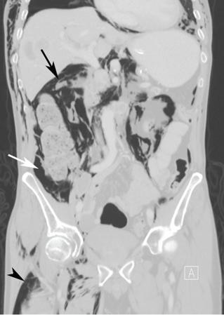
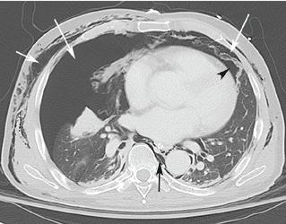
Computed tomography (CT) of the abdomen and pelvis with oral and intravenous contrast revealed widespread pneumoretroperitoneum, pneumoperitoneum, and air along the intermuscular planes in the right lower extremity, with no evidence of extravasation of oral contrast (Figure 1). Also noted were bilateral pneumothorax, pneumomediastinum, pneumopericardium, and extensive subcutaneous emphysema (Figure 2).

FIGURE 1
  • Download figure
  • Open in new tab
  • Download powerpoint
FIGURE 1

Computed tomography with coronal multiplanar reconstruction revealed pneumoperitoneum (black arrow), pneumoretroperitoneum (white arrow), and air along intermuscular planes in the right lower extremity (arrowhead). The image is a lung-window setting, which better demonstrates free air.

FIGURE 2
  • Download figure
  • Open in new tab
  • Download powerpoint
FIGURE 2

Computed tomography with axial imaging through the lungs revealed bilateral pneumothoraces (long white arrows), pneumomediastinum (black arrow), pneumopericardium (arrowhead), and subcutaneous emphysema (short white arrow).

Despite these impressive findings, the patient remained hemodynamically stable and was managed conservatively with broad-spectrum antibiotics, gastric decompression, and bowel rest. But repeat chest radiography 5 hours after admission to the hospital revealed an enlarging right pneumothorax, which was treated with placement of a pigtail catheter. The patient continued to improve with conservative management and was discharged on the 6th day of hospitalization.

PERFORATION DURING ERCP: INCIDENCE AND COMPLICATIONS

Although perforation is an uncommon complication of ERCP, with an incidence of 1%, mortality rates as high as 18% have been reported.1 Older age, longer procedural time, anatomic variations, and diseases of the duodenum and common bile duct can increase the risk of perforation.2

Types of perforation

Stapfer et al1 classified perforation during ERCP into four types, based on etiology and site of perforation. Type 1 is perforation of the lateral or medial duodenal wall caused by excessive pressure from the endoscope or its acute angulation. Type 2 is periampullary injury, often associated with sphincterotomy or difficulty accessing the biliary tree. Type 3 is injury to the common bile duct or pancreatic duct caused by instrumentation. Type 4 is the presence of retroperitoneal free air with no evidence of actual perforation; this is usually an incidental finding and is of little or no clinical consequence.1

In 2015, a review of 18 studies described the distribution of ERCP perforation according to the Stapfer classification: 25% were type 1, 46% were type 2, and 22% were type 3.3

Effects of air insufflation

ERCP requires air insufflation for optimal visualization. During difficult or prolonged procedures, a larger amount of air may be insufflated to maintain bowel lumen visibility. Depending on the site and size of the defect, a variable amount of air can leak under pressure once the perforation occurs. A rapid retroperitoneal air leak can spread to multiple body compartments, including the mediastinum, pleura, neck, subcutaneous tissues, scrotum, and musculature by tracking through various fascial planes. Rarely, rapid ingress of air in these areas can lead to compartment syndrome.4

Small perforations tend to close spontaneously and may remain clinically silent, but large or persistent perforations are known to cause subcutaneous emphysema, sepsis, and respiratory failure.5

Our patient’s type 2 perforation

We presumed that our patient had a type 2 perforation, given the finding of retroperitoneal air. Difficulty cannulating the biliary tree via the friable malignant tissue at the site of the major papilla likely caused punctate perforations, resulting in air leakage into the retroperitoneum. Punctate perforations typically do not allow contrast extravasation, explaining the absence of oral contrast leakage on CT.

TREATMENT OF ENDOSCOPY-RELATED PERFORATION

Conventional supine and upright abdominal radiography is an appropriate initial imaging modality to confirm the diagnosis. However, CT is more sensitive and accurate, especially when air leakage is confined to the retroperitoneum. Intravenous or oral contrast is not necessary but may help localize the perforation and better delineate fluid collections and abscesses.2

Once perforation is suspected, treatment with a broad-spectrum antibiotic, bowel rest, and stomach decompression is imperative.6 Further management depends on the type of perforation and the overall clinical picture. Type 1 perforations usually require immediate surgical intervention. Type 2 perforations often seal spontaneously within 2 to 3 days and thus are managed conservatively (ie, a broad-spectrum antibiotic, gastric decompression, and bowel rest), unless there is a persistent leak or a large fluid collection. Type 3 perforations rarely require surgery since most are very small and close spontaneously, and so they are managed conservatively. Type 4 perforations are the least serious. They result in retroperitoneal free air that is thought be related to the use of compressed air for lumen patency. They require only conservative measures.1

  • Copyright © 2016 The Cleveland Clinic Foundation. All Rights Reserved.

REFERENCES

  1. ↵
    1. Stapfer M,
    2. Selby RR,
    3. Stain SC,
    4. et al
    . Management of duodenal perforation after endoscopic retrograde cholangiopancreatography and sphincterotomy. Ann Surg 2000; 232:191–198.
    OpenUrlCrossRefPubMed
  2. ↵
    1. Enns M,
    2. Eloubeidi K,
    3. Mergener P,
    4. et al
    . ERCP-related perforations: risk factors and management. Endoscopy 2002; 34:293–298.
    OpenUrlCrossRefPubMed
  3. ↵
    1. Vezakis A,
    2. Fragulidis G,
    3. Polydorou A
    . Endoscopic retrograde cholangiopancreatography-related perforations: diagnosis and management. World J Gastrointest Endosc 2015; 7:1135–1341.
    OpenUrl
  4. ↵
    1. Frias Vilaca A,
    2. Reis AM,
    3. Vidal IM
    . The anatomical compartments and their connections as demonstrated by ectopic air. Insights Imaging 2013; 4:759–772.
    OpenUrl
  5. ↵
    1. Machado N
    . Management of duodenal perforation post-endoscopic retrograde cholangiopancreatography. When and whom to operate and what factors determine the outcome? A review article. JOP (Online) 2012; 13:18–25.
    OpenUrl
  6. ↵
    1. Dubecz A,
    2. Ottmann J,
    3. Schweigert M,
    4. et al
    . Management of ERCP-related small bowel perforations: the pivotal role of physical investigation. Can J Surg 2012; 55:99–104.
    OpenUrlPubMed
PreviousNext
Back to top

In this issue

Cleveland Clinic Journal of Medicine: 83 (10)
Cleveland Clinic Journal of Medicine
Vol. 83, Issue 10
1 Oct 2016
  • Table of Contents
  • Table of Contents (PDF)
  • Index by author
Print
Download PDF
Article Alerts
Sign In to Email Alerts with your Email Address
Email Article

Thank you for your interest in spreading the word on Cleveland Clinic Journal of Medicine.

NOTE: We only request your email address so that the person you are recommending the page to knows that you wanted them to see it, and that it is not junk mail. We do not capture any email address.

Enter multiple addresses on separate lines or separate them with commas.
Air leakage in multiple compartments after endoscopy
(Your Name) has sent you a message from Cleveland Clinic Journal of Medicine
(Your Name) thought you would like to see the Cleveland Clinic Journal of Medicine web site.
CAPTCHA
This question is for testing whether or not you are a human visitor and to prevent automated spam submissions.
Citation Tools
Air leakage in multiple compartments after endoscopy
Amanda Grippen Goddard, Bryan McConomy, Girish Bathla, Muhammad Furqan, William B. Silverman
Cleveland Clinic Journal of Medicine Oct 2016, 83 (10) 705-707; DOI: 10.3949/ccjm.83a.15168

Citation Manager Formats

  • BibTeX
  • Bookends
  • EasyBib
  • EndNote (tagged)
  • EndNote 8 (xml)
  • Medlars
  • Mendeley
  • Papers
  • RefWorks Tagged
  • Ref Manager
  • RIS
  • Zotero
Share
Air leakage in multiple compartments after endoscopy
Amanda Grippen Goddard, Bryan McConomy, Girish Bathla, Muhammad Furqan, William B. Silverman
Cleveland Clinic Journal of Medicine Oct 2016, 83 (10) 705-707; DOI: 10.3949/ccjm.83a.15168
Twitter logo Facebook logo Mendeley logo
  • Tweet Widget
  • Facebook Like
  • Linkedin Share Button

Jump to section

  • Article
    • EVIDENCE OF PERFORATION NOTED
    • PERFORATION DURING ERCP: INCIDENCE AND COMPLICATIONS
    • TREATMENT OF ENDOSCOPY-RELATED PERFORATION
    • REFERENCES
  • Figures & Data
  • Info & Metrics
  • PDF

Related Articles

  • No related articles found.
  • PubMed
  • Google Scholar

Cited By...

  • No citing articles found.
  • Google Scholar

More in this TOC Section

  • Tinea incognito
  • Prolonged venous filling time and dependent rubor in a patient with peripheral artery disease
  • Sarcoidosis with diffuse purplish erythematous plaques on the hands
Show more The Clinical Picture

Similar Articles

Subjects

  • Gastroenterology
  • Hepatology
  • Imaging
  • Oncology

Navigate

  • Current Issue
  • Past Issues
  • Supplements
  • Article Type
  • Specialty
  • CME/MOC Articles
  • CME/MOC Calendar
  • Media Kit

Authors & Reviewers

  • Manuscript Submission
  • Authors & Reviewers
  • Subscriptions
  • About CCJM
  • Contact Us
  • Cleveland Clinic Center for Continuing Education
  • Consult QD

Share your suggestions!

Copyright © 2025 The Cleveland Clinic Foundation. All rights reserved. The information provided is for educational purposes only. Use of this website is subject to the website terms of use and privacy policy. 

Powered by HighWire