


Article Figures & Data
Figures
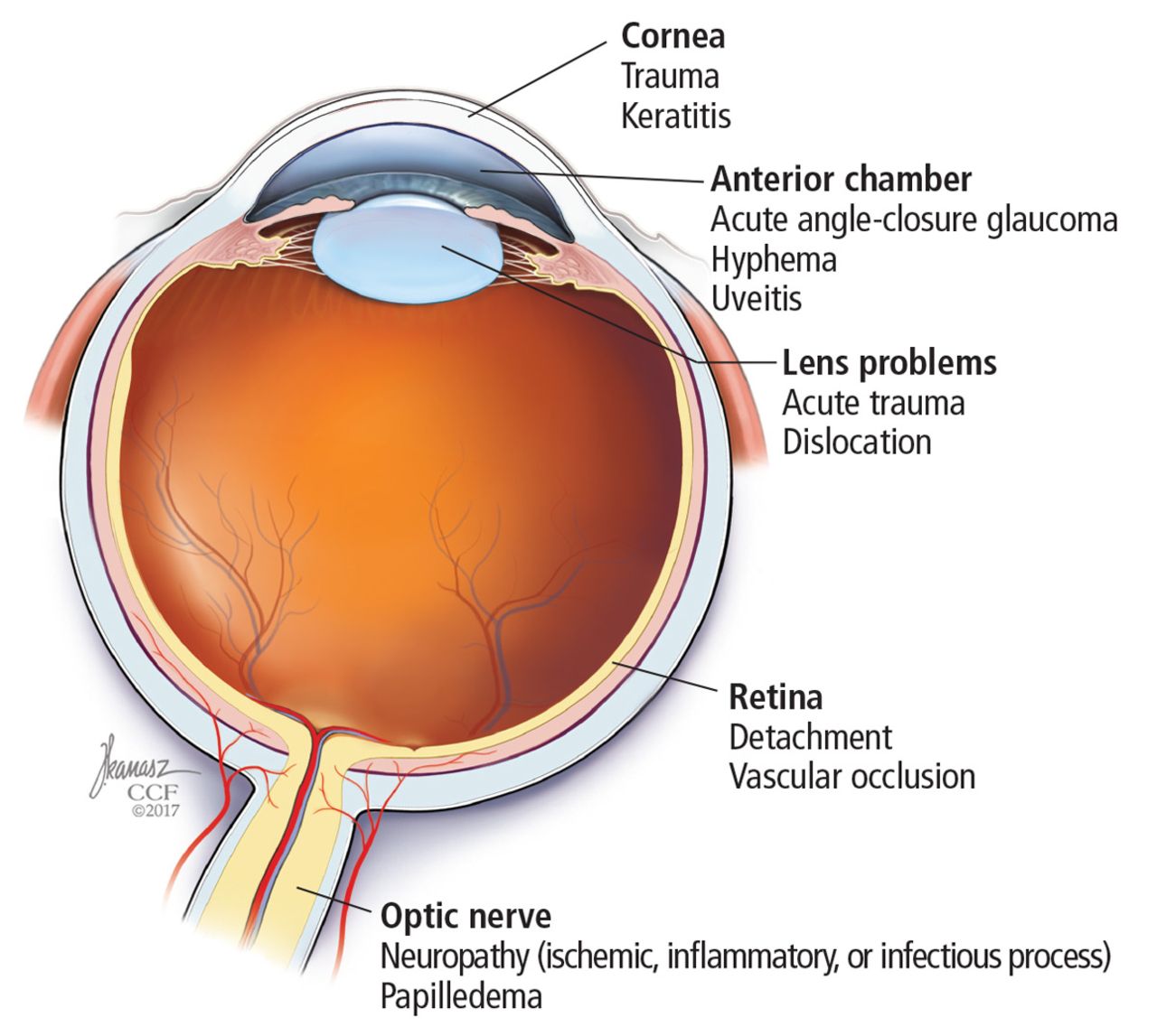
- FIGURE 1
Common causes of monocular vision loss can arise in the media (cornea, anterior chamber, or lens), retina, or optic nerve.
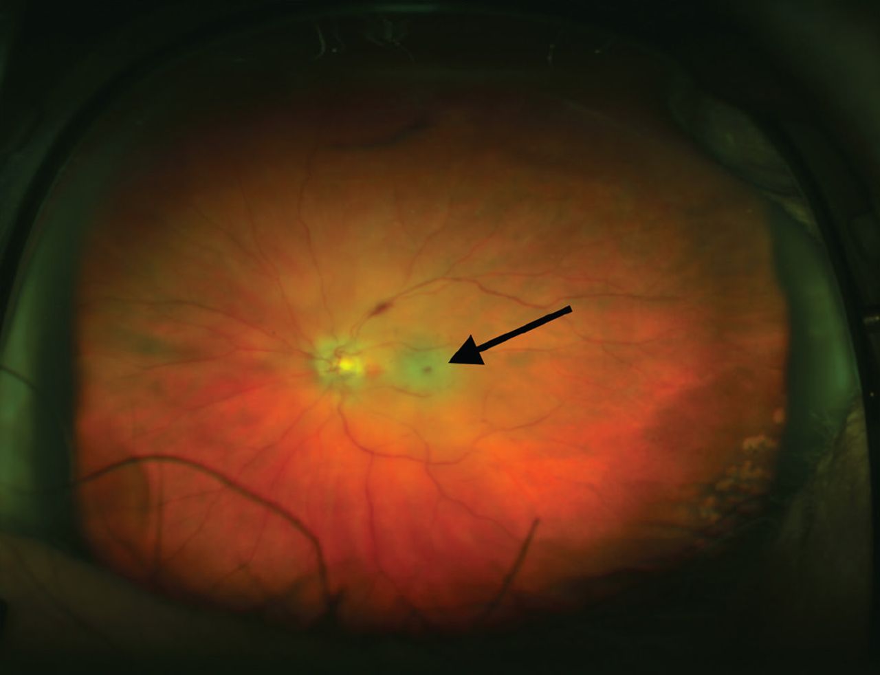
- FIGURE 2
The patient’s funduscopic examination revealed a cherry red spot (arrow), a characteristic finding in central retinal artery occlusion.
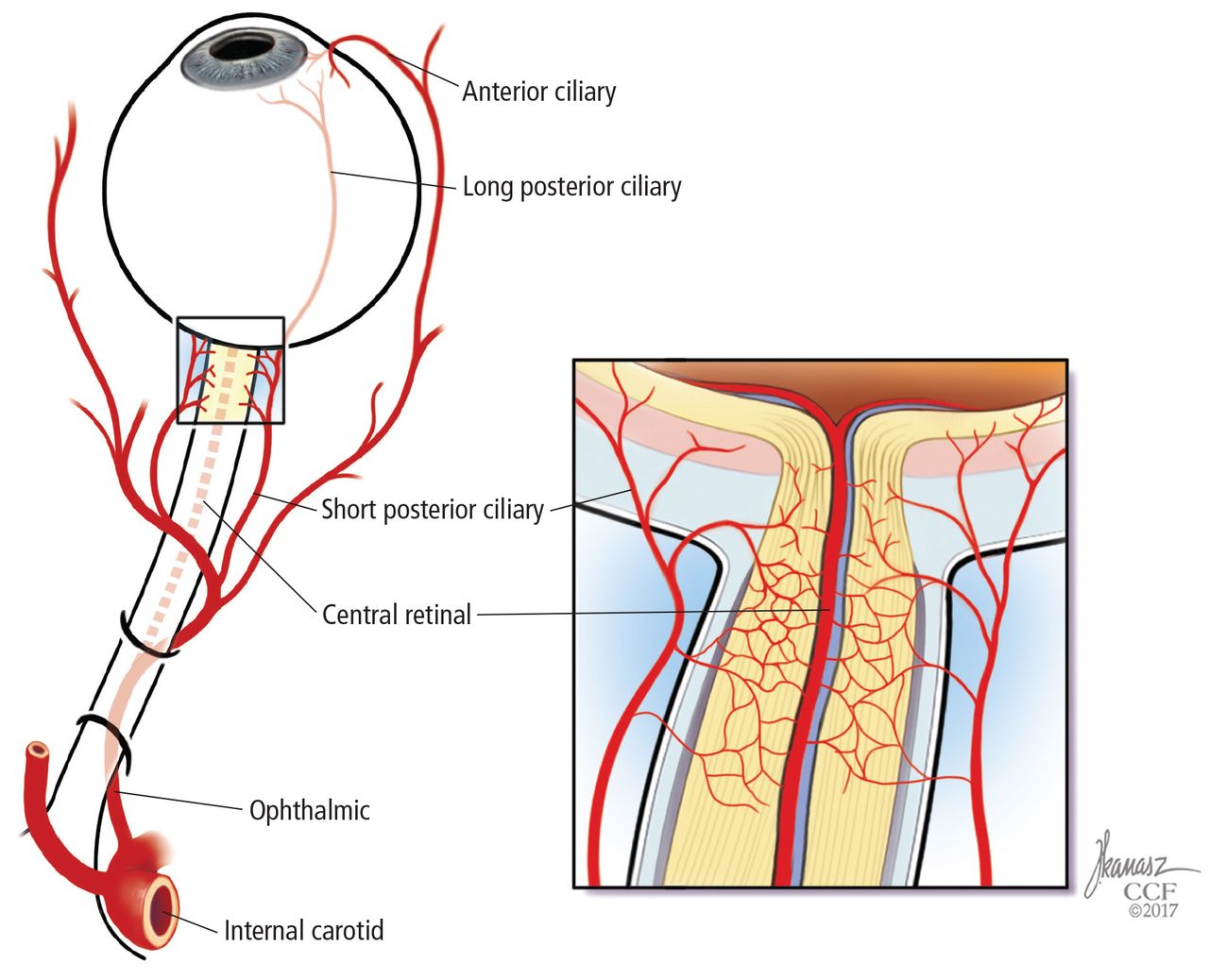
- FIGURE 3
Vascular supply to the eye. The internal carotid artery’s first major branch is the ophthalmic artery. Four major vessels break off from the ophthalmic artery:
Central retinal artery: large-diameter vessel that supplies the retina (vulnerable to embolic disease)
Short and long posterior ciliary arteries: small vessels that supply the optic nerve and macula (susceptible to small-vessel disease)
Anterior ciliary arteries supply the iris and ciliary body.
Information from references 4 and 5.


Tables
Feature Common conditions Additional features Eye pain Acute angle-closure glaucoma Age > 60 and family history
Deep brow, headache with nausea and vomiting
Halos around lightsOptic neuritis Pain worsens with eye movements
Loss of color vision (red desaturation)Keratitis (inflammation of the cornea) Sharp superficial pain (“grittiness”)
Discharge from the eyeConjunctival hyperemia (red eye) Acute angle-closure glaucoma See above Keratitis See above Uveitis Redness prominent at limbus (convergence between cornea and sclera)
Photophobia
Systemic features suggesting autoimmune diseaseHeadache Giant cell arteritis Age > 50
Scalp tenderness (new onset, temporally based headache)
Jaw claudication
Proximal muscle painMigraine Younger patients
Preceded by migraine prodrome
Symptoms resolve within hourPhotopsia (flashes of bright light) Retinal detachment Myopia
Recent history of ophthalmic procedures
Partial loss of peripheral fieldPreceding trauma Keratitis or uveitis Accompanying milder trauma Hyphema (blood in the anterior chamber) History of blunt trauma
Recent history of ophthalmic proceduresLens dislocation or rupture Predisposed by congenital conditions
Associated small, irregular pupilInformation from references 2 and 3.
Physical examination Helpful techniques Results Visual acuity Vision screening apps (eg, EyeChart Vision Screening App by Dok LLC)
Use corrective lens or pinhole occluderPinhole test corrects refractory error by permitting central rays of light into the eye; will not correct underlying neurologic impairment Visual field Monocular assessment
Confrontation visual field testing uses small-amplitude finger movements in all quadrants
Central fields tested by Amsler gridScotoma: discrete area of visual impairment surrounded by intact vision; positive scotoma (seeing something that is not there) may be a sign of retinal damage; negative scotoma may indicate optic nerve dysfunction
Hemianopia: bilateral visual impairment suggesting a lesion posterior to optic chiasmColor testing Use red objects (sharps container or bottle cap) Unilateral color desaturation: optic nerve dysfunction Pupillary examination Examine for size, shape, symmetry
Swinging flashlight examination: paradoxical dilation when stimulating ipsilateral eye after shining light into contralateral eyeAfferent pupillary defect: optic nerve dysfunction Red reflex Performed with ophthalmoscope when standing 1 foot away from patient Loss of reflex: localizes to media and possibly retinal detachment Direct ophthalmoscopy Use dilating drops to enhance the examination
Disc: neuroretinal fibers entering the eye
Macula: located temporally to disc and lacking blood vesselsCherry red macula: ischemic retina from central retinal artery occlusion that contrasts with nourished macula supplied by posterior ciliary arteries
Hollenhorst plaque: cholesterol emboli signifies atherosclerotic disease in carotid or aortic arch
Pale and swollen optic nerve head: ischemic optic neuropathy from posterior ciliary artery obstructionInformation from references 2 and 3.


Jump to section
Related Articles
Cited By...
- No citing articles found.