
Main menu

  • Home
  • Content
    • Current Issue
    • Ahead of Print
    • Past Issues
    • Supplements
    • Article Type
  • Specialty
    • Articles by Specialty
  • CME/MOC
    • Articles
    • Calendar
  • Info For
    • Manuscript Submission
    • Authors & Reviewers
    • Subscriptions
    • About CCJM
    • Contact Us
    • Media Kit
  • Conversations with Leaders
  • Conference Coverage
    • Kidney Week 2024
    • CHEST 2024
    • ACR Convergence 2023
    • Kidney Week 2023
    • ObesityWeek 2023
    • IDWeek 2023
    • CHEST 2023
    • MDS 2023
    • IAS 2023
    • ACP 2023
    • AAN 2023
    • ACC / WCC 2023
    • AAAAI Meeting 2023
    • ACR Convergence 2022
    • Kidney Week 2022
    • AIDS 2022
  • Other Publications
    • www.clevelandclinic.org

User menu

  • Register
  • Log in

Search

  • Advanced search
Cleveland Clinic Journal of Medicine
  • Other Publications
    • www.clevelandclinic.org
  • Register
  • Log in
Cleveland Clinic Journal of Medicine

Advanced Search

  • Home
  • Content
    • Current Issue
    • Ahead of Print
    • Past Issues
    • Supplements
    • Article Type
  • Specialty
    • Articles by Specialty
  • CME/MOC
    • Articles
    • Calendar
  • Info For
    • Manuscript Submission
    • Authors & Reviewers
    • Subscriptions
    • About CCJM
    • Contact Us
    • Media Kit
  • Conversations with Leaders
  • Conference Coverage
    • Kidney Week 2024
    • CHEST 2024
    • ACR Convergence 2023
    • Kidney Week 2023
    • ObesityWeek 2023
    • IDWeek 2023
    • CHEST 2023
    • MDS 2023
    • IAS 2023
    • ACP 2023
    • AAN 2023
    • ACC / WCC 2023
    • AAAAI Meeting 2023
    • ACR Convergence 2022
    • Kidney Week 2022
    • AIDS 2022
The Clinical Picture

A large mass in the right ventricle: Tumor or thrombus?

Soumya Patnaik, MD, Mahek Shah, MD, Saurabh Sharma, MD, Pradhum Ram, MD, Harish Seetha Rammohan, MD and Alexander Rubin, MD
Cleveland Clinic Journal of Medicine July 2017, 84 (7) 517-519; DOI: https://doi.org/10.3949/ccjm.84a.16115
Soumya Patnaik
Department of Medicine, Einstein Medical Center, Philadelphia, PA
  • Find this author on Google Scholar
  • Find this author on PubMed
  • Search for this author on this site
  • For correspondence: [email protected] [email protected]
Mahek Shah
Division of Cardiology, Lehigh Valley Healthcare Network, Allentown, PA
  • Find this author on Google Scholar
  • Find this author on PubMed
  • Search for this author on this site
Saurabh Sharma
Division of Cardiology Einstein Medical Center, Philadelphia, PA
  • Find this author on Google Scholar
  • Find this author on PubMed
  • Search for this author on this site
Pradhum Ram
Department of Medicine, Einstein Medical Center, Philadelphia, PA
  • Find this author on Google Scholar
  • Find this author on PubMed
  • Search for this author on this site
Harish Seetha Rammohan
Division of Cardiology, Bassett Medical Center, Cooperstown, NY
  • Find this author on Google Scholar
  • Find this author on PubMed
  • Search for this author on this site
Alexander Rubin
Pennsylvania Heart and Vascular Group, Jenkintown, PA
  • Find this author on Google Scholar
  • Find this author on PubMed
  • Search for this author on this site
  • Article
  • Figures & Data
  • Info & Metrics
  • PDF
Loading

A 69-year-old woman with hypertension, diabetes mellitus, and chronic kidney disease presented with a 1-month history of worsening episodic dyspnea, lower-extremity edema, and dizziness. Two months earlier, she had been diagnosed with poorly differentiated pelvic adnexal sarcoma associated with a mature teratoma of the left ovary, and she had undergone bilateral salpingo-oophorectomy, pelvic and para-aortic lymph node dissection, and omentectomy.

Examination revealed tachypnea (23 breaths per minute) and bilateral pitting pedal edema. The neck veins were distended. There was no hepatomegaly. Results of laboratory testing were unremarkable.

Chest radiography showed a homogeneous opacity in the lower lobe of the right lung with multiple nodules. Computed tomography (CT) without contrast confirmed the presence of innumerable scattered ground-glass pulmonary nodules, consistent with metastatic disease (Figure 1). Also evident was trace pericardial effusion. Contrast was not used because of her kidney disease.

FIGURE 1
  • Download figure
  • Open in new tab
  • Download powerpoint
FIGURE 1

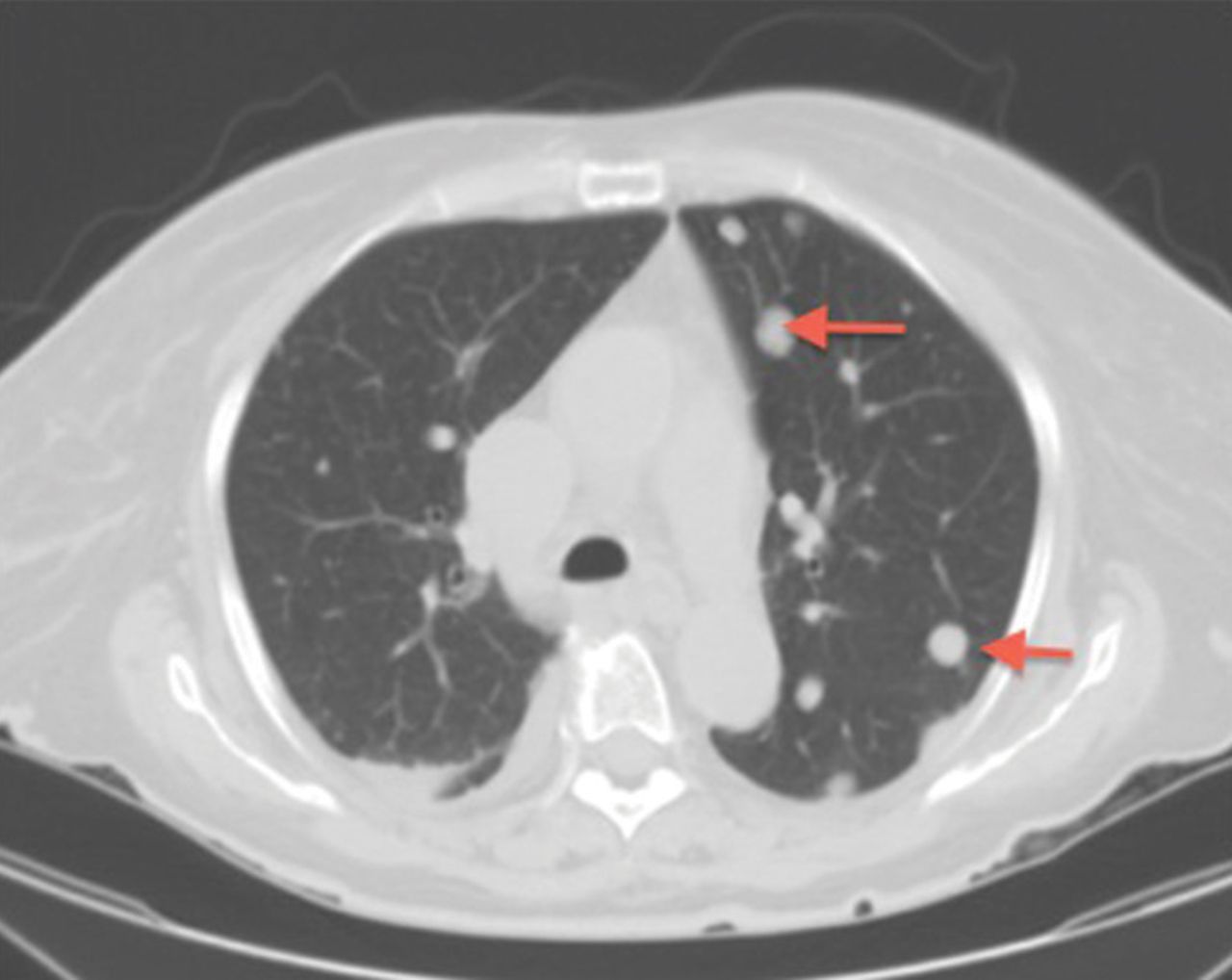
Computed tomography showed scattered solid pulmonary nodules (arrows) consistent with metastasis.

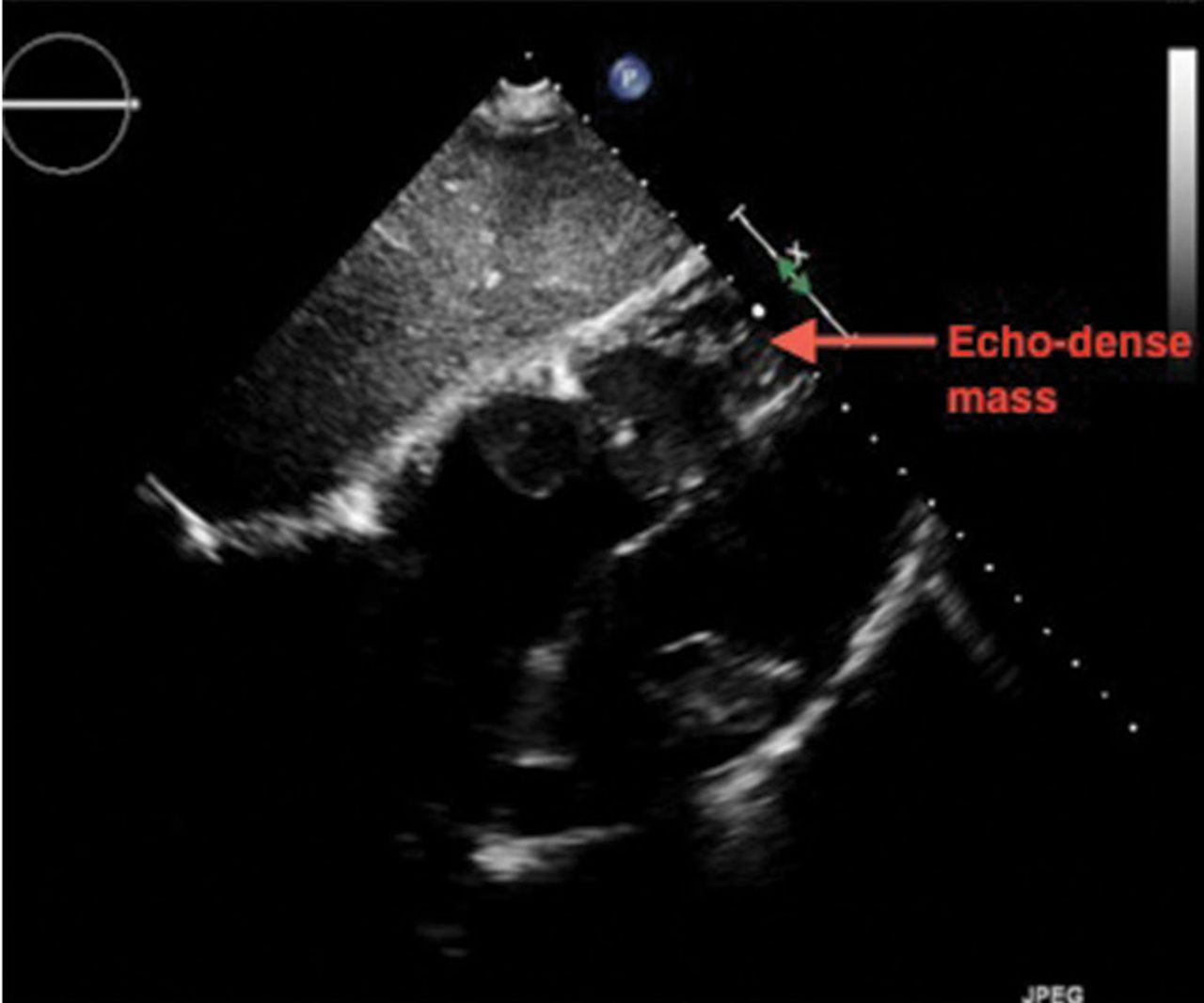
Two-dimensional transthoracic echocardiography performed at the bedside to evaluate her tachypnea and pedal edema demonstrated an echogenic right ventricular mass protruding into a dilated right atrium, with near-complete obstruction of the right ventricle (Figures 2–4). (See video 1 and video 2 at www.ccjm.org.) The tricuspid valve was not visualized. The left ventricle was normal in size and function.

FIGURE 2
  • Download figure
  • Open in new tab
  • Download powerpoint
FIGURE 2

Two-dimensional transthoracic echocardiography (parasternal long-axis view) showed an echo-dense mass in the outflow tract of the right ventricle (RV).

FIGURE 3
  • Download figure
  • Open in new tab
  • Download powerpoint
FIGURE 3

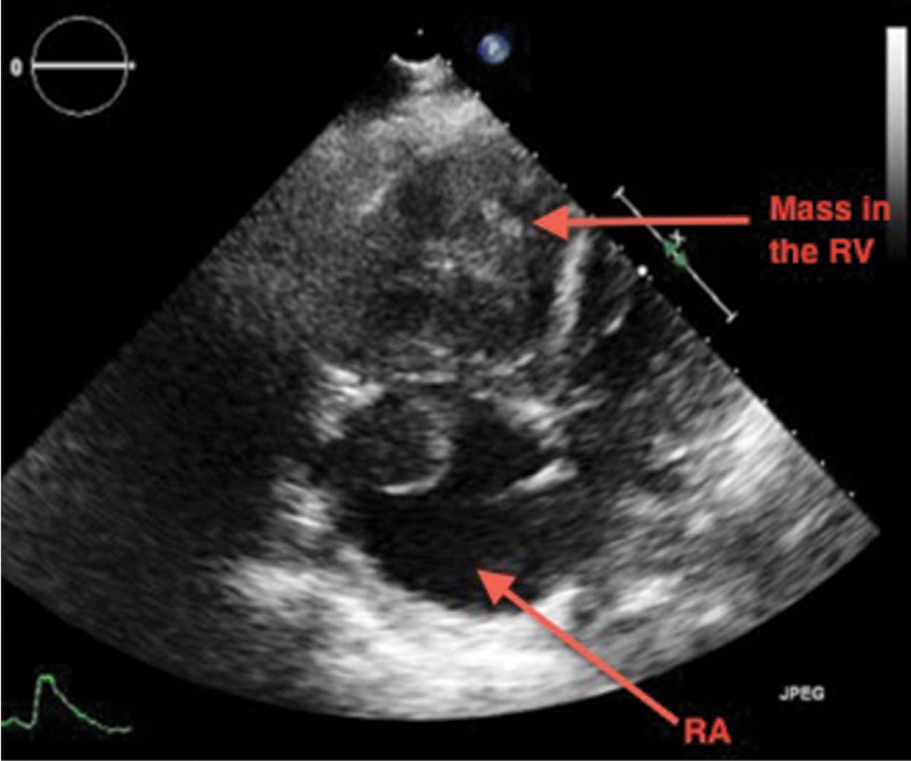
Two-dimensional transthoracic echocardiography (apical 4-chamber view) showed an echodense mass in the right atrium (RA) and right ventricle (RV).

FIGURE 4
  • Download figure
  • Open in new tab
  • Download powerpoint
FIGURE 4

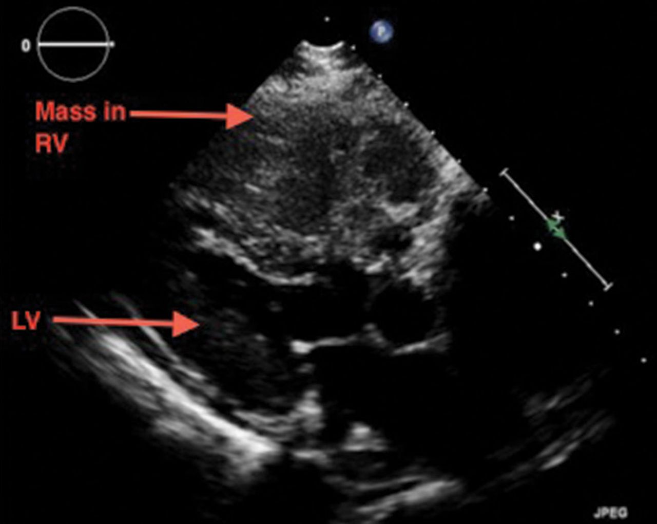
Two-dimensional transthoracic echocardiography (subcostal view) showed an echodense mass in the right atrium and right ventricle (arrow).

This mass was thought to be a metastasis from her ovarian cancer. She was a poor candidate for surgery or chemotherapy and, unfortunately, soon died of respiratory failure.

EVALUATING A CARDIAC MASS

Noninvasive evaluation of cardiac masses includes echocardiography, CT, and magnetic resonance imaging (MRI). Echocardiography can show the anatomic location, the extent, and the physiologic consequences of an intracardiac mass by dynamic assessment during the cardiac cycle. While cardiac masses are often initially detected with transthoracic echocardiography, transesophageal echocardiography shows them better, especially if the mass is located posteriorly.

Thrombus, tumor, or vegetation?

If an intracardiac mass is discovered, we need to determine what it is.

Thrombosis is more likely if contrast echocardiography shows the mass has no stalk (thrombi almost never have a stalk), the atrial chamber is enlarged, cardiac output is low, there is stasis, the mass is avascular, and it responds to thrombolytic therapy. A giant organized thrombus can clinically mimic a tumor if it is immobile, is located close to the wall, and responds poorly to thrombolysis. A wall-motion abnormality adjacent to the mass, global hypokinesis, or a concomitant autoimmune condition such as lupus erythematosus or antiphospholipid antibody syndrome may also favor thrombosis.

Tumors in the heart are uncommon. The prevalence of primary cardiac tumors has been reported as 0.01% to 0.1% in autopsy studies. Metastases to the pericardium, myocardium, coronary arteries, or great vessels have been found at autopsy in 0.7% to 3.5% of the general population and in 9.1% of patients with known malignancy.1

Vegetations from infective endocarditis should also be considered early in the evaluation of an intracardiac mass. They can result from bacterial, fungal, or parasitic infection. Vegetations are generally irregular in appearance, mobile, and attached to a valve. Leftsided valves are generally involved, and a larger mass may indicate fungal origin. Abscess from tuberculosis may need to be considered in the appropriate setting. Whenever feasible, tissue diagnosis is desirable.

Occasionally, there may be an inflammatory component to masses detected in the setting of autoimmune disease.

CT and MRI

If echocardiography cannot clearly distinguish whether the mass is a tumor or a thrombus, MRI with gadolinium contrast is useful. MRI is superior to CT in depicting anatomic details and does not involve radiation.

Cardiac CT is increasingly used when other imaging findings are equivocal or to study a calcified mass. CT with contrast carries a small risk of contrast-induced nephropathy and has lower soft-tissue and temporal resolution. CT without contrast can detect the mass and reveal calcifications within the mass, but contrast is needed to assess the vascularity of the tumor. New-generation CT with electrocardiographic gating nearly matches MRI imaging, and CT is preferred for patients with contraindications to MRI.

CT provides additional information on the global assessment of the chest, lung and vascular structures.2 Cardiac CT and MRI help in precise anatomic delineation, characterization, and preoperative planning of treatment of a large cardiac mass.

TYPES OF CARDIAC TUMORS

Metastases account for most cardiac tumors and are often from primary cancers of the lung, breast, skin, thyroid, and kidney.

Primary cardiac tumors are most often myxomas, which are benign and generally found in the atrial chamber, solitary, with a stalk attached to the area of the fossa ovalis. Other primary cardiac tumors include sarcomas, angiosarcomas, rhabdomyosarcomas, papillary fibroelastomas, lipomas, hemangiomas, mesotheliomas, and rhabdomyomas.

TREATMENT OF CARDIAC TUMORS

For primary and secondary cardiac tumors, complete resection should be considered, provided there is no other organ involvement.3 For suspected lymphomas, image-guided biopsy should be performed before treatment.

For uncertain and diagnostically challenging cases, guided biopsy of the lesions using intracardiac echocardiography or transesophageal echocardiography has been reported to be helpful.4

Most often, the workup and management of cardiac masses calls for a team involving an internist, cardiologist, cardiothoracic surgeon, and vascular medicine specialist. Depending on the nature of the mass, the team may also include an oncologist, radiotherapist, and infectious disease specialist.

Because our patient had significant kidney disease, CT was done without contrast. However, it was not able to clearly delineate the mass in the right ventricle. Cardiac MRI was not performed. Biopsy with transesophageal or intracardiac echocardiographic guidance was not an option, as the patient’s condition was poor.

TAKE-HOME POINTS

The differential diagnosis of an intracardiac mass includes thrombus, benign or malignant tumors, and masses of infectious or inflammatory origin. While noninvasive imaging tests provide clues that can help narrow the differential diagnosis, tissue biopsy with histologic study is necessary to confirm the diagnosis. A team approach is paramount in managing cardiac masses.

  • Copyright © 2017 The Cleveland Clinic Foundation. All Rights Reserved.

REFERENCES

  1. ↵
    1. Goldberg AD,
    2. Blankstein R,
    3. Padera RF
    . Tumors metastatic to the heart. Circulation 2013; 128:1790–1794.
    OpenUrlFREE Full Text
  2. ↵
    1. Exarhos DN,
    2. Tavernaraki EA,
    3. Kyratzi F,
    4. et al
    . Imaging of cardiac tumours and masses. Hospital Chronicles 2010; 5:1–9.
    OpenUrl
  3. ↵
    1. Hoffmeier A,
    2. Sindermann JR,
    3. Scheld HH,
    4. Martens S
    . Cardiac tumors—diagnosis and surgical treatment. Dtsch Arztebl Int 2014; 111:205–211.
    OpenUrlPubMed
  4. ↵
    1. Park K-I,
    2. Kim MJ,
    3. Oh JK,
    4. et al
    . Intracardiac echocardiography to guide biopsy for two cases of intracardiac masses. Korean Circ J 2015; 45:165–168.
    OpenUrl
PreviousNext
Back to top

In this issue

Cleveland Clinic Journal of Medicine: 84 (7)
Cleveland Clinic Journal of Medicine
Vol. 84, Issue 7
1 Jul 2017
  • Table of Contents
  • Table of Contents (PDF)
  • Index by author
Print
Download PDF
Article Alerts
Sign In to Email Alerts with your Email Address
Email Article

Thank you for your interest in spreading the word on Cleveland Clinic Journal of Medicine.

NOTE: We only request your email address so that the person you are recommending the page to knows that you wanted them to see it, and that it is not junk mail. We do not capture any email address.

Enter multiple addresses on separate lines or separate them with commas.
A large mass in the right ventricle: Tumor or thrombus?
(Your Name) has sent you a message from Cleveland Clinic Journal of Medicine
(Your Name) thought you would like to see the Cleveland Clinic Journal of Medicine web site.
CAPTCHA
This question is for testing whether or not you are a human visitor and to prevent automated spam submissions.
Citation Tools
A large mass in the right ventricle: Tumor or thrombus?
Soumya Patnaik, Mahek Shah, Saurabh Sharma, Pradhum Ram, Harish Seetha Rammohan, Alexander Rubin
Cleveland Clinic Journal of Medicine Jul 2017, 84 (7) 517-519; DOI: 10.3949/ccjm.84a.16115

Citation Manager Formats

  • BibTeX
  • Bookends
  • EasyBib
  • EndNote (tagged)
  • EndNote 8 (xml)
  • Medlars
  • Mendeley
  • Papers
  • RefWorks Tagged
  • Ref Manager
  • RIS
  • Zotero
Share
A large mass in the right ventricle: Tumor or thrombus?
Soumya Patnaik, Mahek Shah, Saurabh Sharma, Pradhum Ram, Harish Seetha Rammohan, Alexander Rubin
Cleveland Clinic Journal of Medicine Jul 2017, 84 (7) 517-519; DOI: 10.3949/ccjm.84a.16115
Twitter logo Facebook logo Mendeley logo
  • Tweet Widget
  • Facebook Like
  • Linkedin Share Button

Jump to section

  • Article
    • EVALUATING A CARDIAC MASS
    • TYPES OF CARDIAC TUMORS
    • TREATMENT OF CARDIAC TUMORS
    • TAKE-HOME POINTS
    • REFERENCES
  • Figures & Data
  • Info & Metrics
  • PDF

Related Articles

  • No related articles found.
  • PubMed
  • Google Scholar

Cited By...

  • Cardiac mass: Tumor or thrombus? (July 2017)
  • Google Scholar

More in this TOC Section

  • Tinea incognito
  • Prolonged venous filling time and dependent rubor in a patient with peripheral artery disease
  • Sarcoidosis with diffuse purplish erythematous plaques on the hands
Show more The Clinical Picture

Similar Articles

Subjects

  • Cardiology
  • Imaging
  • Oncology
  • Vascular Medicine
  • Women's Health

Navigate

  • Current Issue
  • Past Issues
  • Supplements
  • Article Type
  • Specialty
  • CME/MOC Articles
  • CME/MOC Calendar
  • Media Kit

Authors & Reviewers

  • Manuscript Submission
  • Authors & Reviewers
  • Subscriptions
  • About CCJM
  • Contact Us
  • Cleveland Clinic Center for Continuing Education
  • Consult QD

Share your suggestions!

Copyright © 2025 The Cleveland Clinic Foundation. All rights reserved. The information provided is for educational purposes only. Use of this website is subject to the website terms of use and privacy policy. 

Powered by HighWire