
Main menu

  • Home
  • Content
    • Current Issue
    • Ahead of Print
    • Past Issues
    • Supplements
    • Article Type
  • Specialty
    • Articles by Specialty
  • CME/MOC
    • Articles
    • Calendar
  • Info For
    • Manuscript Submission
    • Authors & Reviewers
    • Subscriptions
    • About CCJM
    • Contact Us
    • Media Kit
  • Conversations with Leaders
  • Conference Coverage
    • Kidney Week 2024
    • CHEST 2024
    • ACR Convergence 2023
    • Kidney Week 2023
    • ObesityWeek 2023
    • IDWeek 2023
    • CHEST 2023
    • MDS 2023
    • IAS 2023
    • ACP 2023
    • AAN 2023
    • ACC / WCC 2023
    • AAAAI Meeting 2023
    • ACR Convergence 2022
    • Kidney Week 2022
    • AIDS 2022
  • Other Publications
    • www.clevelandclinic.org

User menu

  • Register
  • Log in

Search

  • Advanced search
Cleveland Clinic Journal of Medicine
  • Other Publications
    • www.clevelandclinic.org
  • Register
  • Log in
Cleveland Clinic Journal of Medicine

Advanced Search

  • Home
  • Content
    • Current Issue
    • Ahead of Print
    • Past Issues
    • Supplements
    • Article Type
  • Specialty
    • Articles by Specialty
  • CME/MOC
    • Articles
    • Calendar
  • Info For
    • Manuscript Submission
    • Authors & Reviewers
    • Subscriptions
    • About CCJM
    • Contact Us
    • Media Kit
  • Conversations with Leaders
  • Conference Coverage
    • Kidney Week 2024
    • CHEST 2024
    • ACR Convergence 2023
    • Kidney Week 2023
    • ObesityWeek 2023
    • IDWeek 2023
    • CHEST 2023
    • MDS 2023
    • IAS 2023
    • ACP 2023
    • AAN 2023
    • ACC / WCC 2023
    • AAAAI Meeting 2023
    • ACR Convergence 2022
    • Kidney Week 2022
    • AIDS 2022
The Clinical Picture

A complication of enoxaparin injection

Ahmad Muneer Sharayah, MD, Samrah Zaigham, MD, Noor Hajjaj, MD and Mihir Maniar, DO
Cleveland Clinic Journal of Medicine October 2019, 86 (10) 648-649; DOI: https://doi.org/10.3949/ccjm.86a.19026
Ahmad Muneer Sharayah
Chief Internal Medicine Resident, Monmouth Medical Center, Long Branch, NJ
  • Find this author on Google Scholar
  • Find this author on PubMed
  • Search for this author on this site
  • For correspondence: [email protected]
Samrah Zaigham
Department of Internal Medicine, Monmouth Medical Center, 300 2nd Avenue, Long Branch, NJ
  • Find this author on Google Scholar
  • Find this author on PubMed
  • Search for this author on this site
Noor Hajjaj
Faculty of Medicine, University of Jordan, Amman, Jordan
  • Find this author on Google Scholar
  • Find this author on PubMed
  • Search for this author on this site
Mihir Maniar
Department of Internal Medicine, Monmouth Medical Center, Long Branch, NJ
  • Find this author on Google Scholar
  • Find this author on PubMed
  • Search for this author on this site
  • Article
  • Figures & Data
  • Info & Metrics
  • PDF
Loading

A 78-year-old woman presented to the emergency department with shortness of breath and palpitations and was found to have atrial fibrillation with rapid ventricular response. Medical therapy with drug therapy and cardioversion proved ineffective. She then underwent atrioventricular node ablation and placement of a pacemaker.

At the time of admission, anticoagulation was started with full-dose enoxaparin, injected subcutaneously on the left side of the abdominal wall, as her CHA2DS2-VASc score (http://chadvasc.org) was 5, due to age, female sex, and history of heart failure and hypertension.

Four days after admission, she reported lower abdominal pain, and her urine output was minimal. A bladder scan showed more than 500 mL of residual urine. She was hemodynamically stable, but physical examination revealed mild abdominal distention and tenderness in the suprapubic region. Laboratory testing showed a sharp rise in serum creatinine and a drop in hematocrit.

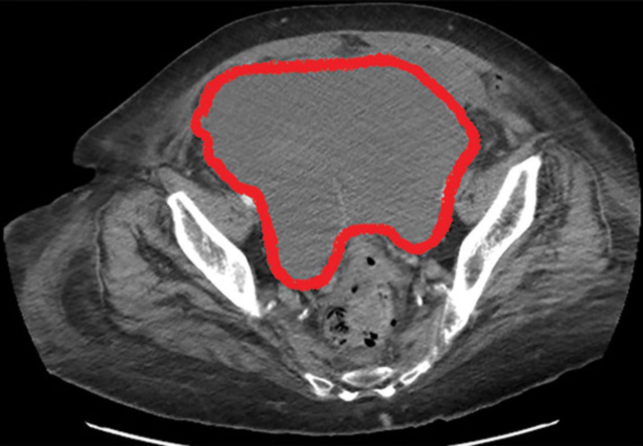
Computed tomography of the abdomen revealed a hematoma measuring 15 by 15 cm within the paracolic gutter and pelvis, compressing the bladder and causing left-sided hydronephrosis (Figure 1). Her laboratory abnormalities were therefore interpreted as postrenal acute kidney injury and anemia due to blood loss.

Figure 1
  • Download figure
  • Open in new tab
  • Download powerpoint
Figure 1

Abdominal computed tomography revealed a hematoma 15 cm × 15 cm within the paracolic gutter and pelvis, causing bladder compression and left-sided hydro-nephrosis.

The patient was initially managed conservatively with serial physical examinations, monitoring of the hematocrit, serial imaging studies, and discontinuation of anticoagulation, but the pain and anuria persisted. Repeat computed tomography 15 days after admission showed that the hematoma had expanded, and she now had hydronephrosis on the right side as well, requiring urologic intervention with bilateral nephrostomy tube placement.

The size of the hematoma was evaluated with serial abdominal and pelvic examinations. After several days, her urine output had improved, the nephrostomy tubes were removed, and she was discharged.

RECTUS SHEATH HEMATOMA

Our patient had a giant pelvic hematoma, probably arising from the rectus sheath. This uncommon problem can arise from trauma, anticoagulation, or increased intra-abdominal pressure, but it can also occur spontaneously.1

In rectus sheath hematoma, a branch of the inferior epigastric artery is injured at its insertion into the rectus abdominis muscle. Symptoms arise if bleeding does not stop spontaneously from a tamponade effect.2

We speculate that in our patient, deep injection of enoxaparin into the abdominal wall injured the inferior epigastric artery, which started the hematoma, and the bleeding was exacerbated by the anticoagulation effect of the enoxaparin.

Another form of pelvic hematoma is retro-peritoneal. It is most commonly caused by trauma but can occur due to rupture of the aorta, compression from tumors, or, infrequently, anticoagulation therapy.3

The role of anticoagulation

Spontaneous pelvic hematoma is usually missed as a cause of abdominal pain in patients on anticoagulation therapy and is mistaken for common acute conditions such as ulcer, diverticulitis, appendicitis, ovarian cyst torsion, and tumor.4 It usually develops within 5 days of starting anticoagulation therapy. Symptoms vary depending on the location of the hematoma and are best diagnosed with abdominal computed tomography, with sensitivity as high as 100%.

MANAGEMENT

Conservative management, reserved for patients in stable condition, includes temporarily stopping and reevaluating the risks and benefits of anticoagulation and antiplatelet agents, giving blood transfusions, and controlling pain. If conservative measures fail, options are arterial embolization, stent grafting, and blood vessel ligation.5 If these measures fail, patients should undergo surgical evacuation of the hematoma and ligation of bleeding vessels.6

TAKE-HOME MESSAGE

Subcutaneous injections, especially of anticoagulants, into the abdominal wall can increase the risk of hematoma. Other risk factors are older age, female sex, and thin body habitus with less abdominal fat.7 Healthcare professionals should avoid deep injections into the abdomen and should counsel patients and their caregivers about this, as well. The deltoid region could be a safer alternative.

  • © 2019 The Cleveland Clinic Foundation. All Rights Reserved.

REFERENCES

  1. ↵
    1. Cherry WB,
    2. Mueller PS
    . Rectus sheath hematoma: review of 126 cases at a single institution. Medicine (Baltimore) 2006; 85(2):105–110. doi: 10.1097/01.md.0000216818.13067.5a
    OpenUrlCrossRefPubMed
  2. ↵
    1. Hatjipetrou A,
    2. Anyfantakis D,
    3. Kastanakis M
    . Rectus sheath hematoma: a review of the literature. Int J Surg 2015; 13267–271. doi: 10.1016/j.ijsu.2014.12.015
    OpenUrlCrossRefPubMed
  3. ↵
    1. Haq MM,
    2. Taimur SDM,
    3. Khan SR,
    4. Rahman MA
    . Retro-peritoneal hematoma following enoxaparin treatment in an elderly woman—a case report. Cardiovasc J 2010; 3(1):94–97. doi: 10.3329/cardio.v3i1.6434
    OpenUrlCrossRef
  4. ↵
    1. Luhmann A,
    2. Williams EV
    . Rectus sheath hematoma: a series of unfortunate events. World J Surg 2006; 30(11):2050–2055. doi: 10.1007/s00268-005-0702-9
    OpenUrlCrossRefPubMed
  5. ↵
    1. Pace F,
    2. Colombo GM,
    3. Del Vecchio LR, et al
    . Low molecular weight heparin and fatal spontaneous extraperitoneal hematoma in the elderly. Geriatr Gerontol Int 2012; 12(1):172–174 doi: 10.1111/j.1447-0594.2011.00742.x
    OpenUrlCrossRefPubMed
  6. ↵
    1. Velicki L,
    2. Cemerlic-Adic N,
    3. Bogdanovic D,
    4. Mrdanin T
    . Rectus sheath haematoma: enoxaparin-related complication. Acta Clin Belg 2013; 68(2):147–149. doi: 10.2143/ACB.68.2.3213
    OpenUrlCrossRef
  7. ↵
    1. Sheth HS,
    2. Kumar R,
    3. DiNella J,
    4. Janov C,
    5. Kaldas H,
    6. Smith RE
    . Evaluation of risk factors for rectus sheath hematoma. Clin Appl Thromb Hemost 2016; 22(3):292–296. doi: 10.1177/1076029614553024
    OpenUrlCrossRefPubMed
PreviousNext
Back to top

In this issue

Cleveland Clinic Journal of Medicine: 86 (10)
Cleveland Clinic Journal of Medicine
Vol. 86, Issue 10
1 Oct 2019
  • Table of Contents
  • Table of Contents (PDF)
  • Index by author
Print
Download PDF
Article Alerts
Sign In to Email Alerts with your Email Address
Email Article

Thank you for your interest in spreading the word on Cleveland Clinic Journal of Medicine.

NOTE: We only request your email address so that the person you are recommending the page to knows that you wanted them to see it, and that it is not junk mail. We do not capture any email address.

Enter multiple addresses on separate lines or separate them with commas.
A complication of enoxaparin injection
(Your Name) has sent you a message from Cleveland Clinic Journal of Medicine
(Your Name) thought you would like to see the Cleveland Clinic Journal of Medicine web site.
CAPTCHA
This question is for testing whether or not you are a human visitor and to prevent automated spam submissions.
Citation Tools
A complication of enoxaparin injection
Ahmad Muneer Sharayah, Samrah Zaigham, Noor Hajjaj, Mihir Maniar
Cleveland Clinic Journal of Medicine Oct 2019, 86 (10) 648-649; DOI: 10.3949/ccjm.86a.19026

Citation Manager Formats

  • BibTeX
  • Bookends
  • EasyBib
  • EndNote (tagged)
  • EndNote 8 (xml)
  • Medlars
  • Mendeley
  • Papers
  • RefWorks Tagged
  • Ref Manager
  • RIS
  • Zotero
Share
A complication of enoxaparin injection
Ahmad Muneer Sharayah, Samrah Zaigham, Noor Hajjaj, Mihir Maniar
Cleveland Clinic Journal of Medicine Oct 2019, 86 (10) 648-649; DOI: 10.3949/ccjm.86a.19026
Twitter logo Facebook logo Mendeley logo
  • Tweet Widget
  • Facebook Like
  • Linkedin Share Button

Jump to section

  • Article
    • RECTUS SHEATH HEMATOMA
    • MANAGEMENT
    • TAKE-HOME MESSAGE
    • REFERENCES
  • Figures & Data
  • Info & Metrics
  • PDF

Related Articles

  • No related articles found.
  • PubMed
  • Google Scholar

Cited By...

  • No citing articles found.
  • Google Scholar

More in this TOC Section

  • Amoxicillin rash in infectious mononucleosis
  • Severe cutaneous reaction induced by allopurinol
  • Tinea incognito
Show more The Clinical Picture

Similar Articles

Subjects

  • Cardiology
  • Drug Therapy
  • Emergency Medicine
  • Hematology
  • Hospital Medicine
  • Imaging
  • Nephrology

Navigate

  • Current Issue
  • Past Issues
  • Supplements
  • Article Type
  • Specialty
  • CME/MOC Articles
  • CME/MOC Calendar
  • Media Kit

Authors & Reviewers

  • Manuscript Submission
  • Authors & Reviewers
  • Subscriptions
  • About CCJM
  • Contact Us
  • Cleveland Clinic Center for Continuing Education
  • Consult QD

Share your suggestions!

Copyright © 2025 The Cleveland Clinic Foundation. All rights reserved. The information provided is for educational purposes only. Use of this website is subject to the website terms of use and privacy policy. 

Powered by HighWire