
Main menu

  • Home
  • Content
    • Current Issue
    • Ahead of Print
    • Past Issues
    • Supplements
    • Article Type
  • Specialty
    • Articles by Specialty
  • CME/MOC
    • Articles
    • Calendar
  • Info For
    • Manuscript Submission
    • Authors & Reviewers
    • Subscriptions
    • About CCJM
    • Contact Us
    • Media Kit
  • Conversations with Leaders
  • Conference Coverage
    • Kidney Week 2024
    • CHEST 2024
    • ACR Convergence 2023
    • Kidney Week 2023
    • ObesityWeek 2023
    • IDWeek 2023
    • CHEST 2023
    • MDS 2023
    • IAS 2023
    • ACP 2023
    • AAN 2023
    • ACC / WCC 2023
    • AAAAI Meeting 2023
    • ACR Convergence 2022
    • Kidney Week 2022
    • AIDS 2022
  • Other Publications
    • www.clevelandclinic.org

User menu

  • Register
  • Log in

Search

  • Advanced search
Cleveland Clinic Journal of Medicine
  • Other Publications
    • www.clevelandclinic.org
  • Register
  • Log in
Cleveland Clinic Journal of Medicine

Advanced Search

  • Home
  • Content
    • Current Issue
    • Ahead of Print
    • Past Issues
    • Supplements
    • Article Type
  • Specialty
    • Articles by Specialty
  • CME/MOC
    • Articles
    • Calendar
  • Info For
    • Manuscript Submission
    • Authors & Reviewers
    • Subscriptions
    • About CCJM
    • Contact Us
    • Media Kit
  • Conversations with Leaders
  • Conference Coverage
    • Kidney Week 2024
    • CHEST 2024
    • ACR Convergence 2023
    • Kidney Week 2023
    • ObesityWeek 2023
    • IDWeek 2023
    • CHEST 2023
    • MDS 2023
    • IAS 2023
    • ACP 2023
    • AAN 2023
    • ACC / WCC 2023
    • AAAAI Meeting 2023
    • ACR Convergence 2022
    • Kidney Week 2022
    • AIDS 2022
Review

Assessing liver fibrosis without biopsy in patients with HCV or NAFLD

Tavankit Singh, MD, Daniela S. Allende, MD and Arthur J. McCullough, MD
Cleveland Clinic Journal of Medicine March 2019, 86 (3) 179-186; DOI: https://doi.org/10.3949/ccjm.86a.17118
Tavankit Singh
Department of Gastroenterology and Hepatology, Cleveland Clinic
  • Find this author on Google Scholar
  • Find this author on PubMed
  • Search for this author on this site
Daniela S. Allende
  • Find this author on Google Scholar
  • Find this author on PubMed
  • Search for this author on this site
Arthur J. McCullough
  • Find this author on Google Scholar
  • Find this author on PubMed
  • Search for this author on this site
  • For correspondence: [email protected]
  • Article
  • Figures & Data
  • Info & Metrics
  • PDF
Loading

ABSTRACT

Staging of liver fibrosis is increasingly done using noninvasive methods, in some cases obviating the need for liver biopsy. Scores based on laboratory values and demographic variables have been developed and validated for assessing fibrosis in patients with hepatitis C virus (HCV) infection and nonalcoholic fatty liver disease (NAFLD), as have several imaging methods that measure shear-wave velocity, a reflection of fibrosis severity.

KEY POINTS
  • Liver biopsy remains the gold standard for determining fibrosis stage but is expensive and entails risk of complications.

  • For patients infected with HCV, fibrosis stage should be determined with transient elastography, a transthoracic ultrasonographic technique that measures shear-wave velocity.

  • For patients with cirrhosis, transient elastography combined with a platelet count can detect developing portal hypertension and determine whether to screen for esophageal varices.

  • For NAFLD, combined elastography and NAFLD fibrosis score—which incorporates patient characteristics and laboratory test results—should be used to determine the need for liver biopsy.

Staging of liver fibrosis, important for determining prognosis in patients with chronic liver disease and for the need to start screening for complications of cirrhosis, was traditionally done only by liver biopsy. While biopsy is still the gold standard method to stage fibrosis, noninvasive methods have been developed that can also assess disease severity.

This article briefly reviews the epidemiology and physiology of chronic liver disease and the traditional role of liver biopsy. Pros and cons of alternative fibrosis assessment methods are discussed, with a focus on their utility for patients with nonalcoholic fatty liver disease (NAFLD) and hepatitis C virus (HCV) infection.

CHRONIC LIVER DISEASE: A HUGE HEALTH BURDEN

Chronic liver disease is associated with enormous health and financial costs in the United States. Its prevalence is about 15%,1 and it is the 12th leading cause of death.2 Hospital costs are estimated at about $4 billion annually.3

The most common causes of chronic liver disease are NAFLD (which may be present in up to one-third of the US population and is increasing with the epidemic of obesity), its aggressive variant, nonalcoholic steatohepatitis (NASH) (present in about 3% of the population), and HCV infection (1%).4,5

Since direct-acting antiviral agents were introduced, HCV infection dropped from being the leading cause of liver transplant to third place.6 But at the same time, the number of patients on the transplant waiting list who have NASH has risen faster than for any other cause of chronic liver disease.7

FIBROSIS: A KEY INDICATOR OF DISEASE SEVERITY

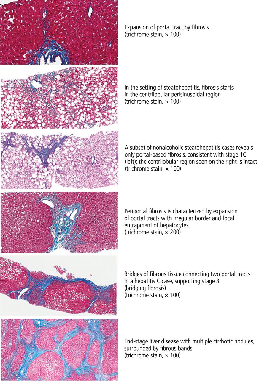
With any form of liver disease, collagen is deposited in hepatic lobules over time, a process called fibrosis. Both HCV infection and NASH involve necroinflammation in the liver, hepatocyte apoptosis, and activation of stellate cells, leading to progressive collagen deposition in hepatic lobules. Fibrosis typically starts in the region of the central vein and portal tracts and eventually extends to other areas of the lobule.

Determining fibrosis severity is critical when a patient is diagnosed with chronic liver disease, as it predicts long-term clinical outcomes and death in HCV8 and NAFLD.9 Different staging systems have been developed to reflect the degree of fibrosis, based on its distribution as seen on liver biopsy (Table 1, Figure 1).

View this table:
  • View inline
  • View popup
TABLE 1

Fibrosis staging systems for HCV and NAFLD

Figure 1
  • Download figure
  • Open in new tab
  • Download powerpoint
Figure 1

Findings on liver biopsy in nonalcoholic fatty liver disease and hepatitis C virus infection.

In HCV infection, advanced fibrosis is defined as either stage 4 to 6 using the Ishak system10 or stage 3 to 4 using the Meta-analysis of Histological Data in Viral Hepatitis (META-VIR) system.11

In NAFLD, advanced fibrosis is defined as stage 3 to 4 using the NASH Clinical Research Network system.12

Staging fibrosis is also important so that patients with cirrhosis can be identified early to begin screening for hepatocellular carcinoma and esophageal varices to reduce the risks of illness and death. In addition, insurance companies often require documentation of fibrosis stage before treating HCV with the new direct-acting antiviral agents.

LIVER BIOPSY IS STILL THE GOLD STANDARD

Although invasive, liver biopsy remains the gold standard for determining fibrosis stage. Liver biopsies were performed “blindly” (without imaging) until the 1990s, but imaging-guided biopsy using ultrasonography was then developed, which entailed less pain and lower complication and hospitalization rates. Slightly more hepatic tissue is obtained with guided liver biopsy, but the difference was deemed clinically insignificant.13 Concern initially arose about the added cost involved with imaging, but imaging-guided biopsy was actually found to be more cost-effective.14

In the 2000s, transjugular liver biopsy via the right internal jugular vein became available. This method was originally used primarily in patients with ascites or significant coagulopathy. At first, there were concerns about the adequacy of specimens obtained to make an accurate diagnosis or establish fibrosis stage, but this limitation was overcome with improved techniques.15,16 Transjugular liver biopsy has the additional advantage of enabling one to measure the hepatic venous pressure gradient, which also has prognostic significance; a gradient greater than 10 mm Hg is associated with worse prognosis.17

Disadvantages of biopsy: Complications, sampling errors

Liver biopsy has disadvantages. Reported rates of complications necessitating hospitalization using the blind method were as high as 6% in the 1970s,18 dropping to 3.2% in a 1993 study.19 Bleeding remains the most worrisome complication. With the transjugular method, major and minor complication rates are less than 1% and 7%, respectively.15,16 Complication rates with imaging-guided biopsy are also low.

Liver biopsy is also prone to sampling error. The number of portal tracts obtained in the biopsy correlates with the accuracy of fibrosis staging, and smaller samples may lead to underestimating fibrosis stage. In patients with HCV, samples more than 15 mm long led to accurate staging diagnosis in 65% of patients, and those longer than 25 mm conferred 75% accuracy.20 Also, different stages can be diagnosed from samples obtained from separate locations in the liver, although rarely is the difference more than a single stage.21

Histologic evaluation of liver biopsies is operator-dependent. Although significant interobserver variation has been reported for degree of inflammation, there tends to be good concordance for fibrosis staging.22,23

STAGING BASED ON DEMOGRAPHIC AND LABORATORY VARIABLES

Several scores based on patient characteristics and laboratory values have been developed for assessing liver fibrosis and have been specifically validated for HCV infection, NAFLD, or both. They can serve as inexpensive initial screening tests for the presence or absence of advanced fibrosis.

FIB-4 index for HCV, NAFLD

The FIB-4 index predicts the presence of advanced fibrosis using, as its name indicates, a combination of 4 factors in fibrosis: age, platelet count, and the levels of aspartate aminotransferase (AST) and alanine aminotransferase (ALT), according to the formula: Embedded Image

The index was derived from data from 832 patients co-infected with HCV and human immunodeficiency virus.24 The Ishak staging system10 for fibrosis on liver biopsy was used for confirmation, with stage 4 to 6 defined as advanced fibrosis. A cutoff value of more than 3.25 had a positive predictive value of 65% for advanced fibrosis, and to exclude advanced fibrosis, a cutoff value of less than 1.45 had a negative predictive value of 90%.

The FIB-4 index has since been validated in patients with HCV infection25 and NAFLD.26 In a subsequent study in 142 patients with NAFLD, the FIB-4 index was more accurate in diagnosing advanced fibrosis than the other noninvasive prediction models discussed below.27

NAFLD fibrosis score

The NAFLD fibrosis score, constructed and validated only in patients with biopsy-confirmed NAFLD, incorporates age, body mass index, presence of diabetes or prediabetes, albumin level, platelet count, and AST and ALT levels.

A group of 480 patients was used to construct the score, and 253 patients were used to validate it. Using the high cutoff value of 0.676, the presence of advanced fibrosis was diagnosed with a positive predictive value of 90% in the group used to construct the model (82% in the validation group). Using the low cutoff score of –1.455, advanced fibrosis could be excluded with a negative predictive value of 93% in the construction group and 88% in the validation group.28 A score between the cutoff values merits liver biopsy to determine fibrosis stage. The score is more accurate in patients with diabetes.29 When used by prima ry care physicians, the NAFLD fibrosis score is more cost-effective than transient elastography and liver biopsy for accurately predicting advanced fibrosis.30

AST-to-platelet ratio index score for HCV, NAFLD

The AST-to-platelet ratio index (APRI) score was developed in 2003 using a cohort of 270 patients with HCV and liver biopsy as the standard. A cutoff value of less than or equal to 0.5 had a negative predictive value of 86% for the absence of significant fibrosis, while a score of more than 1.5 detected the presence of significant fibrosis with a positive predictive value of 88%.31 The APRI score was subsequently validated for NAFLD.27,32

FibroSure uses a patented formula

FibroSure (LabCorp; labcorp.com) uses a patented mathematical formula that takes into account age, sex, and levels of gammaglutamyl transferase, total bilirubin, haptoglobin, apolipoprotein-A, and alpha-2 macroglobulin to assess fibrosis. Developed in 2001 for use in patients with HCV infection, it was reported to have a positive predictive value of greater than 90% and a negative predictive value of 100% for clinically significant fibrosis, defined as stage 2 to 4 based on the METAVIR staging system in the prediction model.33 The use of FibroSure in patients with HCV was subsequently validated in various meta-analyses and systematic reviews.34,35 It is less accurate in patients with normal ALT levels.36

FibroSure also has good accuracy for predicting fibrosis stage in chronic liver disease due to other causes, including NAFLD.37

The prediction models discussed above use routine laboratory tests for chronic liver disease and thus are inexpensive. The high cost of additional testing needed for FibroSure, coupled with the risk of misdiagnosis, makes its cost-effectiveness questionable.38

IMAGING TO PREDICT FIBROSIS STAGE

Conventional ultrasonography (with or without vascular imaging) and computed tomography can detect cirrhosis on the basis of certain imaging characteristics,39,40 including the nodular contour of the liver, caudate lobe hypertrophy, ascites, reversal of blood flow in the portal vein, and splenomegaly. However, they cannot detect fibrosis in its early stages.

The 3 methods discussed below provide more accurate fibrosis staging by measuring the velocity of shear waves sent across hepatic tissue. Because shear-wave velocity increases with liver stiffness, the fibrosis stage can be estimated from this information.41

Transient elastography

Transient elastography uses a special ultrasound transducer. It is highly accurate for predicting advanced fibrosis for almost all causes of chronic liver disease, including HCV infection42,43 and NAFLD.44 The cutoff values of wave velocity to estimate fibrosis stage differ by liver disease etiology.

Transient elastography should not be used to evaluate fibrosis in patients with acute hepatitis, which transiently increases liver stiffness, resulting in a falsely high fibrosis stage diagnosis.45 It is also not a good method for evaluating fibrosis in patients with biliary obstruction or extrahepatic venous congestion. Because liver stiffness can increase after eating,46 the test should be done under fasting conditions.

A significant limitation of transient elastography has been its poor accuracy in patients with obesity.47 This has been largely overcome with the use of a more powerful (XL) probe but is still a limitation for those with morbid obesity.48 Because many patients with NAFLD are obese, this limitation can be significant.

Transient elastography has gained popularity for evaluating fibrosis in patients with chronic liver disease for multiple reasons: it is cost-effective and results are highly reproducible, with low variation in results among different observers and in individual observers.49 Combined with a platelet count, it can also be used to detect the development of clinically significant portal hypertension in patients with cirrhosis, thus determining the need to screen for esophageal varices using endoscopy.50 Screening endoscopy can be avoided in patients whose liver stiffness remains below 20 kPa or whose platelet count is above 150 × 109/L.

Acoustic radiation force imaging

Unlike transient elastography, which requires a separate transducer probe to assess shear-wave velocity, acoustic radiation force imaging uses the same transducer for both this function and imaging. Different image modes are available when testing for liver stiffness, so a region of interest that is optimal for avoiding vascular structures or masses can be selected, increasing accuracy.51

Acoustic radiation force imaging has been tested in different causes of chronic liver disease, including HCV and NAFLD,52 with accuracy similar to that of transient elastog-raphy.53 For overweight and obese patients, acoustic radiation force imaging is more accurate than transient elastography using the XL probe.54 However, this method is still new, and we need more data to support using one method over the other.

Magnetic resonance elastography

Magnetic resonance elastography uses a special transducer placed under the rib cage to transmit shear waves concurrently with magnetic resonance imaging. It has been tested in patients with HCV and NAFLD and has been found to have better diagnostic accuracy than transient elastography and acoustic radiation force imaging.55,56 Patients must be fasting for better diagnostic accuracy57 and must hold their breath while elastography is performed. The need for breath-holding and the high cost limit the use of this method for assessing fibrosis.

BOTTOM LINE FOR ASSESSING FIBROSIS

Although liver biopsy remains the gold standard for accurately determining fibrosis stage, noninvasive methods, especially imaging techniques, are fast evolving. Guidelines recommend using transient elastography to determine fibrosis stage noninvasively in patients with HCV,58 but a similar recommendation cannot be made for NAFLD with available data. For NAFLD, combined elastography and NAFLD fibrosis score are recommended to determine the need for a liver biopsy (Figure 2).59 Currently, we recommend using a combination of the scores discussed above and the imaging tests.

Figure 2
  • Download figure
  • Open in new tab
  • Download powerpoint
Figure 2

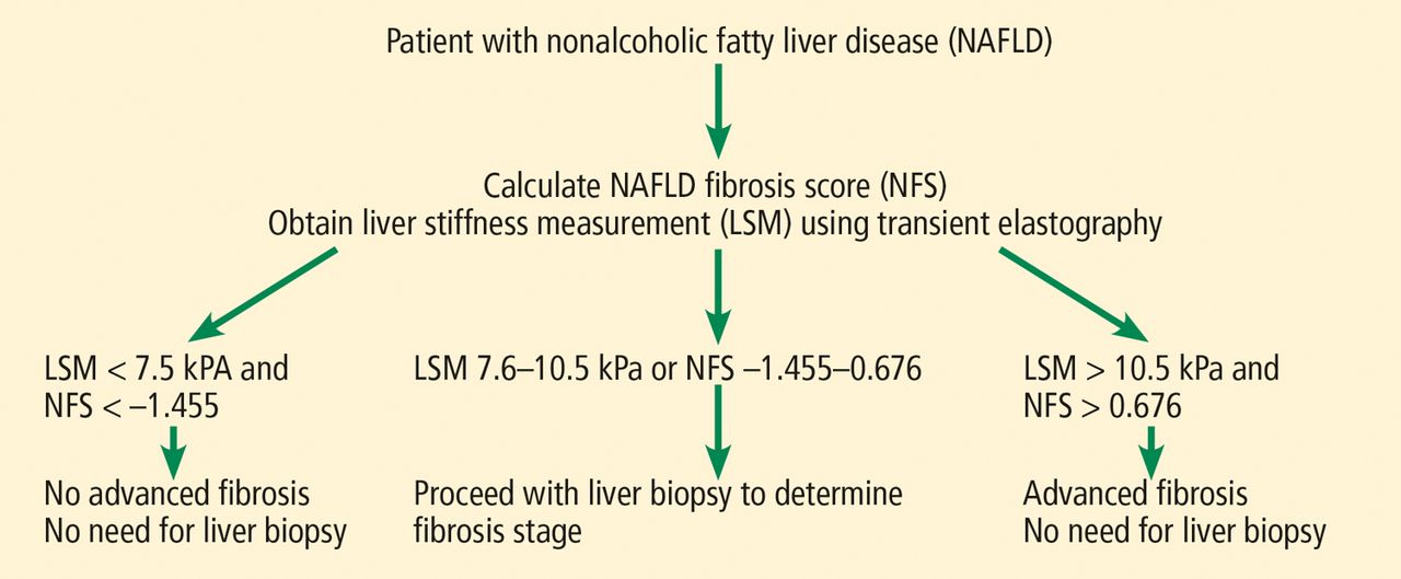
Algorithm to determine fibrosis stage for nonalcoholic fatty liver disease.

  • Copyright © 2019 The Cleveland Clinic Foundation. All Rights Reserved.

REFERENCES

  1. ↵
    1. Younossi ZM,
    2. Stepanova M,
    3. Afendy M,
    4. et al
    . Changes in the prevalence of the most common causes of chronic liver diseases in the United States from 1988 to 2008. Clin Gastroenterol Hepatol 2011; 9(6):524–530.e1. doi:10.1016/j.cgh.2011.03.020
    OpenUrlCrossRefPubMed
  2. ↵
    1. Kochanek KD,
    2. Xu J,
    3. Murphy SL,
    4. Miniño AM,
    5. Kung H-C
    . Deaths: final data for 2009. Natl Vital Stat Rep 2011; 60(3):1–116. pmid:24974587
    OpenUrlPubMed
  3. ↵
    1. Volk ML,
    2. Tocco RS,
    3. Bazick J,
    4. Rakoski MO,
    5. Lok AS
    . Hospital readmissions among patients with decompensated cirrhosis. Am J Gastroenterol 2012; 107(2):247–252. doi:10.1038/ajg.2011.314
    OpenUrlCrossRefPubMed
  4. ↵
    1. Vernon G,
    2. Baranova A,
    3. Younossi ZM
    . Systematic review: the epidemiology and natural history of non-alcoholic fatty liver disease and non-alcoholic steatohepatitis in adults. Aliment Pharmacol Ther 2011; 34(3):274–285. doi:10.1111/j.1365-2036.2011.04724.x
    OpenUrlCrossRefPubMed
  5. ↵
    1. Udompap P,
    2. Kim D,
    3. Kim WR
    . Current and future burden of chronic nonmalignant liver disease. Clin Gastroenterol Hepatol 2015; 13(12):2031–2041. doi:10.1016/j.cgh.2015.08.015
    OpenUrlCrossRef
  6. ↵
    1. Kim WR,
    2. Lake JR,
    3. Smith JM,
    4. et al
    . OPTN/SRTR 2016 annual data report: liver. Am J Transplant 2018; 18(suppl 1):172–253. doi:10.1111/ajt.14559
    OpenUrlCrossRef
  7. ↵
    1. Wong RJ,
    2. Aguilar M,
    3. Cheung R,
    4. et al
    . Nonalcoholic steatohepatitis is the second leading etiology of liver disease among adults awaiting liver transplantation in the United States. Gastroenterology 2015; 148(3):547–555. doi:10.1053/j.gastro.2014.11.039
    OpenUrlCrossRefPubMed
  8. ↵
    1. Ishak K,
    2. Baptista A,
    3. Bianchi L,
    4. et al
    . Histological grading and staging of chronic hepatitis. J Hepatol 1995; 22(6):696–699. pmid:7560864
    OpenUrlCrossRefPubMed
  9. ↵
    1. Bedossa P,
    2. Poynard T
    . An algorithm for the grading of activity in chronic hepatitis C. Hepatology 1996; 24(2):289–293. doi:10.1002/hep.510240201
    OpenUrlCrossRefPubMed
  10. ↵
    1. Kleiner DE,
    2. Brunt EM,
    3. Van Natta M,
    4. et al
    5. Nonalcoholic Steatohepatitis Clinical Research Network
    . Design and validation of a histological scoring system for nonalcoholic fatty liver disease. Hepatology 2005; 41(6):1313–1321. doi:10.1002/hep.20701
    OpenUrlCrossRefPubMed
  11. ↵
    1. Everhart JE,
    2. Wright EC,
    3. Goodman ZD,
    4. et al
    5. HALT-C Trial Group
    . Prognostic value of Ishak fibrosis stage: findings from the hepatitis C antiviral long-term treatment against cirrhosis trial. Hepatology 2010; 51(2):585–594. doi:10.1002/hep.23315
    OpenUrlCrossRefPubMed
  12. ↵
    1. Angulo P,
    2. Kleiner DE,
    3. Dam-Larsen S,
    4. et al
    . Liver fibrosis, but no other histologic features, is associated with long-term outcomes of patients with nonalcoholic fatty liver disease. Gastroenterology 2015; 149(2):389–397.e10. doi:10.1053/j.gastro.2015.04.043
    OpenUrlCrossRefPubMed
  13. ↵
    1. Lindor KD,
    2. Bru C,
    3. Jorgensen RA,
    4. et al
    . The role of ultrasonography and automatic-needle biopsy in outpatient percutaneous liver biopsy. Hepatology 1996; 23(5):1079–1083. doi:0.1002/hep.510230522
    OpenUrlCrossRefPubMed
  14. ↵
    1. Pasha T,
    2. Gabriel S,
    3. Therneau T,
    4. Dickson ER,
    5. Lindor KD
    . Cost-effectiveness of ultrasound-guided liver biopsy. Hepatology 1998; 27(5):1220–1226. doi:10.1002/hep.510270506
    OpenUrlCrossRefPubMed
  15. ↵
    1. Alessandria C,
    2. Debernardi-Venon W,
    3. Rizzetto M,
    4. Marzano A
    . Transjugular liver biopsy: a relatively simple procedure with an indefinite past and an expected brilliant future. J Hepatol 2008; 48(1):171–173. doi:10.1016/j.jhep.2007.10.001
    OpenUrlCrossRefPubMed
  16. ↵
    1. Kalambokis G,
    2. Manousou P,
    3. Vibhakorn S,
    4. et al
    . Transjugular liver biopsy—indications, adequacy, quality of specimens, and complications—a systematic review. J Hepatol 2007; 47(2):284–294. doi:10.1016/j.jhep.2007.05.001
    OpenUrlCrossRefPubMed
  17. ↵
    1. Ripoll C,
    2. Groszmann R,
    3. Garcia-Tsao G,
    4. et al
    5. Portal Hypertension Collaborative Group
    . Hepatic venous pressure gradient predicts clinical decompensation in patients with compensated cirrhosis. Gastroenterology 2007; 133(2):481–488. doi:10.1053/j.gastro.2007.05.024
    OpenUrlCrossRefPubMed
  18. ↵
    1. Perrault J,
    2. McGill DB,
    3. Ott BJ,
    4. Taylor WF
    . Liver biopsy: complications in 1000 inpatients and outpatients. Gastroenterology 1978; 74(1):103–106. pmid:618417
    OpenUrlPubMed
  19. ↵
    1. Janes CH,
    2. Lindor KD
    . Outcome of patients hospitalized for complications after outpatient liver biopsy. Ann Intern Med 1993; 118(2):96–98. pmid:8416324
    OpenUrlCrossRefPubMed
  20. ↵
    1. Bedossa P,
    2. Dargere D,
    3. Paradis V
    . Sampling variability of liver fibrosis in chronic hepatitis C. Hepatology 2003; 38(6):1449–1457. doi:10.1016/j.hep.2003.09.022
    OpenUrlCrossRefPubMed
  21. ↵
    1. Regev A,
    2. Berho M,
    3. Jeffers LJ,
    4. et al
    . Sampling error and intraobserver variation in liver biopsy in patients with chronic HCV infection. Am J Gastroenterol 2002; 97(10):2614–2618. doi:10.1111/j.1572-0241.2002.06038.x
    OpenUrlCrossRefPubMed
  22. ↵
    1. Goldin RD,
    2. Goldin JG,
    3. Burt AD,
    4. et al
    . Intra-observer and inter-observer variation in the histopathological assessment of chronic viral hepatitis. J Hepatol 1996; 25(5):649–654. pmid:8938541
    OpenUrlCrossRefPubMed
  23. ↵
    1. Intraobserver and interobserver variations in liver biopsy interpretation in patients with chronic hepatitis C
    . The French METAVIR Cooperative Study Group. Hepatology 1994; 20(1 Pt 1):15–20. pmid:8020885
    OpenUrlCrossRefPubMed
  24. ↵
    1. Sterling RK,
    2. Lissen E,
    3. Clumeck N,
    4. et al
    5. APRICOT Clinical Investigators
    . Development of a simple noninvasive index to predict significant fibrosis in patients with HIV/HCV coinfection. Hepatology 2006; 43(6):1317–1325. doi:10.1002/hep.21178
    OpenUrlCrossRefPubMed
  25. ↵
    1. Vallet-Pichard A,
    2. Mallet V,
    3. Nalpas B,
    4. et al
    . FIB-4: an inexpensive and accurate marker of fibrosis in HCV infection. comparison with liver biopsy and fibrotest. Hepatology 2007; 46(1):32–36. doi:10.1002/hep.21669
    OpenUrlCrossRefPubMed
  26. ↵
    1. Shah AG,
    2. Lydecker A,
    3. Murray K,
    4. Tetri BN,
    5. Contos MJ,
    6. Sanyal AJ,
    7. Nash Clinical Research Network
    . Comparison of noninvasive markers of fibrosis in patients with nonalcoholic fatty liver disease. Clin Gastroenterol Hepatol 2009; 7(10):1104–1112. doi:10.1016/j.cgh.2009.05.033
    OpenUrlCrossRefPubMed
  27. ↵
    1. McPherson S,
    2. Stewart SF,
    3. Henderson E,
    4. Burt AD,
    5. Day CP
    . Simple non-invasive fibrosis scoring systems can reliably exclude advanced fibrosis in patients with non-alcoholic fatty liver disease. Gut 2010; 59(9):1265–1269. doi:10.1136/gut.2010.216077
    OpenUrlAbstract/FREE Full Text
  28. ↵
    1. Angulo P,
    2. Hui JM,
    3. Marchesini G,
    4. et al
    . The NAFLD fibrosis score: A noninvasive system that identifies liver fibrosis in patients with NAFLD. Hepatology 2007; 45(4):846–854. doi:10.1002/hep.21496
    OpenUrlCrossRefPubMed
  29. ↵
    1. Goh GB,
    2. Pagadala MR,
    3. Dasarathy J,
    4. et al
    . Clinical spectrum of nonalcoholic fatty liver disease in diabetic and non-diabetic patients. BBA Clin 2015; 3:141–145. doi:10.1016/j.bbacli.2014.09.001
    OpenUrlCrossRefPubMed
  30. ↵
    1. Tapper EB,
    2. Hunink MG,
    3. Afdhal NH,
    4. Lai M,
    5. Sengupta N
    . Cost-effectiveness analysis: risk stratification of nonalcoholic fatty liver disease (NAFLD) by the primary care physician using the NAFLD fibrosis score. PLoS One 2016; 11(2):e0147237. doi:10.1371/journal.pone.0147237
    OpenUrlCrossRef
  31. ↵
    1. Wai CT,
    2. Greenson JK,
    3. Fontana RJ,
    4. et al
    . A simple noninvasive index can predict both significant fibrosis and cirrhosis in patients with chronic hepatitis C. Hepatology 2003; 38(2):518–526. doi:10.1053/jhep.2003.50346
    OpenUrlCrossRefPubMed
  32. ↵
    1. Calès P,
    2. Lainé F,
    3. Boursier J,
    4. et al
    . Comparison of blood tests for liver fibrosis specific or not to NAFLD. J Hepatol 2009; 50(1):165–173. doi:10.1016/j.jhep.2008.07.035
    OpenUrlCrossRefPubMed
  33. ↵
    1. Imbert-Bismut F,
    2. Ratziu V,
    3. Pieroni L,
    4. Charlotte F,
    5. Benhamou Y,
    6. Poynard T,
    7. MULTIVIRC Group
    . Biochemical markers of liver fibrosis in patients with hepatitis C virus infection: a prospective study. Lancet 2001; 357(9262):1069–1075. doi:10.1016/S0140-6736(00)04258-6
    OpenUrlCrossRefPubMed
  34. ↵
    1. Shaheen AA,
    2. Wan AF,
    3. Myers RP
    . FibroTest and FibroScan for the prediction of hepatitis C-related fibrosis: a systematic review of diagnostic test accuracy. Am J Gastroenterol 2007; 102(11):2589–2600. doi:10.1111/j.1572-0241.2007.01466.x
    OpenUrlCrossRefPubMed
  35. ↵
    1. Smith JO,
    2. Sterling RK
    . Systematic review: non-invasive methods of fibrosis analysis in chronic hepatitis C. Aliment Pharmacol Ther 2009; 30(6):557–576. doi:10.1111/j.1365-2036.2009.04062.x
    OpenUrlCrossRefPubMed
  36. ↵
    1. Sebastiani G,
    2. Vario A,
    3. Guido M,
    4. Alberti A
    . Performance of noninvasive markers for liver fibrosis is reduced in chronic hepatitis C with normal transaminases. J Viral Hepat 2007; 15(3):212–218. doi:10.1111/j.1365-2893.2007.00932.x
    OpenUrlCrossRef
  37. ↵
    1. Poynard T,
    2. Morra R,
    3. Halfon P,
    4. et al
    . Meta-analyses of FibroTest diagnostic value in chronic liver disease. BMC Gastroenterol 2007; 7:40. doi:10.1186/1471-230X-7-40
    OpenUrlCrossRefPubMed
  38. ↵
    1. Carlson JJ,
    2. Kowdley KV,
    3. Sullivan SD,
    4. Ramsey SD,
    5. Veenstra DL
    . An evaluation of the potential cost-effectiveness of non-invasive testing strategies in the diagnosis of significant liver fibrosis. J Gastroen-terol Hepatol 2009; 24(5):786–791. doi:10.1111/j.1440-1746.2009.05778.x
    OpenUrlCrossRefPubMed
  39. ↵
    1. Aubé C,
    2. Oberti F,
    3. Korali N,
    4. et al
    . Ultrasonographic diagnosis of hepatic fibrosis or cirrhosis. J Hepatol 1999; 30(3):472–478. pmid:10190731
    OpenUrlCrossRefPubMed
  40. ↵
    1. Di Lelio A,
    2. Cestari C,
    3. Lomazzi A,
    4. Beretta L
    . Cirrhosis: diagnosis with sonographic study of the liver surface. Radiology 1989; 172(2):389–392. doi:10.1148/radiology.172.2.2526349
    OpenUrlCrossRefPubMed
  41. ↵
    1. Wong VW,
    2. Chan HL
    . Transient elastography. J Gastroenterol Hepatol 2010; 25(11):1726–1731. doi:10.1111/j.1440-1746.2010.06437.x
    OpenUrlCrossRefPubMed
  42. ↵
    1. Arena U,
    2. Vizzutti F,
    3. Abraldes JG,
    4. et al
    . Reliability of transient elastography for the diagnosis of advanced fibrosis in chronic hepatitis C. Gut 2008; 57(9):1288–1293. doi:10.1136/gut.2008.149708
    OpenUrlAbstract/FREE Full Text
  43. ↵
    1. Ziol M,
    2. Handra-Luca A,
    3. Kettaneh A,
    4. et al
    . Noninvasive assessment of liver fibrosis by measurement of stiffness in patients with chronic hepatitis C. Hepatology 2005; 41(1):48–54. doi:10.1002/hep.20506
    OpenUrlCrossRefPubMed
  44. ↵
    1. Wong VW,
    2. Vergniol J,
    3. Wong GL,
    4. et al
    . Diagnosis of fibrosis and cirrhosis using liver stiffness measurement in nonalcoholic fatty liver disease. Hepatology 2010; 51(2):454–462. doi:10.1002/hep.23312
    OpenUrlCrossRefPubMed
  45. ↵
    1. Sagir A,
    2. Erhardt A,
    3. Schmitt M,
    4. Häussinger D
    . Transient elastography is unreliable for detection of cirrhosis in patients with acute liver damage. Hepatology 2007; 48(2):592–595. doi:10.1002/hep.22056
    OpenUrlCrossRef
  46. ↵
    1. Mederacke I,
    2. Wursthorn K,
    3. Kirschner J,
    4. et al
    . Food intake increases liver stiffness in patients with chronic or resolved hepatitis C virus infection. Liver Int 2009; 29(10):1500–1506. doi:10.1111/j.1478-3231.2009.02100.x
    OpenUrlCrossRefPubMed
  47. ↵
    1. Castéra L,
    2. Foucher J,
    3. Bernard PH,
    4. et al
    . Pitfalls of liver stiffness measurement: a 5-year prospective study of 13,369 examinations. Hepatology 2010; 51(3):828–835. doi:10.1002/hep.23425
    OpenUrlCrossRefPubMed
  48. ↵
    1. Wong VW,
    2. Vergniol J,
    3. Wong GL,
    4. et al
    . Liver stiffness measurement using XL probe in patients with nonalcoholic fatty liver disease. Am J Gastroenterol 2012; 107(12):1862–1871. doi:10.1038/ajg.2012.331
    OpenUrlCrossRefPubMed
  49. ↵
    1. Fraquelli M,
    2. Rigamonti C,
    3. Casazza G,
    4. et al
    . Reproducibility of transient elastography in the evaluation of liver fibrosis in patients with chronic liver disease. Gut 2007; 56(7):968–973. doi:10.1136/gut.2006.111302
    OpenUrlAbstract/FREE Full Text
  50. ↵
    1. de Franchis R,
    2. Baveno VI Faculty
    . Expanding consensus in portal hypertension: report of the Baveno VI Consensus Workshop: stratifying risk and individualizing care for portal hypertension. J Hepatol 2015; 63(3):743–752. doi:10.1016/j.jhep.2015.05.022
    OpenUrlCrossRefPubMed
  51. ↵
    1. Friedrich-Rust M,
    2. Wunder K,
    3. Kriener S,
    4. et al
    . Liver fibrosis in viral hepatitis: noninvasive assessment with acoustic radiation force impulse imaging versus transient elastography. Radiology 2009; 252(2):595–604. doi:10.1148/radiol.2523081928
    OpenUrlCrossRefPubMed
  52. ↵
    1. Yoneda M,
    2. Suzuki K,
    3. Kato S,
    4. et al
    . Nonalcoholic fatty liver disease: US-based acoustic radiation force impulse elastography. Radiology 2010; 256(2):640–647. doi:10.1148/radiol.10091662
    OpenUrlCrossRefPubMed
  53. ↵
    1. Bota S,
    2. Herkner H,
    3. Sporea I,
    4. et al
    . Meta-analysis: ARFI elastography versus transient elastography for the evaluation of liver fibrosis. Liver Int 2013; 33(8):1138–1147. doi:10.1111/liv.12240
    OpenUrlCrossRefPubMed
  54. ↵
    1. Attia D,
    2. Bantel H,
    3. Lenzen H,
    4. Manns MP,
    5. Gebel MJ,
    6. Potthoff A
    . Liver stiffness measurement using acoustic radiation force impulse elastography in overweight and obese patients. Aliment Pharmacol Ther 2016; 44(4):366–379. doi:10.1111/apt.13710
    OpenUrlCrossRef
  55. ↵
    1. Cui J,
    2. Heba E,
    3. Hernandez C,
    4. et al
    . Magnetic resonance elastography is superior to acoustic radiation force impulse for the diagnosis of fibrosis in patients with biopsy-proven nonalcoholic fatty liver disease: a prospective study. Hepatology 2016; 63(2):453–461. doi:10.1002/hep.28337
    OpenUrlCrossRefPubMed
  56. ↵
    1. Huwart L,
    2. Sempoux C,
    3. Vicaut E,
    4. et al
    . Magnetic resonance elastography for the noninvasive staging of liver fibrosis. Gastroenterology 2008; 135(1):32–40. doi:10.1053/j.gastro.2008.03.076
    OpenUrlCrossRefPubMed
  57. ↵
    1. Jajamovich GH,
    2. Dyvorne H,
    3. Donnerhack C,
    4. Taouli B
    . Quantitative liver MRI combining phase contrast imaging, elastography, and DWI: assessment of reproducibility and postprandial effect at 3.0 T. PLoS One 2014; 9(5):e97355. doi:10.1371/journal.pone.0097355
    OpenUrlCrossRef
  58. ↵
    1. Lim JK,
    2. Flamm SL,
    3. Singh S,
    4. Falck-Ytter YT,
    5. Clinical Guidelines Committee of the American Gastroenterological Association
    . American Gastroenterological Association Institute guideline on the role of elastography in the evaluation of liver fibrosis. Gastroenterology 2017; 152(6):1536–1543. doi:10.1053/j.gastro.2017.03.017
    OpenUrlCrossRefPubMed
  59. ↵
    1. Alkhouri N,
    2. Feldstein AE
    . Noninvasive diagnosis of nonalcoholic fatty liver disease: are we there yet? Metabolism 2016; 65(8):1087–1095. doi:10.1016/j.metabol.2016.01.013
    OpenUrlCrossRef
PreviousNext
Back to top

In this issue

Cleveland Clinic Journal of Medicine: 86 (3)
Cleveland Clinic Journal of Medicine
Vol. 86, Issue 3
1 Mar 2019
  • Table of Contents
  • Table of Contents (PDF)
  • Index by author
Print
Download PDF
Article Alerts
Sign In to Email Alerts with your Email Address
Email Article

Thank you for your interest in spreading the word on Cleveland Clinic Journal of Medicine.

NOTE: We only request your email address so that the person you are recommending the page to knows that you wanted them to see it, and that it is not junk mail. We do not capture any email address.

Enter multiple addresses on separate lines or separate them with commas.
Assessing liver fibrosis without biopsy in patients with HCV or NAFLD
(Your Name) has sent you a message from Cleveland Clinic Journal of Medicine
(Your Name) thought you would like to see the Cleveland Clinic Journal of Medicine web site.
CAPTCHA
This question is for testing whether or not you are a human visitor and to prevent automated spam submissions.
Citation Tools
Assessing liver fibrosis without biopsy in patients with HCV or NAFLD
Tavankit Singh, Daniela S. Allende, Arthur J. McCullough
Cleveland Clinic Journal of Medicine Mar 2019, 86 (3) 179-186; DOI: 10.3949/ccjm.86a.17118

Citation Manager Formats

  • BibTeX
  • Bookends
  • EasyBib
  • EndNote (tagged)
  • EndNote 8 (xml)
  • Medlars
  • Mendeley
  • Papers
  • RefWorks Tagged
  • Ref Manager
  • RIS
  • Zotero
Share
Assessing liver fibrosis without biopsy in patients with HCV or NAFLD
Tavankit Singh, Daniela S. Allende, Arthur J. McCullough
Cleveland Clinic Journal of Medicine Mar 2019, 86 (3) 179-186; DOI: 10.3949/ccjm.86a.17118
Twitter logo Facebook logo Mendeley logo
  • Tweet Widget
  • Facebook Like
  • Linkedin Share Button

Jump to section

  • Article
    • ABSTRACT
    • CHRONIC LIVER DISEASE: A HUGE HEALTH BURDEN
    • FIBROSIS: A KEY INDICATOR OF DISEASE SEVERITY
    • LIVER BIOPSY IS STILL THE GOLD STANDARD
    • STAGING BASED ON DEMOGRAPHIC AND LABORATORY VARIABLES
    • IMAGING TO PREDICT FIBROSIS STAGE
    • BOTTOM LINE FOR ASSESSING FIBROSIS
    • REFERENCES
  • Figures & Data
  • Info & Metrics
  • PDF

Related Articles

  • The tests that we order define us
  • PubMed
  • Google Scholar

Cited By...

  • No citing articles found.
  • Google Scholar

More in this TOC Section

  • Alpha-gal syndrome: Recognizing and managing a tick-bite–related meat allergy
  • Managing right ventricular failure in the setting of pulmonary embolism
  • Psychedelics, spirituality, and existential distress in patients at the end of life
Show more Review

Similar Articles

Subjects

  • Gastroenterology
  • Hepatology
  • Imaging
  • Infectious Diseases
  • Obesity
  • Oncology

Navigate

  • Current Issue
  • Past Issues
  • Supplements
  • Article Type
  • Specialty
  • CME/MOC Articles
  • CME/MOC Calendar
  • Media Kit

Authors & Reviewers

  • Manuscript Submission
  • Authors & Reviewers
  • Subscriptions
  • About CCJM
  • Contact Us
  • Cleveland Clinic Center for Continuing Education
  • Consult QD

Share your suggestions!

Copyright © 2025 The Cleveland Clinic Foundation. All rights reserved. The information provided is for educational purposes only. Use of this website is subject to the website terms of use and privacy policy. 

Powered by HighWire