
Main menu

  • Home
  • Content
    • Current Issue
    • Ahead of Print
    • Past Issues
    • Supplements
    • Article Type
  • Specialty
    • Articles by Specialty
  • CME/MOC
    • Articles
    • Calendar
  • Info For
    • Manuscript Submission
    • Authors & Reviewers
    • Subscriptions
    • About CCJM
    • Contact Us
    • Media Kit
  • Conversations with Leaders
  • Conference Coverage
    • Kidney Week 2024
    • CHEST 2024
    • ACR Convergence 2023
    • Kidney Week 2023
    • ObesityWeek 2023
    • IDWeek 2023
    • CHEST 2023
    • MDS 2023
    • IAS 2023
    • ACP 2023
    • AAN 2023
    • ACC / WCC 2023
    • AAAAI Meeting 2023
    • ACR Convergence 2022
    • Kidney Week 2022
    • AIDS 2022
  • Other Publications
    • www.clevelandclinic.org

User menu

  • Register
  • Log in

Search

  • Advanced search
Cleveland Clinic Journal of Medicine
  • Other Publications
    • www.clevelandclinic.org
  • Register
  • Log in
Cleveland Clinic Journal of Medicine

Advanced Search

  • Home
  • Content
    • Current Issue
    • Ahead of Print
    • Past Issues
    • Supplements
    • Article Type
  • Specialty
    • Articles by Specialty
  • CME/MOC
    • Articles
    • Calendar
  • Info For
    • Manuscript Submission
    • Authors & Reviewers
    • Subscriptions
    • About CCJM
    • Contact Us
    • Media Kit
  • Conversations with Leaders
  • Conference Coverage
    • Kidney Week 2024
    • CHEST 2024
    • ACR Convergence 2023
    • Kidney Week 2023
    • ObesityWeek 2023
    • IDWeek 2023
    • CHEST 2023
    • MDS 2023
    • IAS 2023
    • ACP 2023
    • AAN 2023
    • ACC / WCC 2023
    • AAAAI Meeting 2023
    • ACR Convergence 2022
    • Kidney Week 2022
    • AIDS 2022
The Clinical Picture

Giant uric acid stone in the bladder

Alexander E. Sullivan, MD and Suchita Shah Sata, MD
Cleveland Clinic Journal of Medicine January 2020, 87 (1) 14-15; DOI: https://doi.org/10.3949/ccjm.87a.19069
Alexander E. Sullivan
Department of Medicine, Duke University School of Medicine, Durham, NC
  • Find this author on Google Scholar
  • Find this author on PubMed
  • Search for this author on this site
Suchita Shah Sata
Department of Medicine, Duke University School of Medicine, Durham, NC
  • Find this author on Google Scholar
  • Find this author on PubMed
  • Search for this author on this site
  • For correspondence: [email protected]
  • Article
  • Figures & Data
  • Info & Metrics
  • PDF
Loading

A 45-year-old man presented with 2 weeks of dysuria and urinary frequency. He said he had to urinate 30 to 40 times a day, but only a small volume each time. He also said he drank ten 750-mL bottles of wine daily. He had no history of nephrolithiasis, genitourinary infection, or pelvic surgery.

He had no abdominal or costovertebral angle tenderness. The bladder was not palpable, and there were no palpable masses. The prostate was not enlarged on digital rectal examination.

The serum creatinine was 4.3 mg/dL (reference range 0.58–0.96), up from 3.1 mg/dL several weeks earlier. Urinalysis showed a pH of 5.5 (4.5–8.0), 1+ protein (0), 8 red blood cells (0–5 per high-power field), and 42 white blood cells (0–3 per high-power field), with no granular casts. Urine culture was negative. The initial fractional excretion of sodium was 3.9%.

Attempts at urinary catheterization for strict output measurement were abandoned as the procedure caused the patient intense pain, especially during balloon inflation. Bladder scans showed a residual volume of 10 mL. The serum creatinine level peaked at 6.3 mg/dL on hospital day 3. Renal ultrasonography revealed severe bilateral hydronephrosis and a decompressed bladder, and computed tomography showed a radiopaque bladder stone occupying the entirety of the bladder (Figure 1).

Figure 1
  • Download figure
  • Open in new tab
  • Download powerpoint
Figure 1

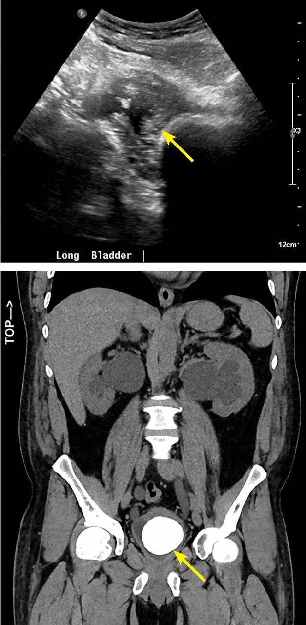
Renal ultrasonography (top) revealed a decompressed bladder (arrow). Computed tomography (bottom) showed a bladder stone occupying the entire bladder (arrow).

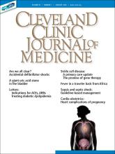
He underwent bilateral percutaneous nephrostomy tube placement followed by open cystolithotomy, and a large calculus (6.9 × 4.8 × 4.5 cm) was removed (Figure 2). Analysis of the stone revealed 100% uric acid composition and no evidence of a foreign body nidus. A 24-hour urine collection showed normal excretion of uric acid, calcium, citrate, phosphorus, and potassium. The acute kidney injury resolved, and his serum creatinine level came down to 1.9 mg/dL.

Figure 2
  • Download figure
  • Open in new tab
  • Download powerpoint
Figure 2

The bladder stone measured 6.9 cm × 4.8 cm × 4.5 cm.

GIANT BLADDER STONES

Giant bladder stones are rare and a rare cause of acute kidney injury.1 They are usually solitary and associated with urinary stasis in the setting of neurologic injury, neobladder reconstruction, or benign prostatic hypertrophy. They can also occur in chronic infections with urease-splitting bacteria or secondary to an iatrogenic foreign body.1,2

While this patient did not have any of these risk factors, computed tomography noted a small bladder diverticulum, a possible nidus of urinary stasis and calculus formation.1

There are case reports of retained surgical material and foreign bodies as a nidus for giant bladder calculi development.1 However, in our patient, no foreign body was identified during stone bisection.

Uric acid bladder stones have been associated with acidic urinary pH.3 Our patient’s initial urine pH was 5.5, which we attributed to chronic ketosis from alcoholism. It increased to 7.0 during his hospitalization with alcohol cessation, but it was again found to be 5.5 on urinalysis at follow-up visits after hospital discharge.

Bladder calculi account for only 5% of all urinary stones; they often arise in the setting of bladder outlet obstruction and are rarely associated with upper tract calculi.1,4 The earliest giant bladder stone was found in the skeleton of a 7,000 year-old Egyptian mummy, and the prevalence of these stones has dramatically decreased in the developed world due to modernization of diets.1 No specific nutritional deficiency has been causally linked to bladder stone formation, though these calculi remain most common in North Africa, the Middle East, and India in chronically undernourished children with diets low in animal protein and phosphate.1,5

Most giant bladder stones are composed of calcium phosphate or struvite, rarely uric acid. Studies suggest that only 7% of bladder calculi in men are composed of uric acid, with far more composed of calcium oxalate or struvite.6

Acknowledgment

The authors thank Dr. Juan Paredes Magaña for his care of this patient.

  • Copyright © 2020 The Cleveland Clinic Foundation. All Rights Reserved.

REFERENCES

  1. ↵
    1. Kavoussi LR,
    2. Partin AW,
    3. Peters C,
    4. Wein AJ
    1. Benway BM,
    2. Bhayani SB
    . Lower urinary tract calculi. In: Kavoussi LR, Partin AW, Peters C, Wein AJ, eds. Campbell-Walsh Urology. 11th ed. Philadelphia, PA: Elsevier; 2016:1291–1299.
  2. ↵
    1. Nguyen S,
    2. Choi C
    . Neobladder stone. N Engl J Med 2017; 377(10):977. doi:10.1056/NEJMicm1611578
    OpenUrlCrossRef
  3. ↵
    1. Torricelli FC,
    2. Chueh SJ,
    3. Shen S,
    4. Monga M
    . Multiple uric acid bladder stones: clinical presentation and endoscopic management. J Endourol Case Rep 2017; 3(1):21–23. doi:10.1089/cren.2016.0134
    OpenUrlCrossRef
  4. ↵
    1. Douenias R,
    2. Rich M,
    3. Badlani G,
    4. Mazor D,
    5. Smith A
    . Predisposing factors in bladder calculi. Review of 100 cases. Urology. 1991; 37(3):240–243. doi:10.1016/0090-4295(91)80293-g
    OpenUrlCrossRefPubMed
  5. ↵
    1. Van Reen R
    . Idiopathic urinary bladder stones of childhood. Aust N Z J Surg 1980; 50(1):18–22. doi:10.1111/j.1445-2197.1980.tb04482.x
    OpenUrlCrossRefPubMed
  6. ↵
    1. Schwartz BF,
    2. Stoller ML
    . The vesical calculus. Urol Clin North Am 2000; 27(2):333–346. pmid:10778475
    OpenUrlCrossRefPubMed
PreviousNext
Back to top

In this issue

Cleveland Clinic Journal of Medicine: 87 (1)
Cleveland Clinic Journal of Medicine
Vol. 87, Issue 1
1 Jan 2020
  • Table of Contents
  • Table of Contents (PDF)
  • Index by author
  • Complete Issue (PDF)
Print
Download PDF
Article Alerts
Sign In to Email Alerts with your Email Address
Email Article

Thank you for your interest in spreading the word on Cleveland Clinic Journal of Medicine.

NOTE: We only request your email address so that the person you are recommending the page to knows that you wanted them to see it, and that it is not junk mail. We do not capture any email address.

Enter multiple addresses on separate lines or separate them with commas.
Giant uric acid stone in the bladder
(Your Name) has sent you a message from Cleveland Clinic Journal of Medicine
(Your Name) thought you would like to see the Cleveland Clinic Journal of Medicine web site.
CAPTCHA
This question is for testing whether or not you are a human visitor and to prevent automated spam submissions.
Citation Tools
Giant uric acid stone in the bladder
Alexander E. Sullivan, Suchita Shah Sata
Cleveland Clinic Journal of Medicine Jan 2020, 87 (1) 14-15; DOI: 10.3949/ccjm.87a.19069

Citation Manager Formats

  • BibTeX
  • Bookends
  • EasyBib
  • EndNote (tagged)
  • EndNote 8 (xml)
  • Medlars
  • Mendeley
  • Papers
  • RefWorks Tagged
  • Ref Manager
  • RIS
  • Zotero
Share
Giant uric acid stone in the bladder
Alexander E. Sullivan, Suchita Shah Sata
Cleveland Clinic Journal of Medicine Jan 2020, 87 (1) 14-15; DOI: 10.3949/ccjm.87a.19069
Twitter logo Facebook logo Mendeley logo
  • Tweet Widget
  • Facebook Like
  • Linkedin Share Button

Jump to section

  • Article
    • GIANT BLADDER STONES
    • Acknowledgment
    • REFERENCES
  • Figures & Data
  • Info & Metrics
  • PDF

Related Articles

  • No related articles found.
  • PubMed
  • Google Scholar

Cited By...

  • No citing articles found.
  • Google Scholar

More in this TOC Section

  • Tinea incognito
  • Prolonged venous filling time and dependent rubor in a patient with peripheral artery disease
  • Sarcoidosis with diffuse purplish erythematous plaques on the hands
Show more The Clinical Picture

Similar Articles

Subjects

  • Emergency Medicine
  • Hospital Medicine
  • Imaging
  • Men's Health
  • Mental Health
  • Nephrology

Navigate

  • Current Issue
  • Past Issues
  • Supplements
  • Article Type
  • Specialty
  • CME/MOC Articles
  • CME/MOC Calendar
  • Media Kit

Authors & Reviewers

  • Manuscript Submission
  • Authors & Reviewers
  • Subscriptions
  • About CCJM
  • Contact Us
  • Cleveland Clinic Center for Continuing Education
  • Consult QD

Share your suggestions!

Copyright © 2025 The Cleveland Clinic Foundation. All rights reserved. The information provided is for educational purposes only. Use of this website is subject to the website terms of use and privacy policy. 

Powered by HighWire