


A 59-year-old man with known chronic obstructive pulmonary disease and poorly controlled diabetes was referred to the hospital by his dermatologist. Two months earlier, he first noticed an itchy “bump” on the cutaneous portion of his upper lip, which then developed into multiple growths that had spread to the tip of his nose.
He reported several episodes of night sweats, dysphagia, and left-sided hand and leg weakness.
He had worked for 30 years in coal mines and sawmills. He was a smoker, with a 25-pack-year history. He lived in eastern Tennessee.
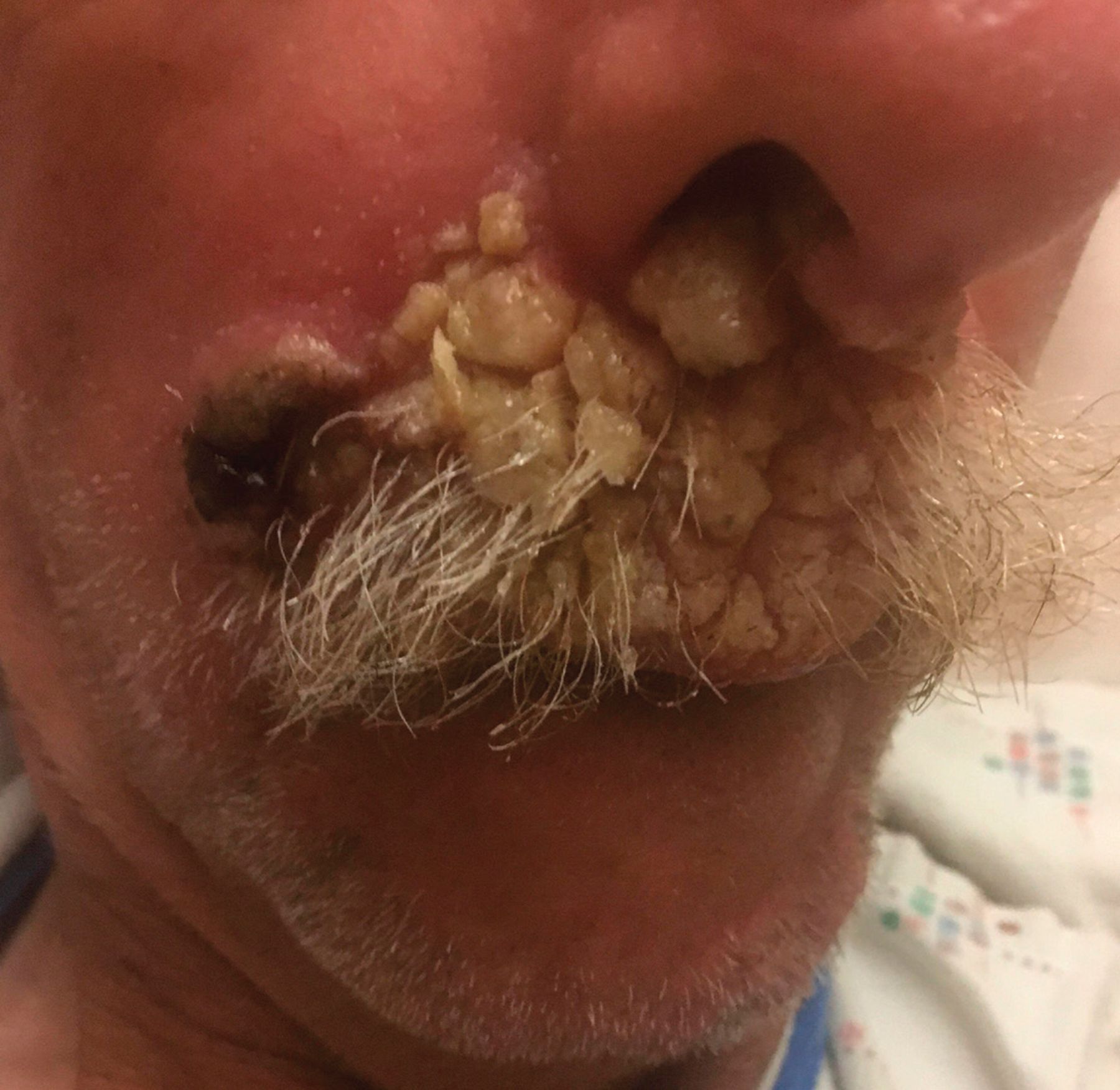
Physical examination confirmed numerous exophytic verrucous plaques on his upper cutaneous lip and nose (Figure 1). Results of serum chemistry, liver enzymes, and complete blood cell count were normal. Testing for human immunodeficiency virus infection was negative.

The patient presented with exophytic verrucous plaques on his upper cutaneous lip and nose.
Biopsy of the facial lesions was performed, and he underwent magnetic resonance imaging of the brain and computed tomography of the chest.
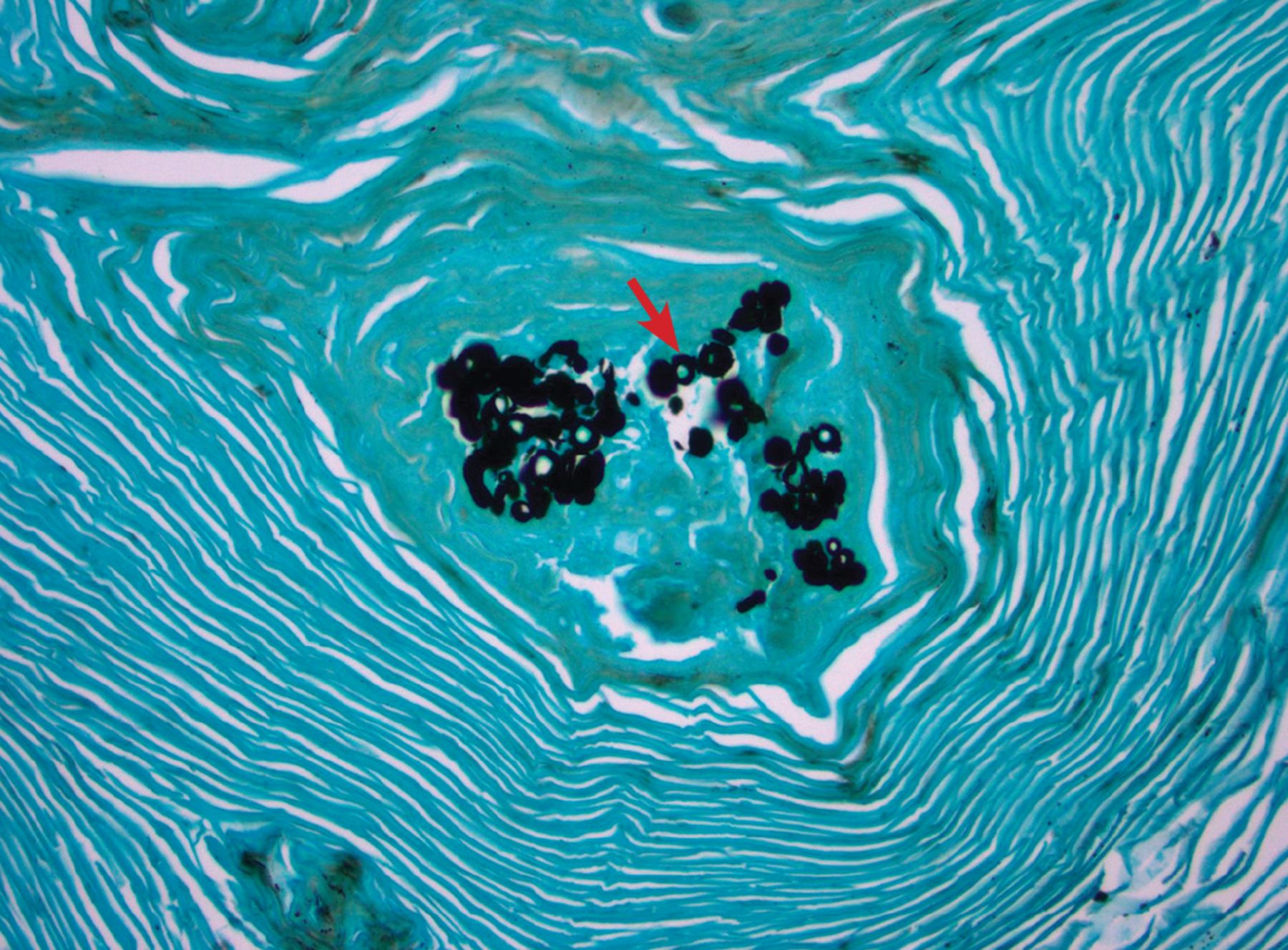
Biopsy study showed pseudoepitheliomatous hyperplasia, and Grocott-Gomori methenamine silver staining (Figure 2) revealed broad-based budding yeasts measuring 8 to 20 μm in diameter and with thickened cell walls, features consistent with blastomycosis. Because of concern for disseminated disease, the patient was admitted for further workup.

Grocott-Gomori methenamine silver staining revealed broad-based budding yeasts with thickened cell walls (arrow).
FURTHER EVALUATION AND DIAGNOSIS
Magnetic resonance imaging of the brain revealed ring-enhancing lesions in the pons and inferior cerebellar peduncle. Computed tomography of the chest showed bilateral upper-lobe scarring and areas of bronchiectasis, as well as nodules in the upper lobes and ground-glass opacities. Urine antigen testing for Blastomyces was positive at 3.07 ng/mL (reference range for negative, 0 ng/mL).
Given these findings and the high likelihood of blastomycosis, sputum cultures and additional serologic testing were not ordered.
The patient was treated with intravenous amphotericin B for 6 weeks and oral voriconazole 200 mg twice daily for 1 year. His skin lesions and neurologic symptoms resolved. Repeat imaging at 4 months showed resolution of brain and chest lesions.
BLASTOMYCOSIS: CLUES TO DIAGNOSIS
The differential diagnosis of cutaneous blastomycosis includes infection with dimorphic fungi (ie, growth forms that change from moldlike to yeastlike), basal cell carcinoma, squamous cell carcinoma, giant keratoacanthoma, lupus vulgaris, scrofuloderma, nocardiosis, atypical Mycobacterium infection, syphilis, bromoderma, iododerma, leishmaniasis, granuloma inguinale, lymphoma, and pyoderma gangrenosum.1–3
In the diagnosis of dimorphic fungal infections—including blastomycosis, histoplasmosis, and coccidioidomycosis—geographic location, epidemiologic factors, and exposure history are key considerations. Blastomycosis and histoplasmosis are endemic in the Ohio and Mississippi River valleys and southeastern areas of the United States (including eastern Tennessee), whereas coccidioidomycosis is endemic in the southwestern United States. All 3 can present as cutaneous lesions with systemic involvement. Culture and histopathology can help distinguish them.
Basal and squamous cell carcinomas and giant keratoacanthoma can also present as cutaneous plaques. These are easily diagnosed with histopathologic study, and systemic involvement would be uncommon.
Most cases of blastomycosis are considered sporadic. But outbreaks have been associated with recreational and occupational activities involving distribution of soil, including construction, underground exploration, tubing, and hunting.4
ROUTES OF TRANSMISSION
Blastomycosis is transmitted by inhalation of spores and can occur in both immunocompromised and immunocompetent individuals. Large series have shown that most cases of disseminated blastomycosis occur in nonimmunosuppressed individuals. Immunosuppressed patients develop more severe infection and are more likely to have symptomatic dissemination.3
B dermatitidis is typically inhaled and affects the lungs, causing pneumonia. However, hematogenous spread has been reported in 25% to 30% of cases.5 The most common sites for hematogenous spread are the skin, bones, and joints. Rarely, primary skin infection may occur through direct inoculation.6,7
In our patient, the presumed source of infection was by inhalation.
Any patient presenting with cutaneous blastomycosis should be investigated for disseminated disease, particularly pulmonary involvement.
DIAGNOSTIC CHALLENGES OF BLASTOMYCOSIS
Diagnosis of blastomycosis can be challenging, and a high index of suspicion is key in endemic areas.
Culture is the gold standard for diagnosis, but Grocott-Gomori methenamine silver stain can show the presence of broad-based budding yeasts. Unfortunately, multiple biopsies are often necessary for diagnosis, as sensitivities of histology and culture have been reported to be as low as 9.7% and 61%, respectively. Cultures may take up to 4 weeks for results.8
Assays are also available for detection of B dermatitidis antigen in the serum or urine and can yield quicker results. The sensitivity of the urine antigen assay has been reported to be as high as 92.9%, whereas the serum assay is less sensitive.9
Cross-reactivity with Histoplasma capsulatum does occur, so a negative result should not preclude a diagnosis of blastomycosis.10
TREATMENT RECOMMENDATIONS
Treatment of blastomycosis depends on severity of illness, central nervous system (CNS) involvement, and the patient’s immune status. For uncomplicated cases, itraconazole therapy is preferred and is curative in 95% of cases.
With CNS involvement or life-threatening infection, liposomal amphotericin-B is given. The recommended course is 1 to 2 weeks for severe infection or 6 weeks for CNS involvement. This can then be transitioned to 1 year of oral azole therapy.
Treatment guidelines recommend itraconazole as the preferred agent for all non-CNS disease. Fluconazole and voriconazole are alternatives in patients unable to tolerate itraconazole.11 Fluconazole must be given at higher dosages and has higher reported failure rates; however, it has good cerebrospinal fluid penetration. Ultimately, further research must be done to determine the efficacy of various azole agents in step-down treatment of CNS blastomycosis.12,13
This patient’s presentation highlights the clinical manifestations of disseminated blastomycosis and stresses the importance of maintaining a high index of suspicion in an endemic region.
- Copyright © 2020 The Cleveland Clinic Foundation. All Rights Reserved.
In this issue


Jump to section
Related Articles
Cited By...
- No citing articles found.