
Main menu

  • Home
  • Content
    • Current Issue
    • Ahead of Print
    • Past Issues
    • Supplements
    • Article Type
  • Specialty
    • Articles by Specialty
  • CME/MOC
    • Articles
    • Calendar
  • Info For
    • Manuscript Submission
    • Authors & Reviewers
    • Subscriptions
    • About CCJM
    • Contact Us
    • Media Kit
  • Conversations with Leaders
  • Conference Coverage
    • Kidney Week 2024
    • CHEST 2024
    • ACR Convergence 2023
    • Kidney Week 2023
    • ObesityWeek 2023
    • IDWeek 2023
    • CHEST 2023
    • MDS 2023
    • IAS 2023
    • ACP 2023
    • AAN 2023
    • ACC / WCC 2023
    • AAAAI Meeting 2023
    • ACR Convergence 2022
    • Kidney Week 2022
    • AIDS 2022
  • Other Publications
    • www.clevelandclinic.org

User menu

  • Register
  • Log in

Search

  • Advanced search
Cleveland Clinic Journal of Medicine
  • Other Publications
    • www.clevelandclinic.org
  • Register
  • Log in
Cleveland Clinic Journal of Medicine

Advanced Search

  • Home
  • Content
    • Current Issue
    • Ahead of Print
    • Past Issues
    • Supplements
    • Article Type
  • Specialty
    • Articles by Specialty
  • CME/MOC
    • Articles
    • Calendar
  • Info For
    • Manuscript Submission
    • Authors & Reviewers
    • Subscriptions
    • About CCJM
    • Contact Us
    • Media Kit
  • Conversations with Leaders
  • Conference Coverage
    • Kidney Week 2024
    • CHEST 2024
    • ACR Convergence 2023
    • Kidney Week 2023
    • ObesityWeek 2023
    • IDWeek 2023
    • CHEST 2023
    • MDS 2023
    • IAS 2023
    • ACP 2023
    • AAN 2023
    • ACC / WCC 2023
    • AAAAI Meeting 2023
    • ACR Convergence 2022
    • Kidney Week 2022
    • AIDS 2022
1-Minute Consult

When is contrast needed for abdominal and pelvic CT?

Mark Radetic, MD, Robert DeVita, MD and John Haaga, MD
Cleveland Clinic Journal of Medicine October 2020, 87 (10) 595-598; DOI: https://doi.org/10.3949/ccjm.87a.19093
Mark Radetic
Department of Internal Medicine, Cleveland Clinic
  • Find this author on Google Scholar
  • Find this author on PubMed
  • Search for this author on this site
  • For correspondence: [email protected]
Robert DeVita
Department of Radiology, University Hospitals Cleveland Medical Center, Cleveland, OH
  • Find this author on Google Scholar
  • Find this author on PubMed
  • Search for this author on this site
John Haaga
Department of Radiology, University Hospitals Cleveland Medical Center, Cleveland, OH
  • Find this author on Google Scholar
  • Find this author on PubMed
  • Search for this author on this site
  • Article
  • Figures & Data
  • Info & Metrics
  • PDF
Loading

Computed tomography (CT) is one of the most frequently utilized imaging modalities in medicine due to its ability to evaluate for a wide range of pathologies. The use of contrast agents, intravenous (IV) or oral, improves image quality by further delineating anatomical structures. However, contrast enhancement is not always necessary and does come with some risks. The appropriateness of contrast enhancement usually depends on the suspected diagnosis. In cases in which the diagnosis is uncertain, administration of contrast is reasonable, although the benefits should be weighed against any potential risks.

INTRAVENOUS CONTRAST

All modern IV contrast agents are iodine-based. The iodine causes increased absorption and scattering of the incoming radiation, which serves to increase the attenuation or “brightness” of the tissue or organ.1 Importantly, the IV contrast used in CT is distinct from the gadolinium-based IV contrast used in magnetic resonance imaging, meaning that there is no cross-reactivity between the two, which is important if the patient is allergic to one of them.

IV contrast is necessary for the evaluation of any kind of vascular disease, since it allows for easy identification of the blood vessel lumen.1 In abdominal imaging, IV contrast is recommended in most cases.

ORAL CONTRAST

The primary benefit of oral contrast is its ability to distend the bowels to help distinguish them from adjacent abdominal structures.

Oral contrast comes in two forms: neutral or positive. Neutral oral contrast consists of water or a dilute, low-attenuation solution that mirrors water. Positive oral contrast is an iodinated (ie, gastrografin) or barium-based solution with high attenuation that further demarcates bowel by opacification.

The advent of multidetector CT, which offers improved resolution, has made it easier to differentiate abdominal structures without the need for the opacification with positive oral contrast. As a result, some have argued that neutral oral contrast may be preferable to positive oral contrast due to similar efficacy, cost-effectiveness, and easier patient tolerability.2

GENERAL INDICATIONS FOR CONTRAST USE IN ABDOMINOPELVIC CT

The decision to use contrast in abdominopelvic CT depends on the diagnosis suspected.

IV contrast is recommended in most cases (Table 1).3 It is useful in the evaluation of infection (appendicitis, colitis, diverticulitis, pyelonephritis) (Figure 1); inflammation (pancreatitis, inflammatory bowel disease), masses and malignancies; and vascular abnormalities (gastrointestinal bleeding, aortic dissection, abdominal aortic aneurysm). However, IV contrast is not necessary to diagnose bowel perforations, nephrolithiasis, or hematomas.

View this table:
  • View inline
  • View popup
TABLE 1

Indications for intravenous contrast in abdominal and pelvic computed tomography

Figure 1
  • Download figure
  • Open in new tab
  • Download powerpoint
Figure 1

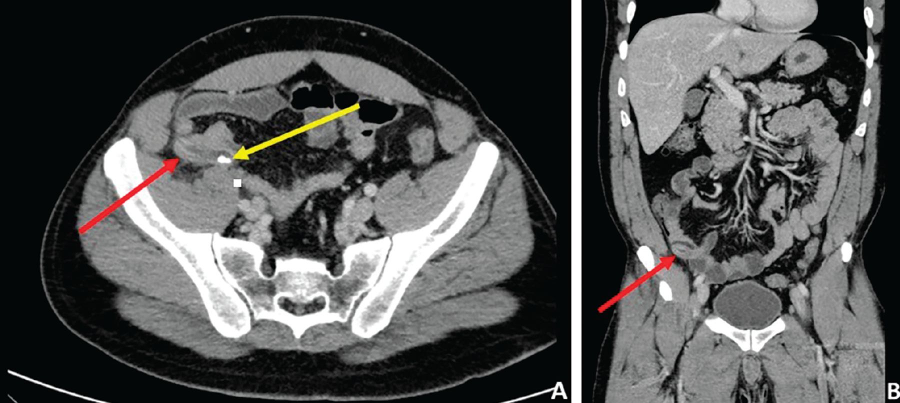
Computed tomography with intravenous contrast enhancement in a patient with right-lower-quadrant abdominal pain. Axial (A) and coronal (B) views reveal diffuse mural and periappendiceal edema, with thickening of the appendix (red arrows). Coarse calcification within the appendix (yellow arrow) likely represents an appendicolith.

In vascular imaging, the study of choice is CT angiography, which is timed so that the image is taken when the IV contrast reaches the arterial system, making it easier to identify active bleeding.

The use of oral contrast is more controversial.4–6 No clear consensus exists on the need for oral contrast, and expert opinion often drives current practices with regard to oral contrast use at academic medical centers.6 In general, when the primary reason for CT is to evaluate the liver, gallbladder, pancreas, spleen, adrenal glands, or urinary tract, oral contrast is unnecessary.

Alternatively, when evaluating the gastrointestinal lumen or bowel wall, oral contrast may be beneficial (Figure 2). However, oral contrast is not needed in the diagnosis of appendicitis or diverticulitis, even though both are luminal disorders.5

Figure 2
  • Download figure
  • Open in new tab
  • Download powerpoint
Figure 2

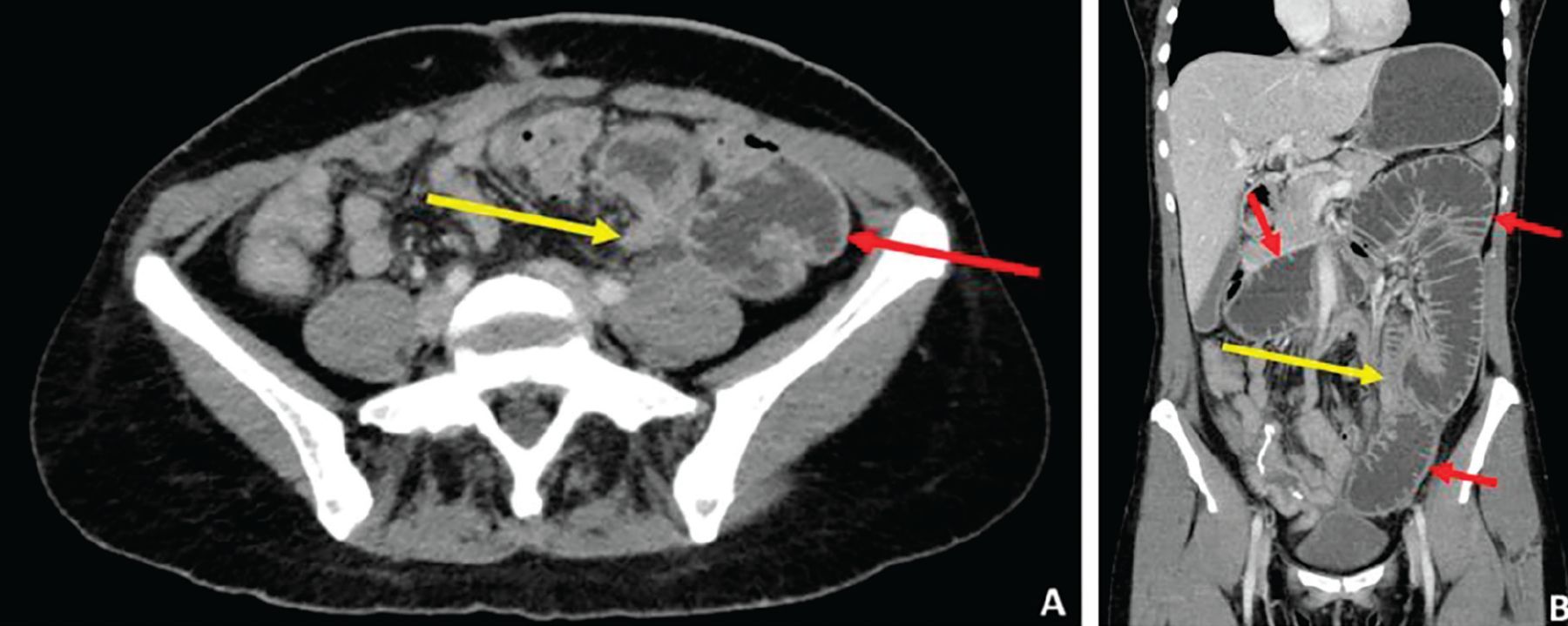
Computed tomography with a neutral oral contrast agent. Axial (A) and coronal (B) views reveal multiple loops of dilated bowel (red arrows) with a transition point—ie, site of sudden luminal narrowing (yellow arrows)—in the left lower quadrant, findings consistent with a small-bowel obstruction.

Bowel “illumination” achieved with positive oral contrast is useful when searching for “breaks” in the bowel wall, such as what would be seen with fistulas and perforations, or for identifying fluid collections or abscesses between loops of opacified bowel.

Conversely, neutral oral contrast is preferred when evaluating for mural abnormalities or a suspected gastrointestinal bleed, as positive contrast will opacify and mask the luminal surface, making it challenging to identify the bleeding source.4

In patients presenting with nonspecific abdominal complaints, some would argue that the addition of the use of oral contrast optimizes the diagnostic yield of abdominal CT.6 Woolen et al found that 89% of patients would prefer oral contrast if it had any diagnostic benefit.7 Thus, in patients undergoing abdominal CT for vague or nonspecific complaints, the addition of oral contrast to the study appears reasonable.

ADVERSE EFFECTS OF CONTRAST

IV contrast carries a risk of an allergic reaction (incidence 0.6%), which can manifest as urticaria, pruritus, bronchospasm with wheezing, or anaphylactic shock.8 Several premedication regimens consisting of a steroid with or without an antihistamine are available for at-risk patients (Table 2).8

View this table:
  • View inline
  • View popup
TABLE 2

Premedication regimens for patients allergic to intravenous contrast

Risk of nephropathy

Another concern with IV contrast is its use in patients with underlying renal disease. These patients are at risk for developing contrast-induced nephropathy (CIN), which is an acute kidney injury (AKI) that develops within 48 hours of IV contrast administration. The diagnosis is fairly controversial, with some studies having found similar rates of AKI in patients undergoing CT with and without IV contrast.9 IV contrast is considered unlikely to cause nephropathy in patients with normal renal function, but can cause CIN in those with impaired renal function.8

Current guidelines for IV contrast administration are based on the estimated glomerular filtration rate (eGFR).8,9 In general, patients with an eGFR of at least 30 mg/dL can receive IV contrast, whereas those with an eGFR less than 30 mg/dL (corresponding to stage 4 chronic kidney disease) are at high risk for renal failure. In these patients, a discussion should be held regarding the high probability of progression to end-stage renal disease, requiring dialysis.

Preventive measures to minimize the risk of contrast-induced nephropathy involve giving IV fluids at 100 mL/hour for 6 hours before and after contrast administration.8,9 Historically, sodium bicarbonate and N-acetylcysteine have been used as adjunctive agents, although there is a lack of evidence supporting their use.8,9

Risks of oral contrast agents

Oral contrast is generally safe and well tolerated, although some patients can experience bothersome symptoms. Neutral contrast agents may contain osmotically active substances that can promote loose stools or diarrhea. Of the positive oral contrast agents, iodinated agents should be avoided in patients at risk for aspiration, as they can cause aspiration pneumonitis. Barium-based oral contrast agents should be avoided in patients with suspected perforations (can cause mediastinitis or peritonitis) or bowel obstruction, as retained barium can harden to form a “barolith,” worsening the obstruction and requiring endoscopic or even surgical removal.10

  • Copyright © 2020 The Cleveland Clinic Foundation. All Rights Reserved.

REFERENCES

  1. ↵
    1. Purysko CP,
    2. Renapurkar R,
    3. Bolen MA
    . When does chest CT require contrast enhancement? Cleve Clin J Med 2016; 83(6):423–426. doi:10.3949/ccjm.83a.15037
    OpenUrlFREE Full Text
  2. ↵
    1. Koo CW,
    2. Shah-Patel LR,
    3. Baer JW,
    4. Frager DH
    . Cost-effectiveness and patient tolerance of low-attenuation oral contrast material: milk versus VoLumen. AJR Am J Roentgenol 2008; 190(5):1307–1313. doi:10.2214/AJR.07.3193
    OpenUrlCrossRefPubMed
  3. ↵
    1. American College of Radiology
    . ACR appropriateness criteria. Accessed September 1, 2020. https://www.acr.org/Clinical-Resources/ACR-Appropriateness-Criteria.
  4. ↵
    1. Kammerer S,
    2. Höink AJ,
    3. Wessling J, et al
    . Abdominal and pelvic CT: is positive enteric contrast still necessary? Results of a retrospective observational study. Eur Radiol 2015; 25(3):669–678. doi:10.1007/s00330-014-3446-9
    OpenUrlCrossRef
  5. ↵
    1. Ramalingam V,
    2. Bates DD,
    3. Buch K, et al
    . Diagnosing acute appendicitis using a nonoral contrast CT protocol in patients with a BMI of less than 25. Emerg Radiol 2016; 23(5):455–462. doi:10.1007/s10140-016-1421-2
    OpenUrlCrossRef
  6. ↵
    1. Pickhardt PJ
    . Positive oral contrast material for abdominal CT: current clinical indications and areas of controversy. AJR Am J Roentgenol 2020: 1–10. doi:10.2214/AJR.19.21989. Epub ahead of print.
    OpenUrlCrossRef
  7. ↵
    1. Woolen SA,
    2. Maturen KE,
    3. Nettles A, et al
    . Patient-centered assessment of the value of oral contrast material. J Am Coll Radiol 2017; 14(12):1626–1631. doi:10.1016/j.jacr.2017.06.036
    OpenUrlCrossRef
  8. ↵
    1. American College of Radiology
    . ACR manual on contrast media. https://www.acr.org/Clinical-Resources/Contrast-Manual. Accessed September 1, 2020.
  9. ↵
    1. Wichmann JL,
    2. Katzberg RW,
    3. Litwin SE, et al
    . Contrast-induced nephropathy. Circulation 2015; 132(20):1931–1936. doi:10.1161/CIRCULATIONAHA.115.014672
    OpenUrlFREE Full Text
  10. ↵
    1. Kurer MA,
    2. Davey C,
    3. Chintapatla S
    . Intestinal obstruction from inspissated barium (barolith): a systematic review of all cases from 1950 to 2006. Colorectal Dis 2008; 10(5):431–439. doi:10.1111/j.1463-1318.2008.01503.x
    OpenUrlCrossRefPubMed
PreviousNext
Back to top

In this issue

Cleveland Clinic Journal of Medicine: 87 (10)
Cleveland Clinic Journal of Medicine
Vol. 87, Issue 10
1 Oct 2020
  • Table of Contents
  • Table of Contents (PDF)
  • Index by author
  • Complete Issue (PDF)
Print
Download PDF
Article Alerts
Sign In to Email Alerts with your Email Address
Email Article

Thank you for your interest in spreading the word on Cleveland Clinic Journal of Medicine.

NOTE: We only request your email address so that the person you are recommending the page to knows that you wanted them to see it, and that it is not junk mail. We do not capture any email address.

Enter multiple addresses on separate lines or separate them with commas.
When is contrast needed for abdominal and pelvic CT?
(Your Name) has sent you a message from Cleveland Clinic Journal of Medicine
(Your Name) thought you would like to see the Cleveland Clinic Journal of Medicine web site.
CAPTCHA
This question is for testing whether or not you are a human visitor and to prevent automated spam submissions.
Citation Tools
When is contrast needed for abdominal and pelvic CT?
Mark Radetic, Robert DeVita, John Haaga
Cleveland Clinic Journal of Medicine Oct 2020, 87 (10) 595-598; DOI: 10.3949/ccjm.87a.19093

Citation Manager Formats

  • BibTeX
  • Bookends
  • EasyBib
  • EndNote (tagged)
  • EndNote 8 (xml)
  • Medlars
  • Mendeley
  • Papers
  • RefWorks Tagged
  • Ref Manager
  • RIS
  • Zotero
Share
When is contrast needed for abdominal and pelvic CT?
Mark Radetic, Robert DeVita, John Haaga
Cleveland Clinic Journal of Medicine Oct 2020, 87 (10) 595-598; DOI: 10.3949/ccjm.87a.19093
Twitter logo Facebook logo Mendeley logo
  • Tweet Widget
  • Facebook Like
  • Linkedin Share Button

Jump to section

  • Article
    • INTRAVENOUS CONTRAST
    • ORAL CONTRAST
    • GENERAL INDICATIONS FOR CONTRAST USE IN ABDOMINOPELVIC CT
    • ADVERSE EFFECTS OF CONTRAST
    • REFERENCES
  • Figures & Data
  • Info & Metrics
  • PDF

Related Articles

  • No related articles found.
  • PubMed
  • Google Scholar

Cited By...

  • No citing articles found.
  • Google Scholar

More in this TOC Section

  • What diagnostic tests should be done after discovering clubbing in a patient without cardiopulmonary symptoms?
  • Does my adult patient need a measles vaccine?
  • Do I need to treat supine hypertension in my hospitalized patient?
Show more 1-Minute Consult

Similar Articles

Subjects

  • Gastroenterology
  • Hepatology
  • Imaging
  • Nephrology
  • Vascular Medicine

Navigate

  • Current Issue
  • Past Issues
  • Supplements
  • Article Type
  • Specialty
  • CME/MOC Articles
  • CME/MOC Calendar
  • Media Kit

Authors & Reviewers

  • Manuscript Submission
  • Authors & Reviewers
  • Subscriptions
  • About CCJM
  • Contact Us
  • Cleveland Clinic Center for Continuing Education
  • Consult QD

Share your suggestions!

Copyright © 2025 The Cleveland Clinic Foundation. All rights reserved. The information provided is for educational purposes only. Use of this website is subject to the website terms of use and privacy policy. 

Powered by HighWire