
Main menu

  • Home
  • Content
    • Current Issue
    • Ahead of Print
    • Past Issues
    • Supplements
    • Article Type
  • Specialty
    • Articles by Specialty
  • CME/MOC
    • Articles
    • Calendar
  • Info For
    • Manuscript Submission
    • Authors & Reviewers
    • Subscriptions
    • About CCJM
    • Contact Us
    • Media Kit
  • Conversations with Leaders
  • Conference Coverage
    • ACC / WCC 2023
    • AAAAI Meeting 2023
    • ACR Convergence 2022
    • Kidney Week 2022
    • AIDS 2022
    • CHEST 2021
    • IDWeek 2021
    • IAS 2021
    • ADA 2021
    • ATS 2021
    • ACC 2021
    • ACP 2021
    • AAN 2021
  • Other Publications
    • www.clevelandclinic.org

User menu

  • Register
  • Log in

Search

  • Advanced search
Cleveland Clinic Journal of Medicine
  • Other Publications
    • www.clevelandclinic.org
  • Register
  • Log in
Cleveland Clinic Journal of Medicine

Advanced Search

  • Home
  • Content
    • Current Issue
    • Ahead of Print
    • Past Issues
    • Supplements
    • Article Type
  • Specialty
    • Articles by Specialty
  • CME/MOC
    • Articles
    • Calendar
  • Info For
    • Manuscript Submission
    • Authors & Reviewers
    • Subscriptions
    • About CCJM
    • Contact Us
    • Media Kit
  • Conversations with Leaders
  • Conference Coverage
    • ACC / WCC 2023
    • AAAAI Meeting 2023
    • ACR Convergence 2022
    • Kidney Week 2022
    • AIDS 2022
    • CHEST 2021
    • IDWeek 2021
    • IAS 2021
    • ADA 2021
    • ATS 2021
    • ACC 2021
    • ACP 2021
    • AAN 2021
The Clinical Picture

Constitutional symptoms, pathologic PET-CT

Julia L. Riera, MD, Jorge L. Musuruana, MD, Fernando Faccio, MD and Javier A. Cavallasca, MD
Cleveland Clinic Journal of Medicine April 2020, 87 (4) 195-197; DOI: https://doi.org/10.3949/ccjm.87a.19053
Julia L. Riera
  • Find this author on Google Scholar
  • Find this author on PubMed
  • Search for this author on this site
Jorge L. Musuruana
Section of Rheumatology and Autoimmune Diseases, Hospital JB Iturraspe, Santa Fe, Argentina
  • Find this author on Google Scholar
  • Find this author on PubMed
  • Search for this author on this site
Fernando Faccio
Section of PET/CT, Sanatorio San Geronimo, Santa Fe, Argentina
  • Find this author on Google Scholar
  • Find this author on PubMed
  • Search for this author on this site
Javier A. Cavallasca
  • Find this author on Google Scholar
  • Find this author on PubMed
  • Search for this author on this site
  • For correspondence: jcavallasca@yahoo.com.ar jcavallasca@gmail.com
  • Article
  • Figures & Data
  • Info & Metrics
  • PDF
Loading

A 74-year-old woman presented with a 6-month history of malaise, generalized fatigue, significant unintentional weight loss, and night sweats, as well as pain, weakness, and stiffness in the shoulder girdle most severe in the morning. She reported no headache, visual disturbances, or jaw claudication.

On examination, blood pressure in both arms was within normal limits, and cardiovascular examination including peripheral pulses was normal without bruits.

Laboratory testing results showed erythrocyte sedimentation rate 142 mm/hour (reference range for age < 30), C-reactive protein 15.80 mg/dL (0–1), hemoglobin 9.9 g/dL (12.0–15.5), and creatine kinase 55 U/L (22– 198); other results were unremarkable.

Initially, malignancy was high on the differential diagnosis, in view of the patient’s advanced age, weight loss, and elevated levels of inflammatory biomarkers. However, computed tomography of the chest, abdomen, and pelvis were unremarkable, and upper endoscopy and colonoscopy showed no abnormalities. Thus, an autoimmune cause was considered.

The patient underwent whole-body fluorine-18 fluorodeoxyglucose positron emission tomography and computed tomography (PET-CT), which showed diffuse hypermetabolism of the great vessel walls, including the aorta and major branches, axillary arteries, common carotid artery, and common iliac artery, consistent with large-vessel giant cell arteritis (GCA) (Figure 1).

Figure 1
  • Download figure
  • Open in new tab
  • Download powerpoint
Figure 1

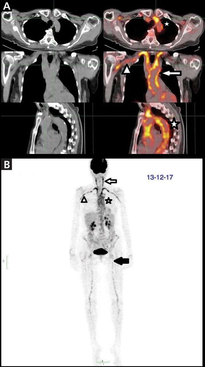
(A) Axial, coronal, sagittal, and (B) frontal positron emission tomography showed highly increased uptake of 18F-fluoro deoxyglucose in the aorta and major branches (star), axillary arteries (arrowheads), carotid arteries (arrows), and femoral arteries (black arrow).

Prednisone 50 mg/day (1 mg/kg/day) was started. This dosing was maintained for 4 weeks, after which it was gradually reduced 10% every 2 weeks. At this time, tocilizumab was not approved by the Argentine regulatory agency for the treatment of GCA, so methotrexate 15 mg weekly was added as a corticosteroid-sparing agent.

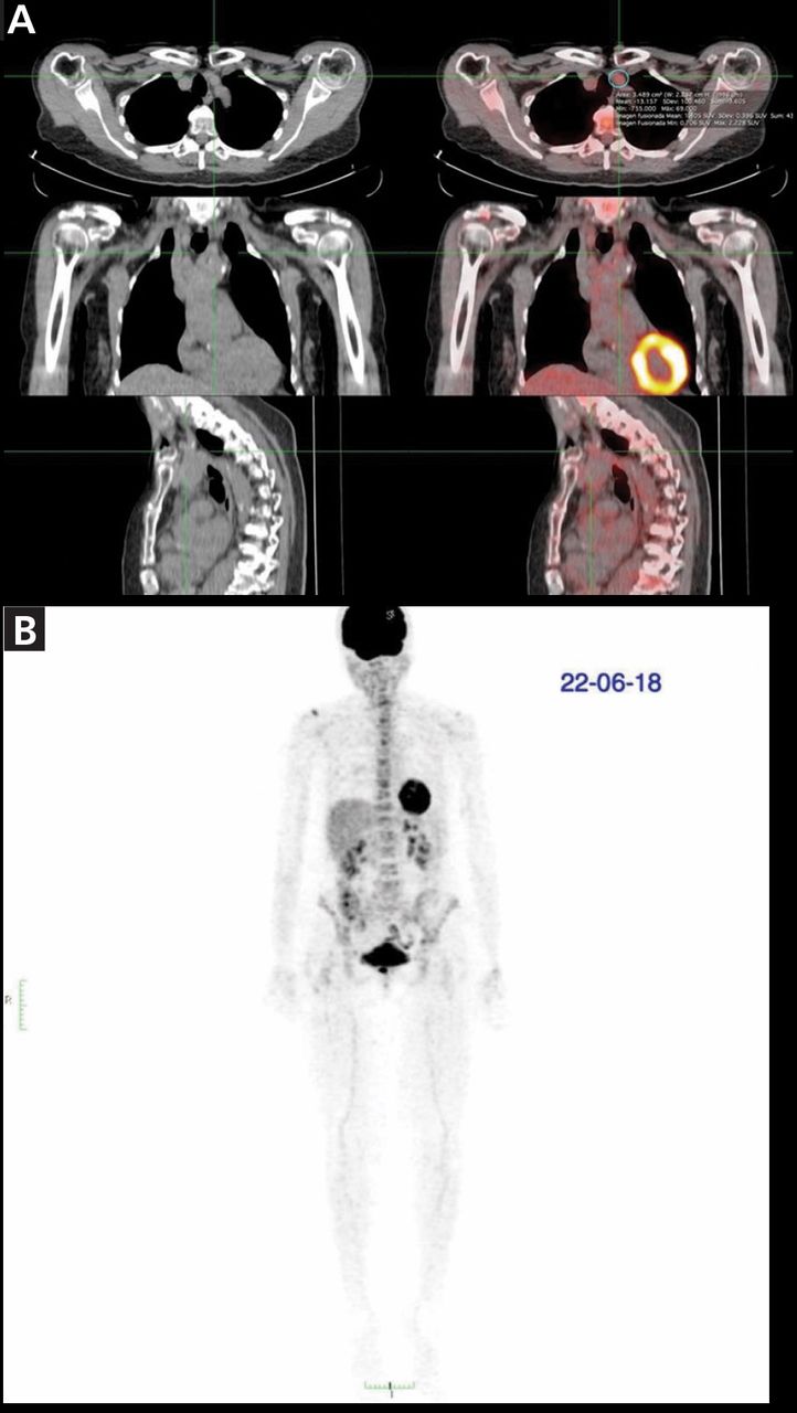
After 6 months of treatment, the patient was free of symptoms and her levels of inflammatory biomarkers had returned to normal; her erythrocyte sedimentation rate was 11 mm/hour and C-reactive protein 0.48 mg/dL. Repeat PET-CT showed significant improvement in vascular hypermetabolism, principally in the aorta and supra-aortic vessels, without metabolic activity in the subclavian arteries or extravascular metabolic hyperactivity (Figure 2).

Figure 2
  • Download figure
  • Open in new tab
  • Download powerpoint
Figure 2

After 6 months of treatment, (A) axial, coronal, sagittal and (B) frontal positron emission tomography showed improvement of uptake in all arterial territories.

LARGE-VESSEL GIANT CELL ARTERITIS

GCA is a granulomatous vasculitis that compromises medium and large arteries. The spectrum encompasses overlapping phenotypes including classic cranial arteritis and large-vessel (extracranial) GCA.1

Like cranial GCA, large-vessel GCA may present with constitutional symptoms such as fever of unknown origin, anorexia, weight loss, symptoms of polymyalgia rheumatica, or elevated inflammatory markers.2

In large-vessel GCA, especially if cranial symptoms are lacking, cross-sectional imaging such as magnetic resonance angiography, CT angiography, or PET-CT may be useful, because biopsy of the extracranial arteries is not feasible. In the Tocilizumab in Giant Cell Arteritis trial (GiACTA), the largest prospective study evaluating treatment in patients with GCA, 119 (86%) of a total of 138 patients who underwent cross-sectional imaging had findings consistent with large-vessel involvement.3

According to the European League Against Rheumatism recommendations for imaging in large-vessel vasculitis, the major advantage of PET in patients with suspected large-vessel vasculitis is the ability to identify GCA along with other serious pathology such as infection or tumor.4 This may be particularly relevant in elderly patients with constitutional symptoms without specific clinical features of GCA or polymyalgia rheumatica.

High cost, limited availability, and radiation exposure limit the use of PET. But combining PET with CT permits the evaluation of wall thickness and luminal changes.4

High doses of glucocorticoids (40–60 mg/day prednisone-equivalent) with gradual tapering are the mainstay of treatment. In patients with refractory disease or at increased risk of glucocorticoid-related adverse effects, methotrexate may be an alternative, glucocorticoid-sparing agent.5

In GiACTA, sustained remission rates at week 52 were 53% with tocilizumab treatment vs 17% with glucocorticoid monotherapy, while the cumulative glucocorticoid dose was reduced by 50% in tocilizumab-treated patients with fewer adverse events than those on glucocorticoids alone.6 These strikingly positive results led to tocilizumab’s approval by the US Food and Drug Administration.

Encouraging results have also been reported with ustekinumab, an interleukin 12 and interleukin 23 antagonist, and with abatacept, a selective T cell costimulation modulator.2 A new era in the treatment of an old disease is coming.

  • Copyright © 2020 The Cleveland Clinic Foundation. All Rights Reserved.

REFERENCES

  1. ↵
    1. Dejaco C,
    2. Duftner C,
    3. Buttgereit F,
    4. Matteson EL,
    5. Dasgupta B
    . The spectrum of giant cell arteritis and polymyalgia rheumatica: revisiting the concept of the disease. Rheumatology (Oxford) 2017; 56(4):506–515. doi:10.1093/rheumatology/kew273
    OpenUrlCrossRefPubMed
  2. ↵
    1. Koster MJ,
    2. Matteson EL,
    3. Warrington KJ
    . Large-vessel giant cell arteritis: diagnosis, monitoring and management. Rheumatology (Oxford) 2018; 57(suppl 2):ii32–ii42. doi:10.1093/rheumatology/kex424
    OpenUrlCrossRefPubMed
  3. ↵
    1. Tuckwell K,
    2. Collinson N,
    3. Klearman M,
    4. Dimonaco S,
    5. Stone JH, on behalf of GiACTA Investigators
    . FRIO377 classification criteria for giant cell arteritis: data from Giacta informing the need for revision. Ann Rheum Dis 2016; 75(suppl 2):571. ard.bmj.com/content/75/Suppl_2/571.1. Accessed February 26, 2020.
    OpenUrlAbstract/FREE Full Text
  4. ↵
    1. Dejaco C,
    2. Ramiro S,
    3. Duftner C, et al
    . EULAR recommendations for the use of imaging in large vessel vasculitis in clinical practice. Ann Rheum Dis 2018; 77(5):636–643. doi:10.1136/annrheumdis-2017-212649
    OpenUrlAbstract/FREE Full Text
  5. ↵
    1. Hellmich B,
    2. Agueda A,
    3. Monti S, et al
    . 2018 Update of the EULAR recommendations for the management of large vessel vasculitis. Ann Rheum Dis 2020; 79(1):19–30. doi:10.1136/annrheumdis-2019-215672
    OpenUrlAbstract/FREE Full Text
  6. ↵
    1. Stone JH,
    2. Tuckwell K,
    3. Dimonaco S, et al
    . Trial of tocilizumab in giant-cell arteritis. N Engl J Med 2017; 377(4):317–328. doi:10.1056/NEJMoa1613849
    OpenUrlCrossRefPubMed
PreviousNext
Back to top

In this issue

Cleveland Clinic Journal of Medicine: 87 (4)
Cleveland Clinic Journal of Medicine
Vol. 87, Issue 4
1 Apr 2020
  • Table of Contents
  • Table of Contents (PDF)
  • Index by author
  • Complete Issue (PDF)
Print
Download PDF
Article Alerts
Sign In to Email Alerts with your Email Address
Email Article

Thank you for your interest in spreading the word on Cleveland Clinic Journal of Medicine.

NOTE: We only request your email address so that the person you are recommending the page to knows that you wanted them to see it, and that it is not junk mail. We do not capture any email address.

Enter multiple addresses on separate lines or separate them with commas.
Constitutional symptoms, pathologic PET-CT
(Your Name) has sent you a message from Cleveland Clinic Journal of Medicine
(Your Name) thought you would like to see the Cleveland Clinic Journal of Medicine web site.
CAPTCHA
This question is for testing whether or not you are a human visitor and to prevent automated spam submissions.
Citation Tools
Constitutional symptoms, pathologic PET-CT
Julia L. Riera, Jorge L. Musuruana, Fernando Faccio, Javier A. Cavallasca
Cleveland Clinic Journal of Medicine Apr 2020, 87 (4) 195-197; DOI: 10.3949/ccjm.87a.19053

Citation Manager Formats

  • BibTeX
  • Bookends
  • EasyBib
  • EndNote (tagged)
  • EndNote 8 (xml)
  • Medlars
  • Mendeley
  • Papers
  • RefWorks Tagged
  • Ref Manager
  • RIS
  • Zotero
Share
Constitutional symptoms, pathologic PET-CT
Julia L. Riera, Jorge L. Musuruana, Fernando Faccio, Javier A. Cavallasca
Cleveland Clinic Journal of Medicine Apr 2020, 87 (4) 195-197; DOI: 10.3949/ccjm.87a.19053
Reddit logo Twitter logo Facebook logo Mendeley logo
  • Tweet Widget
  • Facebook Like
  • Linkedin Share Button

Jump to section

  • Article
    • LARGE-VESSEL GIANT CELL ARTERITIS
    • REFERENCES
  • Figures & Data
  • Info & Metrics
  • PDF

Related Articles

  • No related articles found.
  • PubMed
  • Google Scholar

Cited By...

  • No citing articles found.
  • Google Scholar

More in this TOC Section

  • Mass under the lip
  • Oral leukoplakia and oral cancer
  • Median rhomboid glossitis caused by tongue-brushing
Show more The Clinical Picture

Similar Articles

Subjects

  • Oncology
  • Vascular Medicine
  • Imaging
  • Geriatrics

Navigate

  • Current Issue
  • Past Issues
  • Supplements
  • Article Type
  • Specialty
  • CME/MOC Articles
  • CME/MOC Calendar
  • Media Kit

Authors & Reviewers

  • Manuscript Submission
  • Authors & Reviewers
  • Subscriptions
  • About CCJM
  • Contact Us
  • Cleveland Clinic Center for Continuing Education
  • Consult QD

Share your suggestions!

Copyright © 2023 The Cleveland Clinic Foundation. All rights reserved. The information provided is for educational purposes only. Use of this website is subject to the website terms of use and privacy policy. 

Powered by HighWire