


A 30-year-old man presented following an episode of loss of consciousness. He appeared to be malnourished and was known to have a history of alcohol use disorder complicated by cirrhosis. He also had a 1-year history of widespread pruritic rash.
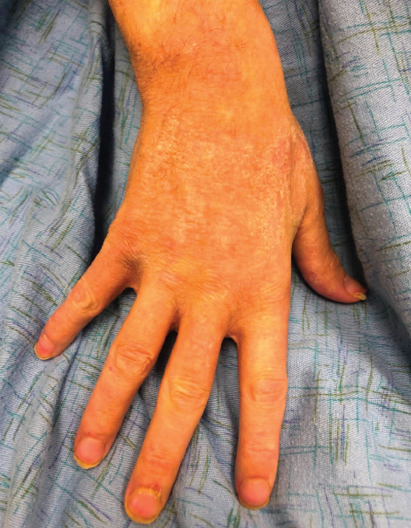
Examination noted well-demarcated pink plaques with cracked scales resembling a riverbed on the scalp, lateral neck, right abdomen, and the dorsal surface of both hands (Figure 1) and forearms, as well as on the feet, inner thighs, and scrotum. There was no involvement of the axillae, interdigital finger and toe web spaces, periungual skin, mons pubis, or umbilicus. Because of the patient’s long-standing history of alcohol use disorder and the presence of chronic rash, malnutrition and nutritional deficiency were suspected.

Sharply demarcated pink plaques on the right dorsal hand with cracked “riverbed” scale.
Laboratory testing revealed the following:
White blood cell count 19.9 × 10⁹/L (reference range 4.5–11)
Hemoglobin 8.6 g/dL (14–18)
Mean corpuscular volume 96.2 fL (80–100)
Serum zinc 35 μg/dL (70–150)
Serum alkaline phosphatase (ALP) 735 IU/L (44–147)
Chromium 0.44 μg/L (≤ 1.4)
Albumin 1.8 g/dL (4.1–5.1)
Serum copper within normal limits
4th-generation human immunodeficiency virus (HIV) antigen/antibody test nonreactive.
The markedly low serum zinc and the characteristic location of the rash in a patient with long-standing alcohol use disorder and cirrhosis confirmed the diagnosis of acrodermatitis enteropathica. The low serum zinc level is diagnostic, but the test may take many days to return a result. ALP is decreased in acrodermatitis enteropathica as zinc is a cofactor for ALP activity. The ALP will return a result sooner than the serum zinc level. In our patient, the ALP was elevated due to his cirrhosis.
TREATMENT
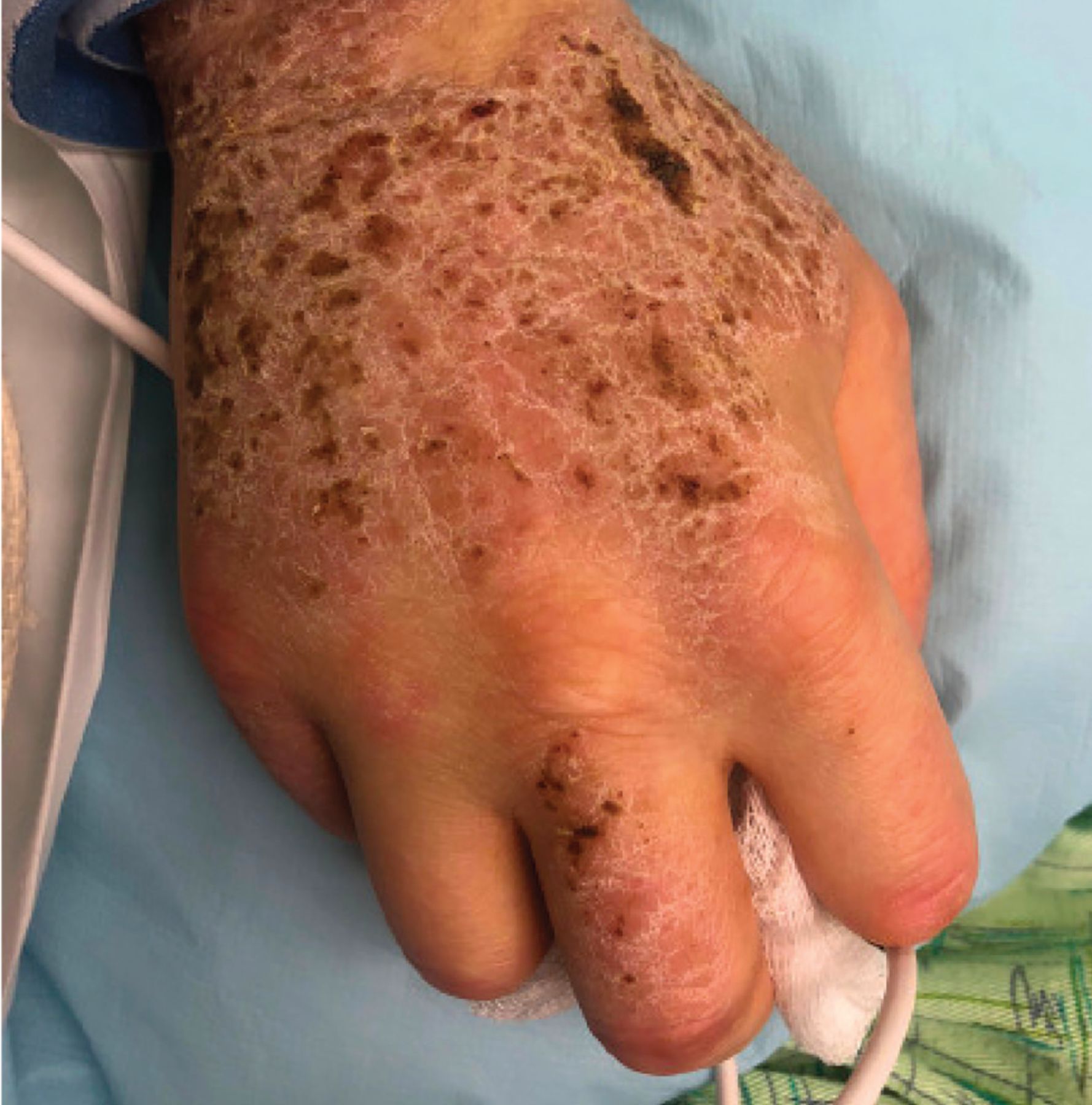
Treatment of acrodermatitis enteropathica typically involves long-term daily zinc supplementation with oral zinc gluconate or zinc sulfate. Our patient was prescribed zinc sulfate 1.5 mg/kg/day. Skin care included topical petrolatum. In 3 weeks, his zinc level had risen from 35 μg/dL to within normal limits at 80 μg/dL, with near resolution of the plaques (Figure 2).
Mild residual light-pink eczematous patches on the right dorsal hand after 3 weeks of oral zinc supplementation.
ACRODERMATITIS ENTEROPATHICA: KEY FEATURES
Acrodermatitis enteropathica is caused by zinc deficiency, and cutaneous findings include sharply demarcated, symmetric erythematous patches and plaques with erosions and scale-crust of the perioral region, genitals, and distal extremities.1 The classically described clinical triad consists of dermatitis, alopecia, and diarrhea, although our patient had no signs of the latter two conditions.
Zinc deficiency may be inherited or acquired.2 Acquired acrodermatitis enteropathica may be secondary to decreased nutritional zinc intake, impaired absorption, or increased gastrointestinal excretion.1 Risk factors for decreased intake include alcoholism, long-term total parental nutrition, vegetarian diets, and diets high in mineral-binding phytates.1,3 Patients with impaired absorption or increased excretion include those with intestinal malabsorption, liver disease, renal disease, Crohn disease, cystic fibrosis, and a history of gastric bypass surgery.2–5
The diagnosis of acquired acrodermatitis enteropathica is largely based on clinical presentation and supported by a serum zinc less than 50 μg/dL. Skin biopsy is rarely necessary but can be diagnostic of a nutritional-deficiency dermatitis that is not specific to acrodermatitis enteropathica. Histopathologic study reveals pallor and ballooning of the keratinocytes in the upper epidermis.6
THE DIFFERENTIAL DIAGNOSIS IN ADULTS
Other diagnoses considered include pellagra, crusted scabies, and an eczematous dermatitis such as atopic or contact dermatitis. Although cracking and fissuring may be seen in patients with chronic eczematous dermatitis, involvement of the distal extremities, face, and groin are classic for acrodermatitis enteropathica. Alcohol use disorder is a common etiology of pellagra, but pellagra typically involves sun-exposed areas, with findings that include a hyperpigmented rash with scale crust and a shiny shellac-like surface along dermatomes C3 and C4 (the Casal collar or necklace).
Crusted scabies is more commonly seen in patients who are immunocompromised, classically in those with HIV. The plaques of crusted scabies are more hyperkeratotic, with a pumice-stone appearance with a distinctive erythema, and they tend to involve the digital interweb spaces, periungual skin, and axillae, which were spared in this patient.
DISCLOSURES
The authors report no relevant financial relationships which, in the context of their contributions, could be perceived as a conflict of interest.
- Copyright © 2022 The Cleveland Clinic Foundation. All Rights Reserved.
In this issue


Jump to section
Related Articles
Cited By...
- No citing articles found.