
Main menu

  • Home
  • Content
    • Current Issue
    • Ahead of Print
    • Past Issues
    • Supplements
    • Article Type
  • Specialty
    • Articles by Specialty
  • CME/MOC
    • Articles
    • Calendar
  • Info For
    • Manuscript Submission
    • Authors & Reviewers
    • Subscriptions
    • About CCJM
    • Contact Us
    • Media Kit
  • Conversations with Leaders
  • Conference Coverage
    • Kidney Week 2024
    • CHEST 2024
    • ACR Convergence 2023
    • Kidney Week 2023
    • ObesityWeek 2023
    • IDWeek 2023
    • CHEST 2023
    • MDS 2023
    • IAS 2023
    • ACP 2023
    • AAN 2023
    • ACC / WCC 2023
    • AAAAI Meeting 2023
    • ACR Convergence 2022
    • Kidney Week 2022
    • AIDS 2022
  • Other Publications
    • www.clevelandclinic.org

User menu

  • Register
  • Log in

Search

  • Advanced search
Cleveland Clinic Journal of Medicine
  • Other Publications
    • www.clevelandclinic.org
  • Register
  • Log in
Cleveland Clinic Journal of Medicine

Advanced Search

  • Home
  • Content
    • Current Issue
    • Ahead of Print
    • Past Issues
    • Supplements
    • Article Type
  • Specialty
    • Articles by Specialty
  • CME/MOC
    • Articles
    • Calendar
  • Info For
    • Manuscript Submission
    • Authors & Reviewers
    • Subscriptions
    • About CCJM
    • Contact Us
    • Media Kit
  • Conversations with Leaders
  • Conference Coverage
    • Kidney Week 2024
    • CHEST 2024
    • ACR Convergence 2023
    • Kidney Week 2023
    • ObesityWeek 2023
    • IDWeek 2023
    • CHEST 2023
    • MDS 2023
    • IAS 2023
    • ACP 2023
    • AAN 2023
    • ACC / WCC 2023
    • AAAAI Meeting 2023
    • ACR Convergence 2022
    • Kidney Week 2022
    • AIDS 2022
The Clinical Picture

Tumoral calcinosis

Hiroyuki Yano, MD and Mitsuyo Kinjo, MD, MPH
Cleveland Clinic Journal of Medicine April 2021, 88 (4) 208-209; DOI: https://doi.org/10.3949/ccjm.88a.20084
Hiroyuki Yano
Division of Rheumatology, Department of Medicine, Okinawa Chubu Hospital, Okinawa, Japan
  • Find this author on Google Scholar
  • Find this author on PubMed
  • Search for this author on this site
Mitsuyo Kinjo
Division of Rheumatology, Department of Medicine, Okinawa Chubu Hospital, Okinawa, Japan
  • Find this author on Google Scholar
  • Find this author on PubMed
  • Search for this author on this site
  • For correspondence: [email protected]
  • Article
  • Figures & Data
  • Info & Metrics
  • PDF
Loading

A 47-year-old woman presented to our hospital with dyspnea and mild hip pain on ambulation. Chest radiography revealed multiple calcified masses on both shoulders.

She had received peritoneal dialysis for 3.5 years because of chronic lupus nephritis. Her prescribed medications included prednisolone 5 mg daily, alfacalcidol 1 μg daily, and evocalcet (a new calcimimetic approved in Japan for secondary hyperparathyroidism in patients undergoing dialysis) 1 mg daily. She had an unremarkable family medical history, and she did not use tobacco or alcohol.

On physical examination, the jugular veins were distended, and heart and lung auscultation revealed an S3 gallop and bibasilar crackles. There were no palpable masses or tenderness of the shoulders, but range of motion of hip joints was limited.

Results of laboratory studies were as follows:

  • Average serum calcium 7.5 mg/dL (reference range 8.8–10.1)

  • Phosphate 4.8 mg/dL (2.7–4.6)

  • Blood urea nitrogen 35 mg/dL (8–20)

  • Serum creatinine 6.9 mg/dL (0.46–0.79)

  • Total protein 5.5 g/dL (6.6–8.1)

  • Albumin 2.7 g/dL (4.1–5.1)

  • Intact parathyroid hormone 390 pg/mL (10–60)

  • Normal white blood cell count.

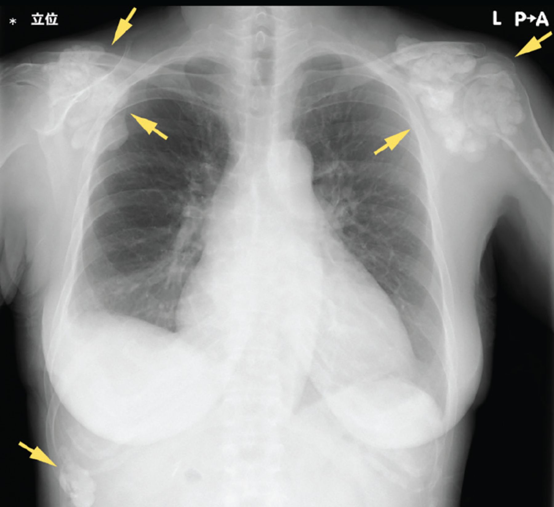
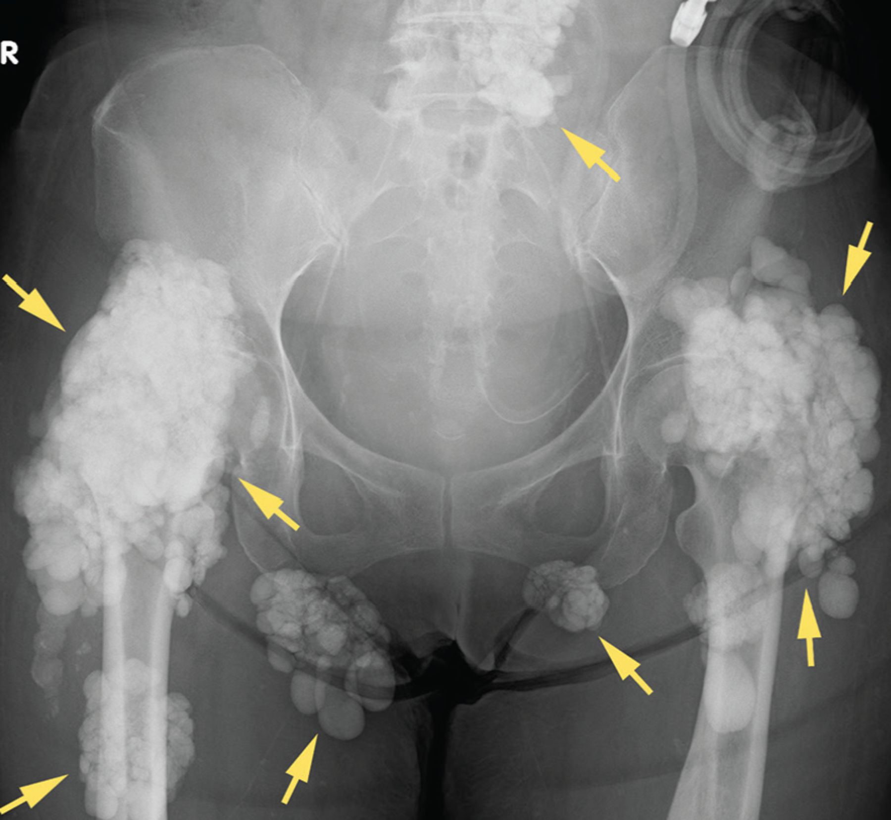
Chest and hip radiographs demonstrated cardiomegaly with pulmonary congestion, as well as large, irregular, calcified masses adjacent to both shoulders, below the right lower ribs (Figure 1), on the left side of the lower lumbar spine, at the hip joints, and superimposed over both ischial tuberosities (Figure 2).

Figure 1
  • Download figure
  • Open in new tab
  • Download powerpoint
Figure 1

Chest radiography showed large, irregular, calcified masses (arrows) adjacent to the shoulders and beneath the right lower ribs.

Figure 2
  • Download figure
  • Open in new tab
  • Download powerpoint
Figure 2

Pelvic radiography showed large,irregular, calcified masses (arrows) adjacent to the left lower lumbar spine, hip joints, and over both ischial tuberosities.

Multiple calcified masses on radiography suggested tumoral calcinosis, the differential diagnosis of which includes calcific tendonitis, synovial osteochondromatosis, and osteosarcoma. Extensive calcification beyond the tendons and synovia of multiple joints made these conditions less likely, and osteosarcoma usually affects children and adolescents.1 In our patient’s case, the diagnosis was tumoral calcinosis associated with chronic kidney disease, along with congestive heart failure.

She began temporary hemodialysis, which improved her heart failure symptoms, and then peritoneal dialysis was reintroduced with an increase in frequency. Her evocalcet was increased to 2 mg daily.

FEATURES OF TUMORAL CALCINOSIS

Tumoral calcinosis is an uncommon disorder characterized by calcium salt deposition around large joints, resulting in irregular, large, lobulated calcified masses.2

Histologic examination of the mass demonstrates extensive amorphous calcium deposits within a fibrous stroma with giant multinucleated foreign-body cells and inflammatory infiltrates.3

Tumoral calcinosis is divided into primary and secondary types, and chronic kidney disease is the leading cause of the secondary type.4 The incidence of secondary tumoral calcinosis in patients undergoing peritoneal dialysis has been estimated as 1.6%.5

Although the precise mechanism is unknown, risk factors include hypercalcemia, hyperphosphatemia, increased calcium-phosphate product, hyperparathyroidism, calcium-containing phosphate binders, and activated vitamin D.6

Common clinical manifestations are joint tenderness, limited range of motion of joints, and skin ulcerations. In one report, 14% of patients (1 of 7) with secondary tumoral calcinosis had no symptoms.5

TREATMENT OPTIONS

Treatment should focus on correcting risk factors to optimize calcium and phosphate homeostasis. Medical management includes restricting dietary calcium and phosphorus, using low-calcium dialysate, and giving non-calcium-containing phosphate binders.

Excision of calcified masses is indicated in cases refractory to conservative medical care.6 Renal transplant has been reported to produce complete remission.5,6 Parathyroidectomy has been tried but reported to be unsatisfactory.

DISCLOSURES

The authors report no relevant financial relationships which, in the context of their contributions, could be perceived as a potential conflict of interest.

  • Copyright © 2021 The Cleveland Clinic Foundation. All Rights Reserved.

REFERENCES

  1. ↵
    1. Misaghi A,
    2. Goldin A,
    3. Awad M,
    4. Kulidjian AA
    . Osteosarcoma: a comprehensive review. SICOT J 2018; 4:12. doi:10.1051/sicotj/2017028
    OpenUrlCrossRef
  2. ↵
    1. Karaca L,
    2. Polat G,
    3. Sade R,
    4. Yüce I,
    5. Ogul H,
    6. Kantarci M
    . Nontraumatic tumoral calcinosis. Joint Bone Spine 2017; 84(4):491. doi:10.1016/j.jbspin.2016.06.009
    OpenUrlCrossRef
  3. ↵
    1. Chi D,
    2. Kamochi H,
    3. Sunaga A, et al
    . Tumoral calcinosis: a giant tumoral mass in the shoulder joint of a dialysis patient. Plast Reconstr Surg Glob Open 2018; 6(4):e1730. doi:10.1097/GOX.0000000000001730
    OpenUrlCrossRef
  4. ↵
    1. Fathi I,
    2. Sakr M
    . Review of tumoral calcinosis: a rare clinicopathological entity. World J Clin Cases 2014; 2(9):409–414. doi:10.12998/wjcc.v2.i9.409
    OpenUrlCrossRefPubMed
  5. ↵
    1. Chu HY,
    2. Chu P,
    3. Lin YF,
    4. Chou HK,
    5. Lin SH
    . Uremic tumoral calcinosis in patients on peritoneal dialysis: clinical, radiologic, and laboratory features. Perit Dial Int 2011; 31(4):430–439. doi:10.3747/pdi.2009.00250
    OpenUrlAbstract/FREE Full Text
  6. ↵
    1. Zhou H,
    2. Yang M,
    3. Zou Y
    . Uraemic tumoral calcinosis in a peritoneal dialysis patient. Intern Med J 2018; 48(12):1544–1546. doi:10.1111/imj.14130
    OpenUrlCrossRef
PreviousNext
Back to top

In this issue

Cleveland Clinic Journal of Medicine: 88 (4)
Cleveland Clinic Journal of Medicine
Vol. 88, Issue 4
1 Apr 2021
  • Table of Contents
  • Table of Contents (PDF)
  • Index by author
  • Complete Issue (PDF)
Print
Download PDF
Article Alerts
Sign In to Email Alerts with your Email Address
Email Article

Thank you for your interest in spreading the word on Cleveland Clinic Journal of Medicine.

NOTE: We only request your email address so that the person you are recommending the page to knows that you wanted them to see it, and that it is not junk mail. We do not capture any email address.

Enter multiple addresses on separate lines or separate them with commas.
Tumoral calcinosis
(Your Name) has sent you a message from Cleveland Clinic Journal of Medicine
(Your Name) thought you would like to see the Cleveland Clinic Journal of Medicine web site.
CAPTCHA
This question is for testing whether or not you are a human visitor and to prevent automated spam submissions.
Citation Tools
Tumoral calcinosis
Hiroyuki Yano, Mitsuyo Kinjo
Cleveland Clinic Journal of Medicine Apr 2021, 88 (4) 208-209; DOI: 10.3949/ccjm.88a.20084

Citation Manager Formats

  • BibTeX
  • Bookends
  • EasyBib
  • EndNote (tagged)
  • EndNote 8 (xml)
  • Medlars
  • Mendeley
  • Papers
  • RefWorks Tagged
  • Ref Manager
  • RIS
  • Zotero
Share
Tumoral calcinosis
Hiroyuki Yano, Mitsuyo Kinjo
Cleveland Clinic Journal of Medicine Apr 2021, 88 (4) 208-209; DOI: 10.3949/ccjm.88a.20084
Twitter logo Facebook logo Mendeley logo
  • Tweet Widget
  • Facebook Like
  • Linkedin Share Button

Jump to section

  • Article
    • FEATURES OF TUMORAL CALCINOSIS
    • TREATMENT OPTIONS
    • DISCLOSURES
    • REFERENCES
  • Figures & Data
  • Info & Metrics
  • PDF

Related Articles

  • No related articles found.
  • PubMed
  • Google Scholar

Cited By...

  • Familial tumoral calcinosis: a rare autosomal recessive disease
  • Extraosseous calcification in kidney disease
  • Google Scholar

More in this TOC Section

  • Tinea incognito
  • Prolonged venous filling time and dependent rubor in a patient with peripheral artery disease
  • Sarcoidosis with diffuse purplish erythematous plaques on the hands
Show more The Clinical Picture

Similar Articles

Subjects

  • Cardiology
  • Endocrinology
  • Imaging
  • Nephrology
  • Pulmonology
  • Rheumatology

Navigate

  • Current Issue
  • Past Issues
  • Supplements
  • Article Type
  • Specialty
  • CME/MOC Articles
  • CME/MOC Calendar
  • Media Kit

Authors & Reviewers

  • Manuscript Submission
  • Authors & Reviewers
  • Subscriptions
  • About CCJM
  • Contact Us
  • Cleveland Clinic Center for Continuing Education
  • Consult QD

Share your suggestions!

Copyright © 2025 The Cleveland Clinic Foundation. All rights reserved. The information provided is for educational purposes only. Use of this website is subject to the website terms of use and privacy policy. 

Powered by HighWire