
Main menu

  • Home
  • Content
    • Current Issue
    • Ahead of Print
    • Past Issues
    • Supplements
    • Article Type
  • Specialty
    • Articles by Specialty
  • CME/MOC
    • Articles
    • Calendar
  • Info For
    • Manuscript Submission
    • Authors & Reviewers
    • Subscriptions
    • About CCJM
    • Contact Us
    • Media Kit
  • Conversations with Leaders
  • Conference Coverage
    • Kidney Week 2024
    • CHEST 2024
    • ACR Convergence 2023
    • Kidney Week 2023
    • ObesityWeek 2023
    • IDWeek 2023
    • CHEST 2023
    • MDS 2023
    • IAS 2023
    • ACP 2023
    • AAN 2023
    • ACC / WCC 2023
    • AAAAI Meeting 2023
    • ACR Convergence 2022
    • Kidney Week 2022
    • AIDS 2022
  • Other Publications
    • www.clevelandclinic.org

User menu

  • Register
  • Log in

Search

  • Advanced search
Cleveland Clinic Journal of Medicine
  • Other Publications
    • www.clevelandclinic.org
  • Register
  • Log in
Cleveland Clinic Journal of Medicine

Advanced Search

  • Home
  • Content
    • Current Issue
    • Ahead of Print
    • Past Issues
    • Supplements
    • Article Type
  • Specialty
    • Articles by Specialty
  • CME/MOC
    • Articles
    • Calendar
  • Info For
    • Manuscript Submission
    • Authors & Reviewers
    • Subscriptions
    • About CCJM
    • Contact Us
    • Media Kit
  • Conversations with Leaders
  • Conference Coverage
    • Kidney Week 2024
    • CHEST 2024
    • ACR Convergence 2023
    • Kidney Week 2023
    • ObesityWeek 2023
    • IDWeek 2023
    • CHEST 2023
    • MDS 2023
    • IAS 2023
    • ACP 2023
    • AAN 2023
    • ACC / WCC 2023
    • AAAAI Meeting 2023
    • ACR Convergence 2022
    • Kidney Week 2022
    • AIDS 2022
The Clinical Picture

A large, painless bulla on the right foot

Jorge Camilo Mora, MD, MPH and Amalia Galindez, MD, FACP
Cleveland Clinic Journal of Medicine January 2022, 89 (1) 10-12; DOI: https://doi.org/10.3949/ccjm.89a.21051
Jorge Camilo Mora
Associate Professor, Associate Dean for Faculty Affairs, Director of Geriatric Medicine, Department of Translational Medicine, Herbert Wertheim FIU College of Medicine, Miami, FL
  • Find this author on Google Scholar
  • Find this author on PubMed
  • Search for this author on this site
  • For correspondence: [email protected]
Amalia Galindez
Clerkship Director and Chief, Division of Internal Medicine; Interim Chair, Department of Translational Medicine, Herbert Wertheim FIU College of Medicine, Miami, FL
  • Find this author on Google Scholar
  • Find this author on PubMed
  • Search for this author on this site
  • Article
  • Figures & Data
  • Info & Metrics
  • PDF
Loading

A 71-year-old woman with a history of diabetic neuropathy presented with a large, painless bulla on her right foot. She said it had started as a small vesicle but had slowly enlarged over the past 3 days. She had been working in her yard for several days prior to the development of the vesicle but did not sustain any insect bites or local trauma. She reported no fever or chills, and she had no history of skin vesicles or bullae or similar lesions on other parts of her body. No new medications had been recently prescribed.

Her medications at the time of presentation included metformin extended-release 500 mg once daily, insulin lispro protamine–insulin lispro (75/25) 35 units twice daily, anastrozole 1 mg daily, and gabapentin 600 mg twice daily. She was not employed, mostly stayed at home, and had no history of smoking or alcohol or drug use. Comorbidities included breast cancer treated with chemotherapy and radiotherapy 1 year previously, type 2 diabetes mellitus with peripheral neuropathy, and hyper lipidemia.

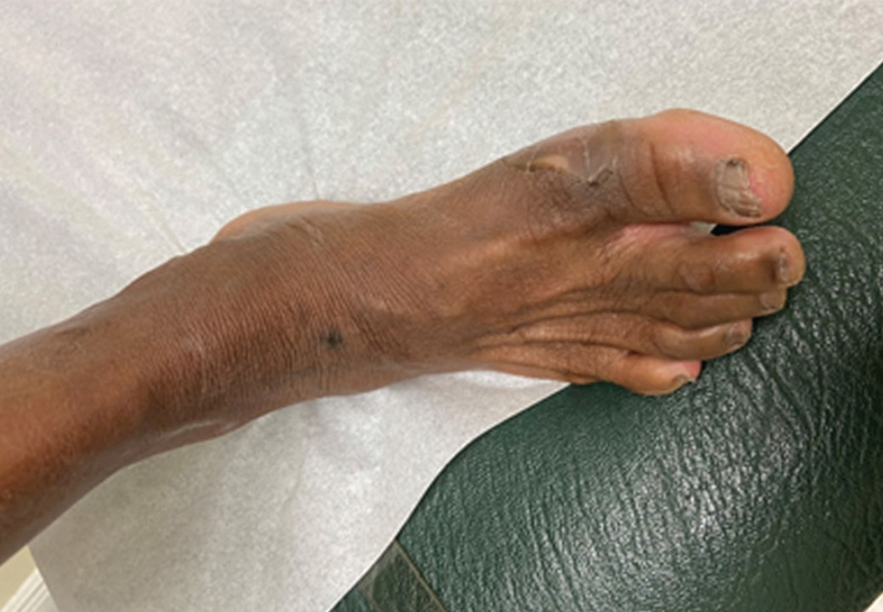
On physical examination, the patient’s vital signs were normal, and examination of the extremities revealed a large, tense, noninflammatory bulla at the base of the first metatarsal joint of the right foot, 3.75 cm in diameter and containing clear fluid (Figure 1). There were no other lesions on the extremities or trunk and no signs of local inflammation or spontaneous drainage. She had decreased perception to monofilament on both feet with normal peripheral pulses. As the bulla contained clear fluid, it was presumed to be sterile and thus, to avoid the risk of secondary infection, aspiration was not attempted. At a 4-week follow-up visit, the lesion had completely resolved, and the patient did not report any recurrence (Figure 2).

Figure 1
  • Download figure
  • Open in new tab
  • Download powerpoint
Figure 1

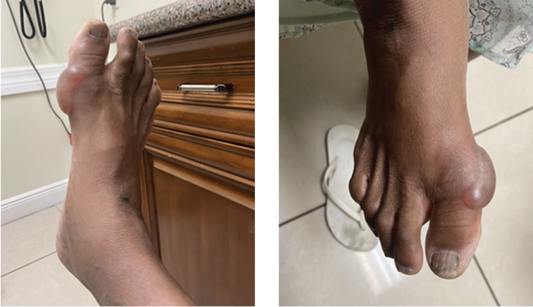
A large, tense, noninflammatory bulla 3.75 cm in diameter at the base of the first metatarsal joint of the right foot. The bulla contained clear fluid.

Figure 2
  • Download figure
  • Open in new tab
  • Download powerpoint
Figure 2

At a 4-week follow-up visit, the lesion had completely resolved with no signs of recurrence.

Laboratory testing during the follow-up visit revealed a hemoglobin A1c of 10.3% (reference range < 5.7%), serum creatinine 0.87 mg/dL (reference range 0.60–0.93 mg/dL), and albumin-to-creatinine ratio 12 μg/mg creatinine (reference range < 30 μg/creatinine). Results of other laboratory testing were normal, including complete blood cell count, alanine transaminase, aspartate transaminase, total protein, and albumin.

BULLOUS DISEASE OF DIABETES

Bullosis diabeticorum is a noninflammatory blistering condition that affects patients with prediabetes and diabetes.1 The condition was first reported by Kramer in 1930, and the name was coined by Cantwell and Martz in 1967.2 The blisters are tense with serous content. They are recurrent and spontaneous around acral regions, particularly on the lower extremities, including the toes and plantar area of the feet. They can also be present on the hands and forearms. They seldom present on the trunk.3–7 Vesicles and bullae can range in size from 0.5 cm to several centimeters. It is a rare disease, affecting approximately 0.5% of diabetic patients. Men are twice as likely as women to develop the condition.

Because bullosis diabeticorum mostly affects patients with diabetes mellitus who have associated nephropathy and neuropathy, it has been postulated that microangiopathy and disturbances in carbohydrate metabolism may lead to premature aging of connective tissue and other structural alterations in connective tissue,8 increasing the risk of spontaneous appearance of these lesions.2 The lesions are usually self-limited and heal within 4 to 6 weeks without scarring.5

Treatment is conservative, and the blister should be kept clean and protected to avoid secondary infection. No antibiotic therapy is required, although fluid aspiration and culture with use of oral antibiotics may be considered if secondary bacterial infection is suspected.6 Signs of possible infection include erythema, warmth, edema with surrounding inflammation, the presence of purulent fluid or drainage, and fever. The etiology is unclear.

The differential diagnosis for bullosis diabeticorum includes insect bite reaction, contact dermatitis with bullae, poison ivy dermatitis, bullous drug eruption, bullous impetigo, edema bullae, burns, and friction blister.7 Unlike friction blisters, bullosis diabeticorum often develops overnight and in the absence of known trauma.2 Friction blisters are generally associated with high levels of exercise and activity and are often seen in active military personnel or athletes with no underlying health issues, whereas bullosis diabeticorum commonly presents in the absence of foot trauma or pressure in vulnerable populations. Although rare, recurrent episodes leading to severe infection and ulceration have been described, with some bullae recurring years after the initial appearance.9

TAKE-HOME POINTS

Bullosis diabeticorum may be the first clinical presentation of hyperglycemia.2 Bullosis diabeticorum in patients with diabetes often signals poor diabetes control or the existence of associated neuropathy or nephropathy. Patients with this condition should be routinely screened for diabetes and for microscopic proteinuria and assessed for findings of peripheral neuropathy. Supportive care of the lesions with monitoring for signs of secondary infection is advised.

DISCLOSURES

The authors report no relevant financial relationships which, in the context of their contributions, could be perceived as a potential conflict of interest.

  • Copyright © 2022 The Cleveland Clinic Foundation. All Rights Reserved.

REFERENCES

  1. ↵
    1. Lopez PR,
    2. Leicht S,
    3. Sigmon JR,
    4. Stigall L
    . Bullosis diabeticorum associated with a prediabetic state. South Med J 2009; 102(6):643–644. doi:10.1097/SMJ.0b013e3181a506d6
    OpenUrlCrossRefPubMed
  2. ↵
    1. Chouk C,
    2. Litaiem N
    . Bullosis diabeticorum. In: StatPearls. Treasure Island, FL: StatPearls Publishing; 2021. https://www.ncbi.nlm.nih.gov/books/NBK539872/. Accessed December 7, 2021.
  3. ↵
    1. Mota AN,
    2. Nery NS,
    3. Barcaui CB
    . Case for diagnosis: bullosis diabeticorum. An Bras Dermatol 2013; 88(4):652–654. doi:10.1590/abd1806-4841.20132114
    OpenUrlCrossRef
    1. Oursler JR,
    2. Goldblum OM
    . Blistering eruption in a diabetic. Bullosis diabeticorum. Arch Dermatol 1991; 127(2):247–248. doi:10.1001/archderm.1991.01680020115017
    OpenUrlCrossRefPubMed
  4. ↵
    1. Bernstein JE,
    2. Medenica M,
    3. Soltani K,
    4. Griem SF
    . Bullous eruption of diabetes mellitus. Arch Dermatol 1979; 115(3):324–325. pmid:373635
    OpenUrlCrossRefPubMed
  5. ↵
    1. Lipsky BA,
    2. Baker PD,
    3. Ahroni JH
    . Diabetic bullae: 12 cases of a purportedly rare cutaneous disorder. Int J Dermatol 2000; 39(3): 196–200. doi:10.1046/j.1365-4362.2000.00947.x
    OpenUrlCrossRefPubMed
  6. ↵
    1. Goldsmith LA
    1. Zampella J,
    2. Craft N,
    3. Fox L,
    4. Goldsmith L
    . Bullosis diabeticorum. In: Goldsmith LA, ed. VisualDx. Rochester, NY: VisualDx; 2010.
  7. ↵
    1. Braverman IM,
    2. Keh-Yen A
    . Ultrastructural abnormalities of the microvasculature and elastic fibers in the skin of juvenile diabetics. J Invest Dermatol 1984; 82(3):270–274. doi:10.1111/1523-1747.ep12260279
    OpenUrlCrossRefPubMed
  8. ↵
    1. Bhutani R,
    2. Walton S
    . Diabetic bullae. Br J Diabetes Vasc Dis 2015; 15(1):8–10. http://dx.doi.org/10.15277/bjdvd.2015.004
    OpenUrl
PreviousNext
Back to top

In this issue

Cleveland Clinic Journal of Medicine: 89 (1)
Cleveland Clinic Journal of Medicine
Vol. 89, Issue 1
1 Jan 2022
  • Table of Contents
  • Table of Contents (PDF)
  • Index by author
  • Complete Issue (PDF)
Print
Download PDF
Article Alerts
Sign In to Email Alerts with your Email Address
Email Article

Thank you for your interest in spreading the word on Cleveland Clinic Journal of Medicine.

NOTE: We only request your email address so that the person you are recommending the page to knows that you wanted them to see it, and that it is not junk mail. We do not capture any email address.

Enter multiple addresses on separate lines or separate them with commas.
A large, painless bulla on the right foot
(Your Name) has sent you a message from Cleveland Clinic Journal of Medicine
(Your Name) thought you would like to see the Cleveland Clinic Journal of Medicine web site.
CAPTCHA
This question is for testing whether or not you are a human visitor and to prevent automated spam submissions.
Citation Tools
A large, painless bulla on the right foot
Jorge Camilo Mora, Amalia Galindez
Cleveland Clinic Journal of Medicine Jan 2022, 89 (1) 10-12; DOI: 10.3949/ccjm.89a.21051

Citation Manager Formats

  • BibTeX
  • Bookends
  • EasyBib
  • EndNote (tagged)
  • EndNote 8 (xml)
  • Medlars
  • Mendeley
  • Papers
  • RefWorks Tagged
  • Ref Manager
  • RIS
  • Zotero
Share
A large, painless bulla on the right foot
Jorge Camilo Mora, Amalia Galindez
Cleveland Clinic Journal of Medicine Jan 2022, 89 (1) 10-12; DOI: 10.3949/ccjm.89a.21051
Twitter logo Facebook logo Mendeley logo
  • Tweet Widget
  • Facebook Like
  • Linkedin Share Button

Jump to section

  • Article
    • BULLOUS DISEASE OF DIABETES
    • TAKE-HOME POINTS
    • DISCLOSURES
    • REFERENCES
  • Figures & Data
  • Info & Metrics
  • PDF

Related Articles

  • Megaloblastic anemia due to severe vitamin B12 deficiency
  • PubMed
  • Google Scholar

Cited By...

  • No citing articles found.
  • Google Scholar

More in this TOC Section

  • Tinea incognito
  • Prolonged venous filling time and dependent rubor in a patient with peripheral artery disease
  • Sarcoidosis with diffuse purplish erythematous plaques on the hands
Show more The Clinical Picture

Similar Articles

Subjects

  • Dermatology
  • Diabetes
  • Endocrinology
  • Geriatrics
  • Neurology
  • Preventive Care

Navigate

  • Current Issue
  • Past Issues
  • Supplements
  • Article Type
  • Specialty
  • CME/MOC Articles
  • CME/MOC Calendar
  • Media Kit

Authors & Reviewers

  • Manuscript Submission
  • Authors & Reviewers
  • Subscriptions
  • About CCJM
  • Contact Us
  • Cleveland Clinic Center for Continuing Education
  • Consult QD

Share your suggestions!

Copyright © 2025 The Cleveland Clinic Foundation. All rights reserved. The information provided is for educational purposes only. Use of this website is subject to the website terms of use and privacy policy. 

Powered by HighWire