
Main menu

  • Home
  • Content
    • Current Issue
    • Ahead of Print
    • Past Issues
    • Supplements
    • Article Type
  • Specialty
    • Articles by Specialty
  • CME/MOC
    • Articles
    • Calendar
  • Info For
    • Manuscript Submission
    • Authors & Reviewers
    • Subscriptions
    • About CCJM
    • Contact Us
    • Media Kit
  • Conversations with Leaders
  • Conference Coverage
    • ACC / WCC 2023
    • AAAAI Meeting 2023
    • ACR Convergence 2022
    • Kidney Week 2022
    • AIDS 2022
    • CHEST 2021
    • IDWeek 2021
    • IAS 2021
    • ADA 2021
    • ATS 2021
    • ACC 2021
    • ACP 2021
    • AAN 2021
  • Other Publications
    • www.clevelandclinic.org

User menu

  • Register
  • Log in

Search

  • Advanced search
Cleveland Clinic Journal of Medicine
  • Other Publications
    • www.clevelandclinic.org
  • Register
  • Log in
Cleveland Clinic Journal of Medicine

Advanced Search

  • Home
  • Content
    • Current Issue
    • Ahead of Print
    • Past Issues
    • Supplements
    • Article Type
  • Specialty
    • Articles by Specialty
  • CME/MOC
    • Articles
    • Calendar
  • Info For
    • Manuscript Submission
    • Authors & Reviewers
    • Subscriptions
    • About CCJM
    • Contact Us
    • Media Kit
  • Conversations with Leaders
  • Conference Coverage
    • ACC / WCC 2023
    • AAAAI Meeting 2023
    • ACR Convergence 2022
    • Kidney Week 2022
    • AIDS 2022
    • CHEST 2021
    • IDWeek 2021
    • IAS 2021
    • ADA 2021
    • ATS 2021
    • ACC 2021
    • ACP 2021
    • AAN 2021
The Clinical Picture

Oral leukoplakia and oral cancer

Takeshi Onda, DDS, PhD, Kamichika Hayashi, DDS, PhD, Akira Katakura, DDS, PhD and Masayuki Takano, DDS, PhD
Cleveland Clinic Journal of Medicine February 2023, 90 (2) 79-80; DOI: https://doi.org/10.3949/ccjm.90a.22044
Takeshi Onda
Department of Oral and Maxillofacial Surgery, Tokyo Dental College, Tokyo, Japan
  • Find this author on Google Scholar
  • Find this author on PubMed
  • Search for this author on this site
  • For correspondence: ondatake@tdc.ac.jp
Kamichika Hayashi
Department of Oral and Maxillofacial Surgery, Tokyo Dental College, Tokyo, Japan
  • Find this author on Google Scholar
  • Find this author on PubMed
  • Search for this author on this site
Akira Katakura
Department of Oral Pathological Science and Surgery, Tokyo Dental College, Tokyo, Japan
  • Find this author on Google Scholar
  • Find this author on PubMed
  • Search for this author on this site
Masayuki Takano
Department of Oral and Maxillofacial Surgery, Tokyo Dental College, Tokyo, Japan
  • Find this author on Google Scholar
  • Find this author on PubMed
  • Search for this author on this site
  • Article
  • Figures & Data
  • Info & Metrics
  • PDF
Loading

A 56-year-old male was referred by his dentist with noticeable worsening of vitiligo of the tongue, which had been diagnosed as oral leukoplakia 3 years earlier and had been monitored at the dental clinic.

The patient had no significant medical history. He had a continuous smoking habit of more than 36 years but did not consume alcohol. Examination confirmed oral leukoplakia, with an uneven and rough surface, and a white papillary mass was noted within the area of leukoplakia on the right edge of the tongue (Figure 1). No lymph node swelling in the neck was noted. Biopsy of the lesion confirmed it to be squamous cell carcinoma. Computed tomography, magnetic resonance imaging, and ultrasonography showed that the mass was confined to the surface of the tongue.

Figure 1
  • Download figure
  • Open in new tab
  • Download powerpoint
Figure 1

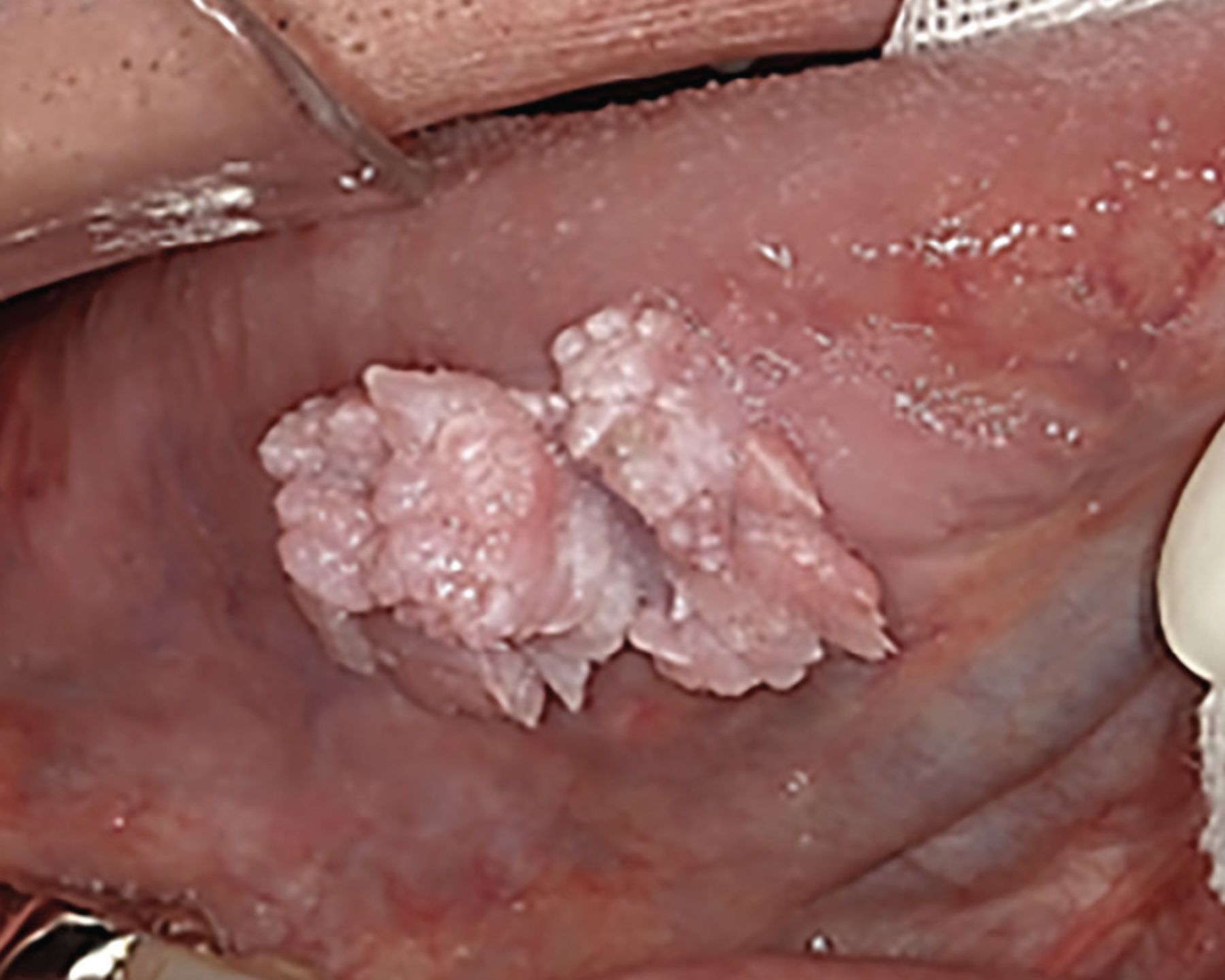
White lesions were observed on the right tongue edge. Papillary cell proliferation was noted in the same area.

TREATMENT AND FOLLOW-UP

Resection of the mass with at least 10 mm of surrounding tissue was performed under general anesthesia. During surgery, rapid pathological diagnosis was performed to confirm that no tumor cells remained, and the wound was subsequently sutured. Postoperatively, a slight deformation of the tongue and scarring were noted, affecting the patient’s eating, swallowing, and pronunciation, but these functions gradually improved. At 5 years postoperatively, the patient’s clinical course was favorable, without recurrence.

PREMALIGNANT ORAL LESIONS

Tongue cancer accounts for 60% of all oral cancers, and usually originates from the tongue border.1 Nearly 90% of all oral cancers are squamous cell carcinoma, and major risk factors are chronic irritation, smoking, and alcohol consumption.2

Most oral cancers have a premalignant lesion stage.3 Regular monitoring for progression of premalignant lesions is critical for the early detection and treatment of oral cancer. Oral leukoplakia, the most common potentially cancerous oral lesion, progresses to squamous cell carcinoma at a rate ranging from 0.1% to 36.4%.4 This transformation depends on factors such as sex, age, clinical type, locus, onset mode, and the presence or absence of epithelial atypia,5 although the mechanism remains unclear to date.

Currently, no clear guidelines exist as to whether aggressive resection or progression monitoring produces better outcomes. Consequently, there is an urgent and unmet need for molecular biological investigation of leukoplakia.

IMPORTANCE OF REGULAR FOLLOW-UP

Our report illustrates the importance of regular follow-up of leukoplakia. Before our patient presented, he had been followed regularly by his dentist, and this led to earlier recognition of possible malignant transformation, resulting in earlier resection of the cancer and better prognosis.

DISCLOSURES

The authors report no relevant financial relationships which, in the context of their contributions, could be perceived as a potential conflict of interest.

  • Copyright © 2023 The Cleveland Clinic Foundation. All Rights Reserved.

REFERENCES

  1. ↵
    1. Omura K
    . Current status of oral cancer treatment strategies: surgical treatments for oral squamous cell carcinoma. Int J Clin Oncol 2014; 19(3):423–430. doi:10.1007/s10147-014-0689-z
    OpenUrlCrossRefPubMed
  2. ↵
    1. Abati S,
    2. Bramati C,
    3. Bondi S,
    4. Lissoni A,
    5. Trimarchi M
    . Oral cancer and precancer: a narrative review on the relevance of early diagnosis. Int J Environ Res Public Health 2020; 17(24):9160. doi:10.3390/ijerph17249160
    OpenUrlCrossRefPubMed
  3. ↵
    1. Yang SW,
    2. Lee YS,
    3. Chang LC,
    4. Yang CH,
    5. Luo CM,
    6. Wu PW
    . Oral tongue leukoplakia: analysis of clinicopathological characteristics, treatment outcomes, and factors related to recurrence and malignant transformation. Clin Oral Investig 2021; 25(6):4045–4058. doi:10.1007/s00784-020-03735-1
    OpenUrlCrossRef
  4. ↵
    1. Evren I,
    2. Brouns ER,
    3. Poell JB, et al
    . Associations between clinical and histopathological characteristics in oral leukoplakia. Oral Dis 2021; 10.1111/odi.14038. doi:10.1111/odi.14038
    OpenUrlCrossRef
  5. ↵
    1. Speight PM,
    2. Khurram SA,
    3. Kujan O
    . Oral potentially malignant disorders: risk of progression to malignancy. Oral Surg Oral Med Oral Pathol Oral Radiol 2018; 125(6):612–627. doi:10.1016/j.oooo.2017.12.011
    OpenUrlCrossRefPubMed
PreviousNext
Back to top

In this issue

Cleveland Clinic Journal of Medicine: 90 (2)
Cleveland Clinic Journal of Medicine
Vol. 90, Issue 2
1 Feb 2023
  • Table of Contents
  • Table of Contents (PDF)
  • Index by author
  • Complete Issue (PDF)
Print
Download PDF
Article Alerts
Sign In to Email Alerts with your Email Address
Email Article

Thank you for your interest in spreading the word on Cleveland Clinic Journal of Medicine.

NOTE: We only request your email address so that the person you are recommending the page to knows that you wanted them to see it, and that it is not junk mail. We do not capture any email address.

Enter multiple addresses on separate lines or separate them with commas.
Oral leukoplakia and oral cancer
(Your Name) has sent you a message from Cleveland Clinic Journal of Medicine
(Your Name) thought you would like to see the Cleveland Clinic Journal of Medicine web site.
CAPTCHA
This question is for testing whether or not you are a human visitor and to prevent automated spam submissions.
Citation Tools
Oral leukoplakia and oral cancer
Takeshi Onda, Kamichika Hayashi, Akira Katakura, Masayuki Takano
Cleveland Clinic Journal of Medicine Feb 2023, 90 (2) 79-80; DOI: 10.3949/ccjm.90a.22044

Citation Manager Formats

  • BibTeX
  • Bookends
  • EasyBib
  • EndNote (tagged)
  • EndNote 8 (xml)
  • Medlars
  • Mendeley
  • Papers
  • RefWorks Tagged
  • Ref Manager
  • RIS
  • Zotero
Share
Oral leukoplakia and oral cancer
Takeshi Onda, Kamichika Hayashi, Akira Katakura, Masayuki Takano
Cleveland Clinic Journal of Medicine Feb 2023, 90 (2) 79-80; DOI: 10.3949/ccjm.90a.22044
Reddit logo Twitter logo Facebook logo Mendeley logo
  • Tweet Widget
  • Facebook Like
  • Linkedin Share Button

Jump to section

  • Article
    • TREATMENT AND FOLLOW-UP
    • PREMALIGNANT ORAL LESIONS
    • IMPORTANCE OF REGULAR FOLLOW-UP
    • DISCLOSURES
    • REFERENCES
  • Figures & Data
  • Info & Metrics
  • PDF

Related Articles

  • No related articles found.
  • PubMed
  • Google Scholar

Cited By...

  • No citing articles found.
  • Google Scholar

More in this TOC Section

  • Mass under the lip
  • Median rhomboid glossitis caused by tongue-brushing
Show more The Clinical Picture

Similar Articles

Subjects

  • Physical Exam
  • Oncology
  • Dermatology

Navigate

  • Current Issue
  • Past Issues
  • Supplements
  • Article Type
  • Specialty
  • CME/MOC Articles
  • CME/MOC Calendar
  • Media Kit

Authors & Reviewers

  • Manuscript Submission
  • Authors & Reviewers
  • Subscriptions
  • About CCJM
  • Contact Us
  • Cleveland Clinic Center for Continuing Education
  • Consult QD

Share your suggestions!

Copyright © 2023 The Cleveland Clinic Foundation. All rights reserved. The information provided is for educational purposes only. Use of this website is subject to the website terms of use and privacy policy. 

Powered by HighWire