
Main menu

  • Home
  • Content
    • Current Issue
    • Ahead of Print
    • Past Issues
    • Supplements
    • Article Type
  • Specialty
    • Articles by Specialty
  • CME/MOC
    • Articles
    • Calendar
  • Info For
    • Manuscript Submission
    • Authors & Reviewers
    • Subscriptions
    • About CCJM
    • Contact Us
    • Media Kit
  • Conversations with Leaders
  • Conference Coverage
    • Kidney Week 2024
    • CHEST 2024
    • ACR Convergence 2023
    • Kidney Week 2023
    • ObesityWeek 2023
    • IDWeek 2023
    • CHEST 2023
    • MDS 2023
    • IAS 2023
    • ACP 2023
    • AAN 2023
    • ACC / WCC 2023
    • AAAAI Meeting 2023
    • ACR Convergence 2022
    • Kidney Week 2022
    • AIDS 2022
  • Other Publications
    • www.clevelandclinic.org

User menu

  • Register
  • Log in

Search

  • Advanced search
Cleveland Clinic Journal of Medicine
  • Other Publications
    • www.clevelandclinic.org
  • Register
  • Log in
Cleveland Clinic Journal of Medicine

Advanced Search

  • Home
  • Content
    • Current Issue
    • Ahead of Print
    • Past Issues
    • Supplements
    • Article Type
  • Specialty
    • Articles by Specialty
  • CME/MOC
    • Articles
    • Calendar
  • Info For
    • Manuscript Submission
    • Authors & Reviewers
    • Subscriptions
    • About CCJM
    • Contact Us
    • Media Kit
  • Conversations with Leaders
  • Conference Coverage
    • Kidney Week 2024
    • CHEST 2024
    • ACR Convergence 2023
    • Kidney Week 2023
    • ObesityWeek 2023
    • IDWeek 2023
    • CHEST 2023
    • MDS 2023
    • IAS 2023
    • ACP 2023
    • AAN 2023
    • ACC / WCC 2023
    • AAAAI Meeting 2023
    • ACR Convergence 2022
    • Kidney Week 2022
    • AIDS 2022
Symptoms to Diagnosis

Unilateral green pleural effusion in a 22-year-old woman

Sauradeep Sarkar, MD, Utsav Shrestha, MD, Huda Elzahrany, MD and Bathmapriya Balakrishnan, BMedSci, BMBS, FCCP
Cleveland Clinic Journal of Medicine August 2023, 90 (8) 491-498; DOI: https://doi.org/10.3949/ccjm.90a.22097
Sauradeep Sarkar
Pulmonary and Critical Care Fellow, Department of Medicine, Section of Pulmonary, Critical Care, and Sleep Medicine, West Virginia University School of Medicine, Morgantown, WV
  • Find this author on Google Scholar
  • Find this author on PubMed
  • Search for this author on this site
Utsav Shrestha
Pulmonary and Critical Care Fellow, Department of Medicine, Section of Pulmonary, Critical Care, and Sleep Medicine, West Virginia University School of Medicine, Morgantown, WV
  • Find this author on Google Scholar
  • Find this author on PubMed
  • Search for this author on this site
Huda Elzahrany
Pathology Resident, Anatomy and Laboratory Medicine, West Virginia University School of Medicine, Morgantown, WV
  • Find this author on Google Scholar
  • Find this author on PubMed
  • Search for this author on this site
Bathmapriya Balakrishnan
Assistant Professor, Department of Medicine, Section of Pulmonary, Critical Care, and Sleep Medicine, West Virginia University School of Medicine, Morgantown, WV
  • Find this author on Google Scholar
  • Find this author on PubMed
  • Search for this author on this site
  • For correspondence: [email protected]
  • Article
  • Figures & Data
  • Info & Metrics
  • PDF
Loading

Article Figures & Data

Figures

  • Tables
  • Figure 1
    • Download figure
    • Open in new tab
    • Download powerpoint
    Figure 1

    Posterior-anterior chest radiography showed a widened mediastinum (yellow arrow) and right unilateral pleural effusion (red arrow).
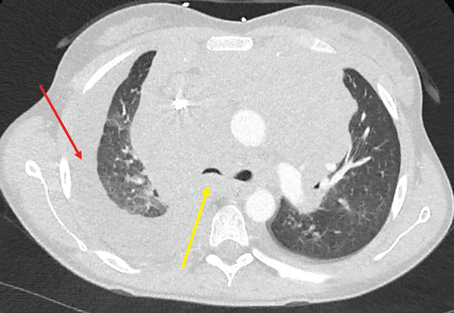
  • Figure 2
    • Download figure
    • Open in new tab
    • Download powerpoint
    Figure 2

    Computed tomography of the chest with contrast showed atelectasis of the right upper and middle lobes secondary to extrinsic compression of the right mainstem by mediastinal lymphadenopathy (yellow arrow) and right unilateral pleural effusion (red arrow).
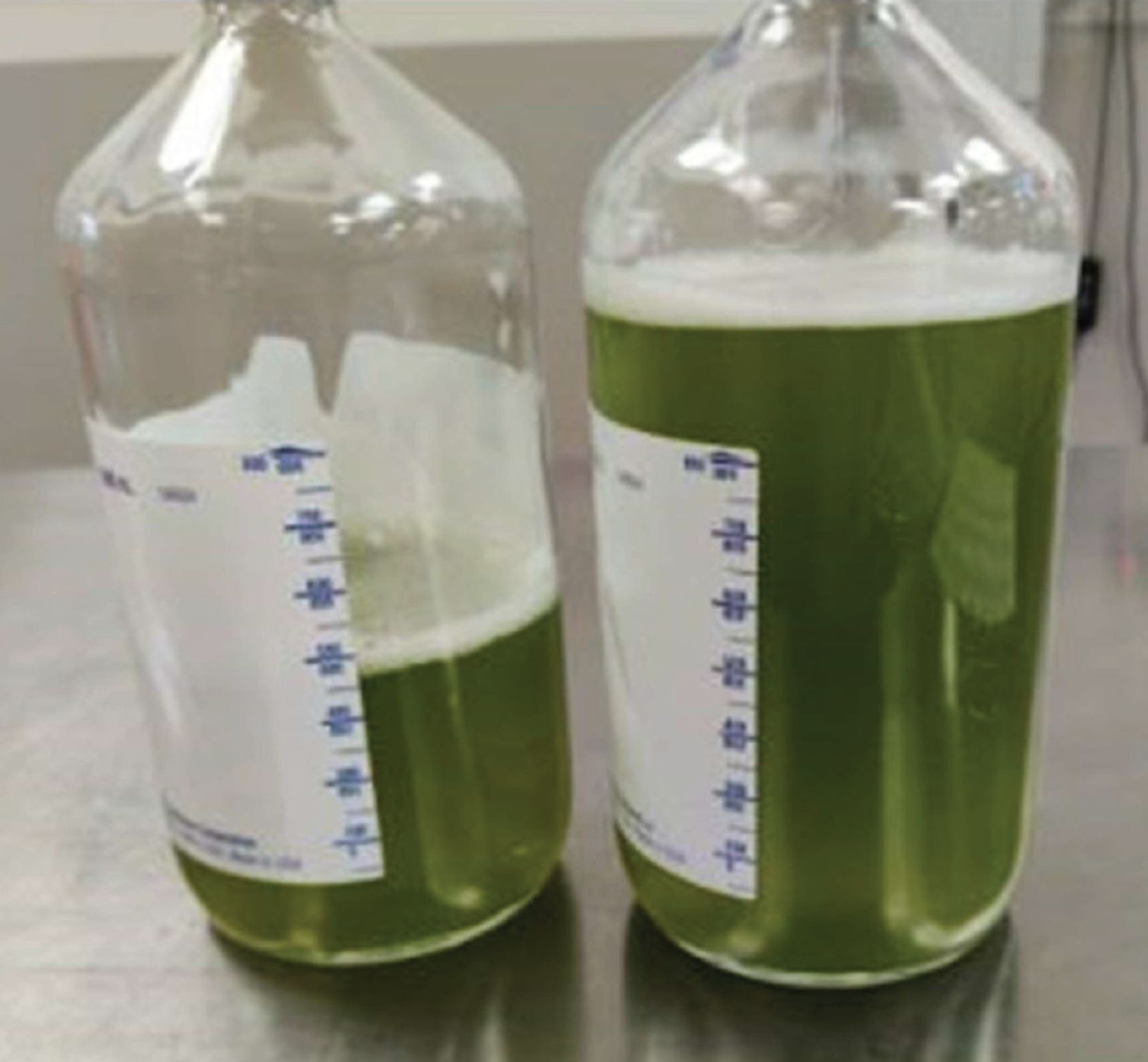
  • Figure 3
    • Download figure
    • Open in new tab
    • Download powerpoint
    Figure 3

    Green pleural fluid.

  • Figure 4
    • Download figure
    • Open in new tab
    • Download powerpoint
    Figure 4

    Pictograph of the whole lymph node illustrating the complete effacement of the normal lymph node architecture by atypical polymorphous infiltrates.
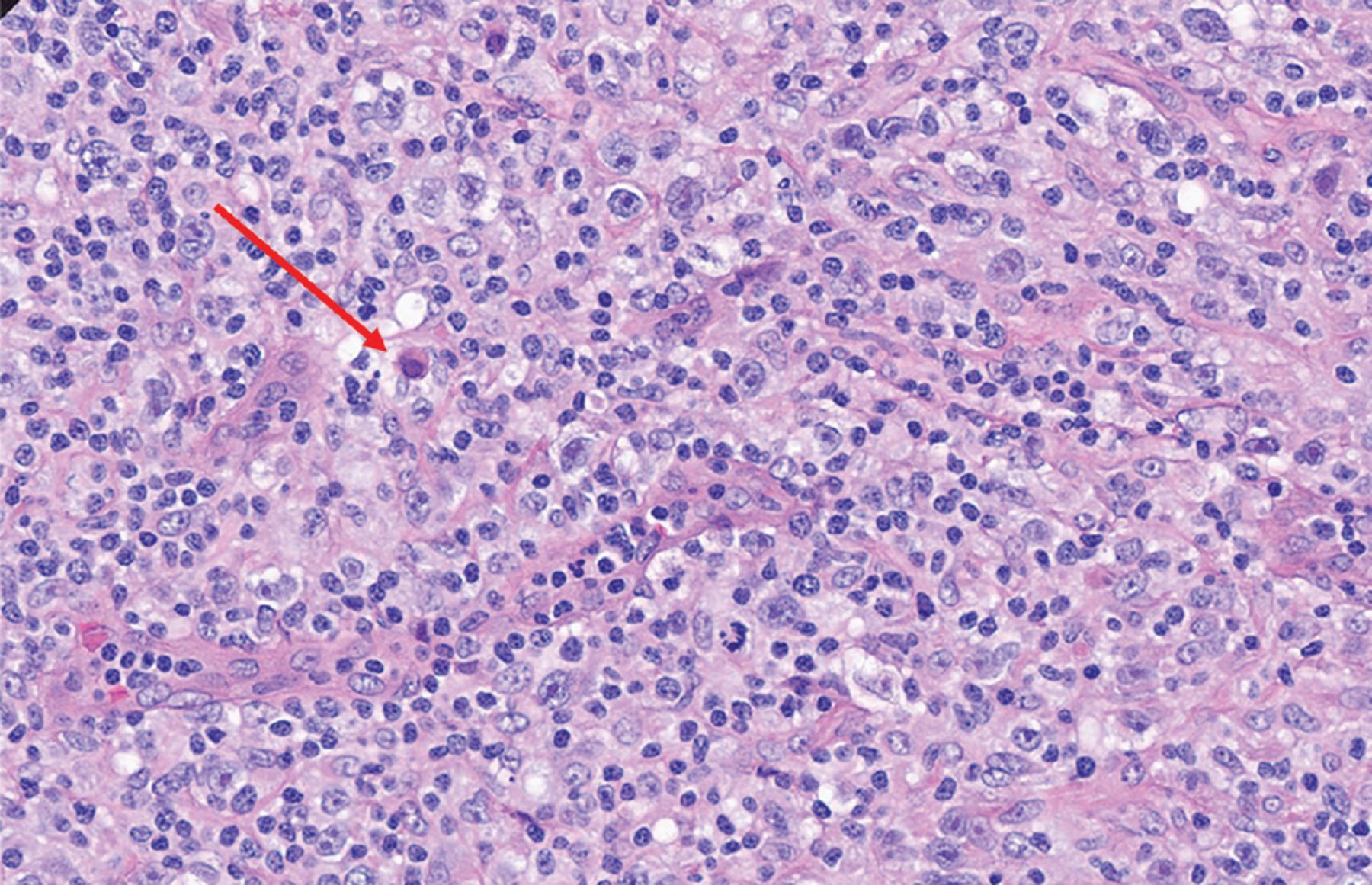
  • Figure 5
    • Download figure
    • Open in new tab
    • Download powerpoint
    Figure 5

    The characteristic Reed-Sternberg cell (red arrow) on excisional lymph node biopsy study (hematoxylin and eosin, magnification × 40).

Tables

  • Figures
    • View popup
    TABLE 1

    Differential diagnosis of pleural effusion

    DiagnosisRisk factorsClinical presentationPhysical examination findingsLaboratory and imaging characteristicsAlternative diagnosis
    Decompensated heart failureHistory of ischemic or nonischemic cardiomyopathy, heart failureExertional dyspnea, dyspnea at rest, orthopnea, PNDElevated JVD, pedal edema, crackles on lung auscultationTransudative pleural effusion; usually bilateral simple pleural effusionLack of prior diagnosis of heart failure, orthopnea, PND
    Malignancy (solid-organ or hematopoietic)Known malignancy of lungs, metastasis to the lungB symptoms (fatigue, weight loss, anorexia, night sweats, chills, fevers)Lymph node enlargementExudative effusion; unilateral simple pleural effusionLack of personal or family history of malignancy; lack of risk factors such as smoking
    InfectionImmunocompromised statusFever, chills, productive cough, sweatingBronchial breaths sound on auscultationExudative effusion; consolidation associated with parapneumonic effusion; loculated effusion; empyema; ground-glass opacities or lobular consolidation on chest CTSymptoms for a few weeks, no documented fevers
    Pulmonary embolismHistory of venous thromboembolism, active malignancy, hypercoagulable statePleuritic chest painSinus tachycardiaExudative effusion; normal chest radiograph; filling defect on chest CT with contrast; may be associated with small pleural effusionNo known risk factors for venous thromboembolism; large pleural effusion
    LymphangioleiomyomatosisYoung females in reproductive age groupChronic dyspnea, fatigue, spontaneous pneumothorax, pleural effusionsAssociated with axillary and mediastinal lymphadenopathyChylous, exudative pleural effusion; cystic lung disease; ground-glass opacities and septal thickening on chest CT; renal angiomyolipomaLack of history of spontaneous pneumothorax
    CirrhosisAlcoholic or nonalcoholic liver diseaseJaundice, ascites, fatigue, weight lossFluid thrill, shifting dullness on abdominal examinationTransudative pleural effusion; cirrhotic morphology of liver, ascitesLack of history, risk factors for liver disease
    ChylothoraxHistory of thoracic surgery, trauma, congenital disordersDyspnea, fatigue, yellow nailsDecreased breath sounds at site of pleural effusion, lymphadenopathyChylous pleural effusion; unilateral pleural effusionLack of thoracic duct injury due to surgery, trauma
    • CT = computed tomography; JVD = jugular venous distention; PND = paroxysmal nocturnal dyspnea

    • View popup
    TABLE 2

    Pleural fluid analysis and rationale

    Blood cell count and differentialA neutrophilic-predominant pleural effusion would make bacterial infection the most likely cause
    Lymphocytic pleural effusion is mainly encountered in conditions like rheumatoid arthritis-associated pleural effusion and fungal or tuberculosis-associated pleural effusion
    Cholesterol levelCholesterol levels > 60 mg/dL are seen in exudative pleural effusion
    CytologyPathologic analysis of pleural fluid allows for detection of malignancy
    Culture and Gram stainAllows for the speciation of the pathogenic organism
    Culture for bacteria, fungal, and acid-fast bacilli can be sent
    Antimicrobial resistance can be determined by sensitivity data
    Amylase levelElevated levels seen in acute pancreatitis-associated exudative pleural effusion
    Triglyceride levelElevated (> 110 mg/dL) in chylothorax
    Bilirubin levelElevated in biliothorax
    Albumin levelPleural fluid albumin and serum albumin gradient allow for the determination of pseudoexudative and exudative effusions in the setting of diuretic use
    HematocritPleural fluid hematocrit > 50% is pathognomonic for hemothorax
    pHLow pH pleural fluid seen in empyema or rheumatoid arthritis-associated pleural effusion
    Light criteriaDifferentiates between exudative or transudative pleural effusion; if at least 1 of the following criteria is met, the pleural effusion is exudative:
    1. Ratio of pleural fluid protein to serum protein concentration > 0.5

    2. Pleural fluid LDH greater than two-thirds of the upper limit of normal for serum LDH

    3. Ratio of pleural fluid LDH to serum LDH concentration > 0.6

    • LDH = lactate dehydrogenase

    • Data from references 2–9.

PreviousNext
Back to top

In this issue

Cleveland Clinic Journal of Medicine: 90 (8)
Cleveland Clinic Journal of Medicine
Vol. 90, Issue 8
1 Aug 2023
  • Table of Contents
  • Table of Contents (PDF)
  • Index by author
  • Complete Issue (PDF)
Print
Download PDF
Article Alerts
Sign In to Email Alerts with your Email Address
Email Article

Thank you for your interest in spreading the word on Cleveland Clinic Journal of Medicine.

NOTE: We only request your email address so that the person you are recommending the page to knows that you wanted them to see it, and that it is not junk mail. We do not capture any email address.

Enter multiple addresses on separate lines or separate them with commas.
Unilateral green pleural effusion in a 22-year-old woman
(Your Name) has sent you a message from Cleveland Clinic Journal of Medicine
(Your Name) thought you would like to see the Cleveland Clinic Journal of Medicine web site.
CAPTCHA
This question is for testing whether or not you are a human visitor and to prevent automated spam submissions.
Citation Tools
Unilateral green pleural effusion in a 22-year-old woman
Sauradeep Sarkar, Utsav Shrestha, Huda Elzahrany, Bathmapriya Balakrishnan
Cleveland Clinic Journal of Medicine Aug 2023, 90 (8) 491-498; DOI: 10.3949/ccjm.90a.22097

Citation Manager Formats

  • BibTeX
  • Bookends
  • EasyBib
  • EndNote (tagged)
  • EndNote 8 (xml)
  • Medlars
  • Mendeley
  • Papers
  • RefWorks Tagged
  • Ref Manager
  • RIS
  • Zotero
Share
Unilateral green pleural effusion in a 22-year-old woman
Sauradeep Sarkar, Utsav Shrestha, Huda Elzahrany, Bathmapriya Balakrishnan
Cleveland Clinic Journal of Medicine Aug 2023, 90 (8) 491-498; DOI: 10.3949/ccjm.90a.22097
Twitter logo Facebook logo Mendeley logo
  • Tweet Widget
  • Facebook Like
  • Linkedin Share Button

Jump to section

  • Article
    • IMAGING FINDINGS
    • DIFFERENTIAL DIAGNOSES
    • STAGING
    • TREATMENT
    • PLEURAL EFFUSIONS IN HODGKIN LYMPHOMA
    • TAKE-HOME POINTS
    • DISCLOSURES
    • Acknowledgment
    • REFERENCES
  • Figures & Data
  • Info & Metrics
  • PDF

Related Articles

  • No related articles found.
  • PubMed
  • Google Scholar

Cited By...

  • No citing articles found.
  • Google Scholar

More in this TOC Section

  • Thyroid obstacle course: Many challenges from a single gland
  • Shortness of breath in a 52-year-old man with HIV and severe mitral regurgitation
  • A 74-year-old woman with purple toes
Show more Symptoms to Diagnosis

Similar Articles

Subjects

  • Hematology
  • Oncology
  • Pulmonology

Navigate

  • Current Issue
  • Past Issues
  • Supplements
  • Article Type
  • Specialty
  • CME/MOC Articles
  • CME/MOC Calendar
  • Media Kit

Authors & Reviewers

  • Manuscript Submission
  • Authors & Reviewers
  • Subscriptions
  • About CCJM
  • Contact Us
  • Cleveland Clinic Center for Continuing Education
  • Consult QD

Share your suggestions!

Copyright © 2025 The Cleveland Clinic Foundation. All rights reserved. The information provided is for educational purposes only. Use of this website is subject to the website terms of use and privacy policy. 

Powered by HighWire