
Main menu

  • Home
  • Content
    • Current Issue
    • Ahead of Print
    • Past Issues
    • Supplements
    • Article Type
  • Specialty
    • Articles by Specialty
  • CME/MOC
    • Articles
    • Calendar
  • Info For
    • Manuscript Submission
    • Authors & Reviewers
    • Subscriptions
    • About CCJM
    • Contact Us
    • Media Kit
  • Conversations with Leaders
  • Conference Coverage
    • ACC / WCC 2023
    • AAAAI Meeting 2023
    • ACR Convergence 2022
    • Kidney Week 2022
    • AIDS 2022
    • CHEST 2021
    • IDWeek 2021
    • IAS 2021
    • ADA 2021
    • ATS 2021
    • ACC 2021
    • ACP 2021
    • AAN 2021
  • Other Publications
    • www.clevelandclinic.org

User menu

  • Register
  • Log in

Search

  • Advanced search
Cleveland Clinic Journal of Medicine
  • Other Publications
    • www.clevelandclinic.org
  • Register
  • Log in
Cleveland Clinic Journal of Medicine

Advanced Search

  • Home
  • Content
    • Current Issue
    • Ahead of Print
    • Past Issues
    • Supplements
    • Article Type
  • Specialty
    • Articles by Specialty
  • CME/MOC
    • Articles
    • Calendar
  • Info For
    • Manuscript Submission
    • Authors & Reviewers
    • Subscriptions
    • About CCJM
    • Contact Us
    • Media Kit
  • Conversations with Leaders
  • Conference Coverage
    • ACC / WCC 2023
    • AAAAI Meeting 2023
    • ACR Convergence 2022
    • Kidney Week 2022
    • AIDS 2022
    • CHEST 2021
    • IDWeek 2021
    • IAS 2021
    • ADA 2021
    • ATS 2021
    • ACC 2021
    • ACP 2021
    • AAN 2021
The Clinical Picture

Polypoid melanoma mistaken for verruca vulgaris

Andrea Tan, MD, Daniel Gutierrez, MD, Nooshin K. Brinster, MD and Jennifer A. Stein, MD, PhD
Cleveland Clinic Journal of Medicine September 2020, 87 (9) 534-536; DOI: https://doi.org/10.3949/ccjm.87a.19079
Andrea Tan
The Ronald O. Perelman Department of Dermatology, New York University Grossman School of Medicine, New York, NY; University of Texas Southwestern Medical Center, Dallas, TX
  • Find this author on Google Scholar
  • Find this author on PubMed
  • Search for this author on this site
Daniel Gutierrez
The Ronald O. Perelman Department of Dermatology, New York University Grossman School of Medicine, New York, NY
  • Find this author on Google Scholar
  • Find this author on PubMed
  • Search for this author on this site
  • For correspondence: Daniel.Gutierrez@nyumc.org
Nooshin K. Brinster
The Ronald O. Perelman Department of Dermatology, New York University Grossman School of Medicine, New York, NY
  • Find this author on Google Scholar
  • Find this author on PubMed
  • Search for this author on this site
Jennifer A. Stein
The Ronald O. Perelman Department of Dermatology, New York University Grossman School of Medicine, New York, NY
  • Find this author on Google Scholar
  • Find this author on PubMed
  • Search for this author on this site
  • Article
  • Figures & Data
  • Info & Metrics
  • PDF
Loading

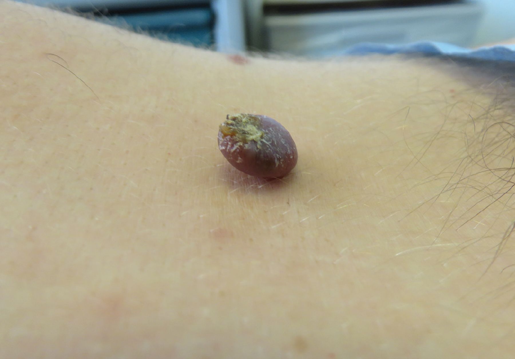
A 30-year-old man presented with a 6-month history of an exophytic mass growing on the lower mid-back (Figure 1). It was initially suspected to be verruca vulgaris, and he had been referred to a specialty clinic for sexually transmitted infections to confirm it. The lesion was occasionally tender to palpation and bled spontaneously.

Figure 1
  • Download figure
  • Open in new tab
  • Download powerpoint
Figure 1

A solitary ulcerated, purple-red polypoid nodule with overlying serous crust on the lower mid-back.

Physical examination revealed a 1.5-cm ulcerated, violaceous-to-erythematous polypoid nodule with overlying serous crust on the lower back. No pigmentation was noted within the mass or adjacent to the base of the lesion.

Histopathologic examination of a shave biopsy specimen revealed:

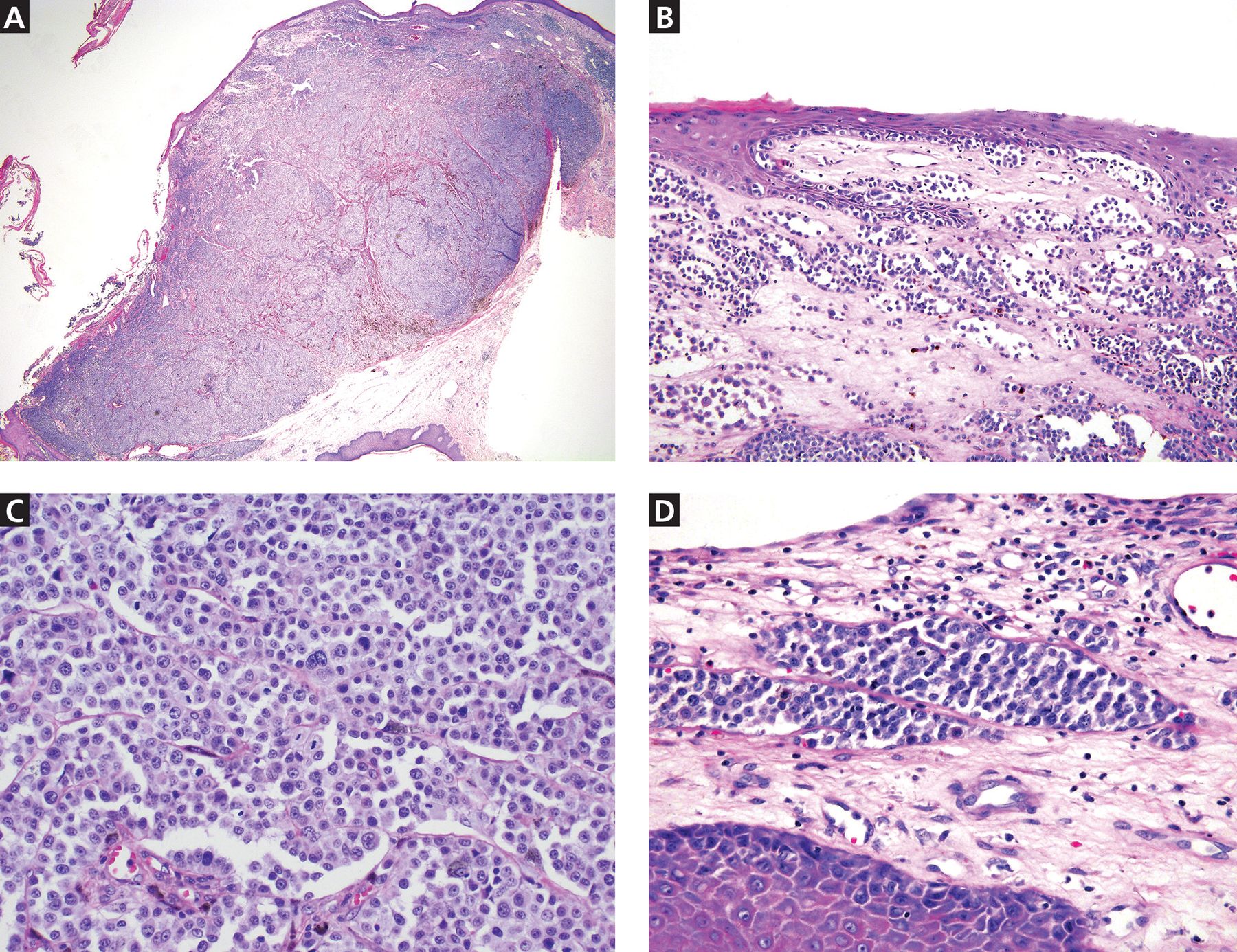
  • An exophytic polypoid lesion with broad ulceration, composed of markedly atypical melanocytes in a sheet-like pattern throughout the dermis (Figure 2A)

  • Focal epidermal contiguity with atypical melanocytes arranged in single cells and nests in the epidermis (Figure 2B)

  • A mitotic rate of greater than 30 mitoses/mm2 (Figure 2C)

  • Lymphovascular invasion (Figure 2D)

  • Melanocytes highlighted by S-100 protein on immunohistochemical staining.

Figure 2
  • Download figure
  • Open in new tab
  • Download powerpoint
Figure 2

Histopathologic images show (A) an exophytic polyp with ulceration and atypical melanocytes arranged in a sheet-like pattern throughout the dermis (hematoxylin and eosin [H&E] stain, original magnification × 2) and (B) focal epidermal contiguity with atypical melanocytes arranged in single cells and nests in the epidermis (H&E stain, original magnification × 20). High-power images (H&E stain, original magnification × 40) show (C) confluent, atypical melanocytes with conspicuous mitoses and focal cytoplasmic melanin, and (D) lymphovascular invasion at the periphery of the lesion.

These findings were diagnostic of melanoma, specifically the polypoid variant, with a tumor thickness of 5 mm.

The patient was referred for wide local excision with sentinel lymph node biopsy, which demonstrated inguinal node involvement and BRAF mutation on immunostaining. Positron-emission tomography–computed tomography, magnetic resonance imaging, and computed tomography of the head, chest, abdomen, and pelvis were unrevealing. The formal diagnosis was stage IIIC disease (T4bN1M0). The patient began immunotherapy with nivolumab. He was treated with this drug for 1 year and now is on active surveillance.

IMPORTANCE OF ACCURATE DIAGNOSIS

Polypoid melanoma is a rare clinical variant of nodular melanoma characterized by an exophytic mass with frequent ulceration, young age at onset (20 to 39 years), and poor prognosis.1,2 It has been reported to account for 2% to 43% of all melanomas, with the wide variability attributed to discrepancies in clinicopathologic criteria used in different reports.1

Lesions can affect the mucosa of the upper respiratory tract, esophagus, and anorectal junction, although cutaneous lesions are most frequently on the back.2–5 Polypoid melanomas have a propensity for ulceration, rapid progression over several weeks to months, and early metastasis to the lymph nodes, followed by possible metastasis to distant sites such as the skin, brain, liver, and subcutaneous soft tissue.2,4

Of the melanoma variants, polypoid melanoma has the poorest prognosis, given the risk of regional lymphatic and distal micro-metastatic involvement, often attributed to increased tumor thickness and ulceration at presentation.2–4 The 5-year survival rate for the polypoid nodular variant ranges from 32% to 42%, compared with 57% for nonpolypoid nodular melanoma and 77% for superficial spreading melanoma.2,5

Potential for misdiagnosis

Polypoid melanoma is often misdiagnosed, as it may be confused with benign skin conditions such as verruca vulgaris, leading to inappropriate treatment with cryotherapy or electrodessication and curettage. Other conditions to consider in the differential diagnosis include pyogenic granuloma, keratoacanthoma, and infarcted intradermal nevi or acrochordons, as these lesion types may similarly present with small protruding or dome-shaped papules.

Although these lesions often have distinguishing features, such as central hyperkeratosis in keratoacanthomas or the characteristic collarette of acanthotic epidermis at the base of pyogenic granulomas, polypoid melanomas may still be difficult to diagnose clinically, given that both pedunculated and sessile forms exist, as well as both pigmented and amelanotic variants. Additionally, ulceration may have many causes and can be seen in both benign and malignant growths. It may also obscure clinical presentation of cutaneous neoplasms, and tissue biopsy with histopathologic review should be considered for diagnostic guidance in such cases.

Given the highly aggressive clinical behavior and poor prognosis of polypoid melanoma, clinicians should maintain a low threshold for removal of rapidly growing pedunculated lesions with histopathologic evaluation, especially if the patient is relatively young and the lesion has progressed quickly or has ulcerated.

MANAGEMENT

Because polypoid melanoma may closely mimic benign lesions, patients with suspect lesions should be referred to dermatology for evaluation and biopsy as soon as possible. If timely dermatologic care is unavailable, priority should be given to biopsy techniques yielding adequate material for histopathologic analysis, such as a shave biopsy. Destructive treatments such as cryotherapy, used to manage benign lesions with similar appearances, should be avoided, as inappropriate treatment may delay accurate diagnosis and management.

Management of polypoid melanoma begins with prompt surgical excision. If it is not diagnosed early, sentinel lymph node biopsy and imaging studies may be needed to assess for disease progression. Additional therapy depends on extent of the disease and can include immunotherapy with immune checkpoint inhibitors or targeted therapy, such as BRAF and MEK inhibitors.

  • Copyright © 2020 The Cleveland Clinic Foundation. All Rights Reserved.

REFERENCES

  1. ↵
    1. Cutler K,
    2. Chu P,
    3. Levin M,
    4. Wallack M,
    5. Don PC,
    6. Weinberg JM
    . Pedunculated malignant melanoma. Dermatol Surg 2000; 26(2):127–129. doi:10.1046/j.1524-4725.2000.99092.x
    OpenUrlCrossRefPubMed
  2. ↵
    1. Dini M,
    2. Quercioli F,
    3. Caldarella V,
    4. Gaetano M,
    5. Franchi A,
    6. Agostini T
    . Head and neck polypoid melanoma. J Craniofac Surg 2012; 23(1):e23–e25. doi:10.1097/SCS.0b013e3182420801
    OpenUrlCrossRefPubMed
    1. McGovern VJ,
    2. Shaw HM,
    3. Milton GW
    . Prognostic significance of a polypoid configuration in malignant melanoma. Histopathology 1983;7(5):663–672. doi:10.1111/j.1365-2559.1983.tb02278.x
    OpenUrlCrossRefPubMed
  3. ↵
    1. Plotnick H,
    2. Rachmaninoff N,
    3. VandenBerg HJ Jr.
    . Polypoid melanoma: a virulent variant of nodular melanoma. Report of three cases and literature review. J Am Acad Dermatol 1990; 23(5 pt 1):880–884. doi:10.1016/0190-9622(90)70309-6
    OpenUrlCrossRefPubMed
  4. ↵
    1. Manci EA,
    2. Balch CM,
    3. Murad TM,
    4. Soong SJ
    . Polypoid melanoma, a virulent variant of the nodular growth pattern. Am J Clin Pathol 1981; 75(6):810–815. doi:10.1093/ajcp/75.6.810
    OpenUrlCrossRefPubMed
PreviousNext
Back to top

In this issue

Cleveland Clinic Journal of Medicine: 87 (9)
Cleveland Clinic Journal of Medicine
Vol. 87, Issue 9
1 Sep 2020
  • Table of Contents
  • Table of Contents (PDF)
  • Index by author
  • Complete Issue (PDF)
Print
Download PDF
Article Alerts
Sign In to Email Alerts with your Email Address
Email Article

Thank you for your interest in spreading the word on Cleveland Clinic Journal of Medicine.

NOTE: We only request your email address so that the person you are recommending the page to knows that you wanted them to see it, and that it is not junk mail. We do not capture any email address.

Enter multiple addresses on separate lines or separate them with commas.
Polypoid melanoma mistaken for verruca vulgaris
(Your Name) has sent you a message from Cleveland Clinic Journal of Medicine
(Your Name) thought you would like to see the Cleveland Clinic Journal of Medicine web site.
CAPTCHA
This question is for testing whether or not you are a human visitor and to prevent automated spam submissions.
Citation Tools
Polypoid melanoma mistaken for verruca vulgaris
Andrea Tan, Daniel Gutierrez, Nooshin K. Brinster, Jennifer A. Stein
Cleveland Clinic Journal of Medicine Sep 2020, 87 (9) 534-536; DOI: 10.3949/ccjm.87a.19079

Citation Manager Formats

  • BibTeX
  • Bookends
  • EasyBib
  • EndNote (tagged)
  • EndNote 8 (xml)
  • Medlars
  • Mendeley
  • Papers
  • RefWorks Tagged
  • Ref Manager
  • RIS
  • Zotero
Share
Polypoid melanoma mistaken for verruca vulgaris
Andrea Tan, Daniel Gutierrez, Nooshin K. Brinster, Jennifer A. Stein
Cleveland Clinic Journal of Medicine Sep 2020, 87 (9) 534-536; DOI: 10.3949/ccjm.87a.19079
Reddit logo Twitter logo Facebook logo Mendeley logo
  • Tweet Widget
  • Facebook Like
  • Linkedin Share Button

Jump to section

  • Article
    • IMPORTANCE OF ACCURATE DIAGNOSIS
    • MANAGEMENT
    • REFERENCES
  • Figures & Data
  • Info & Metrics
  • PDF

Related Articles

  • No related articles found.
  • PubMed
  • Google Scholar

Cited By...

  • No citing articles found.
  • Google Scholar

More in this TOC Section

  • Mass under the lip
  • Oral leukoplakia and oral cancer
  • Median rhomboid glossitis caused by tongue-brushing
Show more The Clinical Picture

Similar Articles

Subjects

  • Infectious Diseases
  • Oncology
  • Drug Therapy
  • Dermatology

Navigate

  • Current Issue
  • Past Issues
  • Supplements
  • Article Type
  • Specialty
  • CME/MOC Articles
  • CME/MOC Calendar
  • Media Kit

Authors & Reviewers

  • Manuscript Submission
  • Authors & Reviewers
  • Subscriptions
  • About CCJM
  • Contact Us
  • Cleveland Clinic Center for Continuing Education
  • Consult QD

Share your suggestions!

Copyright © 2023 The Cleveland Clinic Foundation. All rights reserved. The information provided is for educational purposes only. Use of this website is subject to the website terms of use and privacy policy. 

Powered by HighWire