
Main menu

  • Home
  • Content
    • Current Issue
    • Ahead of Print
    • Past Issues
    • Supplements
    • Article Type
  • Specialty
    • Articles by Specialty
  • CME/MOC
    • Articles
    • Calendar
  • Info For
    • Manuscript Submission
    • Authors & Reviewers
    • Subscriptions
    • About CCJM
    • Contact Us
    • Media Kit
  • Conversations with Leaders
  • Conference Coverage
    • Kidney Week 2024
    • CHEST 2024
    • ACR Convergence 2023
    • Kidney Week 2023
    • ObesityWeek 2023
    • IDWeek 2023
    • CHEST 2023
    • MDS 2023
    • IAS 2023
    • ACP 2023
    • AAN 2023
    • ACC / WCC 2023
    • AAAAI Meeting 2023
    • ACR Convergence 2022
    • Kidney Week 2022
    • AIDS 2022
  • Other Publications
    • www.clevelandclinic.org

User menu

  • Register
  • Log in

Search

  • Advanced search
Cleveland Clinic Journal of Medicine
  • Other Publications
    • www.clevelandclinic.org
  • Register
  • Log in
Cleveland Clinic Journal of Medicine

Advanced Search

  • Home
  • Content
    • Current Issue
    • Ahead of Print
    • Past Issues
    • Supplements
    • Article Type
  • Specialty
    • Articles by Specialty
  • CME/MOC
    • Articles
    • Calendar
  • Info For
    • Manuscript Submission
    • Authors & Reviewers
    • Subscriptions
    • About CCJM
    • Contact Us
    • Media Kit
  • Conversations with Leaders
  • Conference Coverage
    • Kidney Week 2024
    • CHEST 2024
    • ACR Convergence 2023
    • Kidney Week 2023
    • ObesityWeek 2023
    • IDWeek 2023
    • CHEST 2023
    • MDS 2023
    • IAS 2023
    • ACP 2023
    • AAN 2023
    • ACC / WCC 2023
    • AAAAI Meeting 2023
    • ACR Convergence 2022
    • Kidney Week 2022
    • AIDS 2022
The Clinical Picture

Revisiting the ‘great masquerader’

Raihan Ashraf, MD, Rahul Mahajan, MD, Debajyoti Chatterjee, DM and Kollabathula Arpitha, MD
Cleveland Clinic Journal of Medicine October 2021, 88 (10) 536-537; DOI: https://doi.org/10.3949/ccjm.88a.20203
Raihan Ashraf
Department of Dermatology, Venereology, and Leprology, Postgraduate Institute of Medical Education and Research, Chandigarh, India
  • Find this author on Google Scholar
  • Find this author on PubMed
  • Search for this author on this site
Rahul Mahajan
Associate Professor, Department of Dermatology, Venereology, and Leprology, Postgraduate Institute of Medical Education and Research, Chandigarh, India
  • Find this author on Google Scholar
  • Find this author on PubMed
  • Search for this author on this site
  • For correspondence: [email protected]
Debajyoti Chatterjee
Assistant Professor, Department of Histopathology, Postgraduate Institute of Medical Education and Research, Chandigarh, India
  • Find this author on Google Scholar
  • Find this author on PubMed
  • Search for this author on this site
Kollabathula Arpitha
Department of Histopathology, Postgraduate Institute of Medical Education and Research, Chandigarh, India
  • Find this author on Google Scholar
  • Find this author on PubMed
  • Search for this author on this site
  • Article
  • Figures & Data
  • Info & Metrics
  • PDF
Loading

A 51-year-old man presented with a 3-month history of reddish-brown lesions on his body and scaly brown lesions on the palms and soles and a 1-month history of mildly painful oral erosions. He had also noticed a buzzing sound in the right ear for the past 20 days.

Review of his medical record showed that he had engaged in unprotected sexual contact with a commercial sex worker 1 year earlier. He had human immunodeficiency virus (HIV) infection and had been taking antiretroviral therapy consisting of zidovudine, lamivudine, and nevirapine for the last 10 years, and his CD4 cell count was 706/μL.

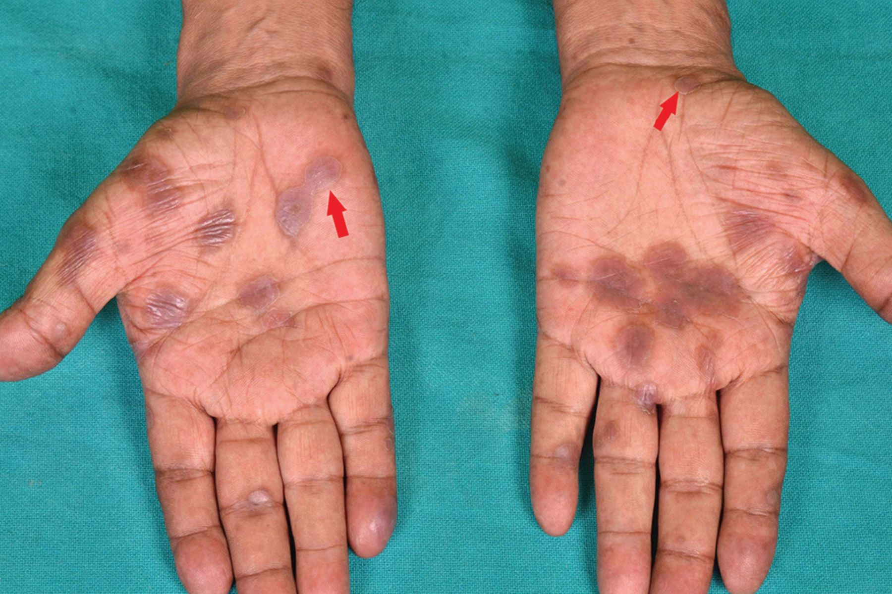
Cutaneous examination revealed well-defined dusky to erythematous papules and plaques on his face, neck, back, and hands. There were well-defined ulcers on the tip of the tongue and split papules on both angles of the mouth. There were discoid plaques with peripheral scales on the palms (Figure 1) and soles (Figure 2). Other mucosae were normal, as were the results of the nervous system and ophthalmologic examinations.

Figure 1
  • Download figure
  • Open in new tab
  • Download powerpoint
Figure 1

Well-defined dusky, discoid patches and plaques with peripheral collarette of scales (red arrows) on the palms.

Figure 2
  • Download figure
  • Open in new tab
  • Download powerpoint
Figure 2

Well-defined dusky, discoid plaques with peripheral desquamative scaling on the instep of the patient’s soles. Some of the lesions appear psoriasiform.

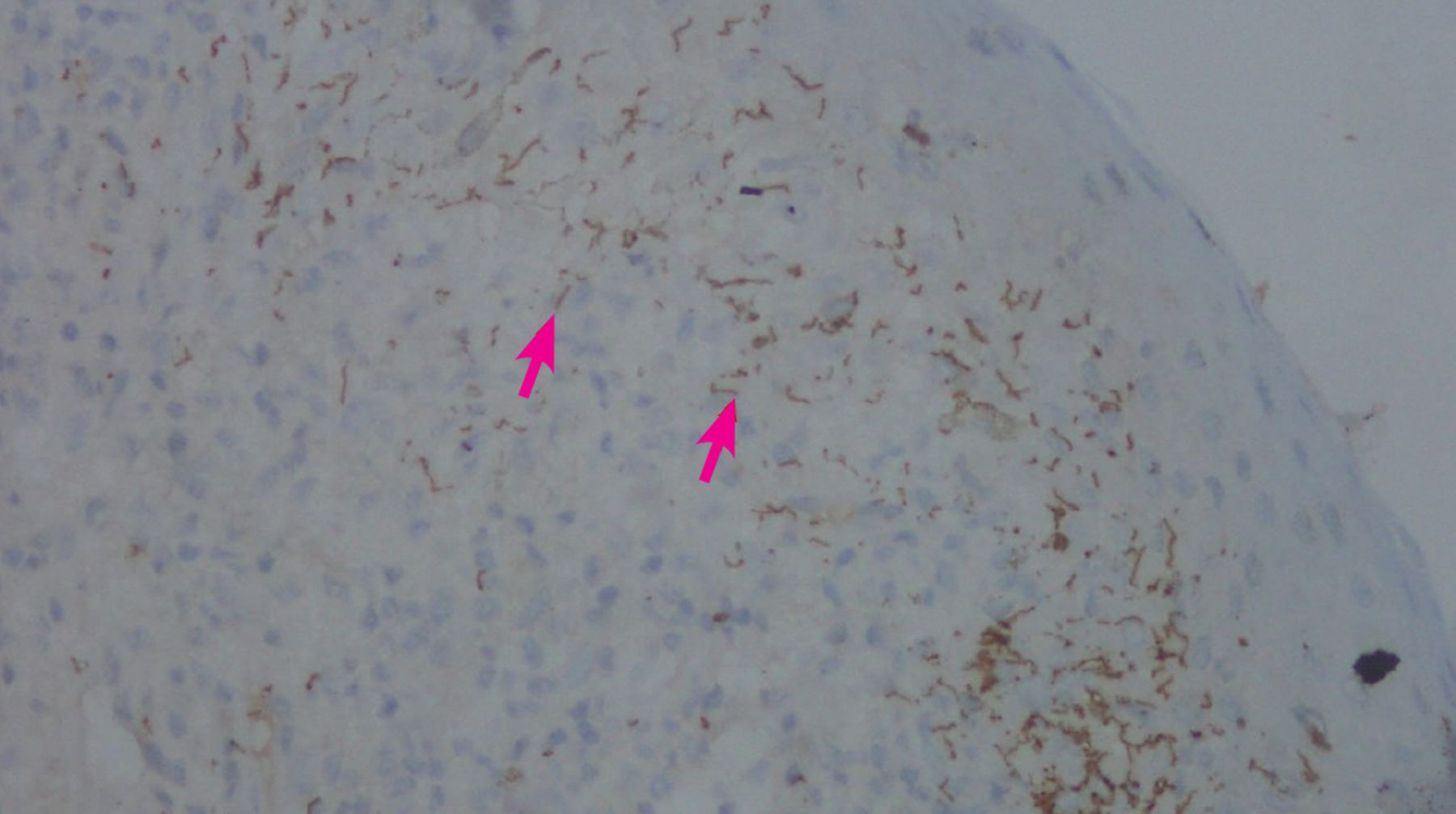
Skin biopsy of a cutaneous papule revealed dermis with periadnexal and perivascular plasma-cell-rich inflammatory infiltrate. Immunostain for Treponema further revealed helically coiled spirochetes present diffusely in the dermis (Figure 3).

Figure 3
  • Download figure
  • Open in new tab
  • Download powerpoint
Figure 3

Immunostain for Treponema showed helically coiled spirochetes, stained brown (red arrows), scattered throughout the dermis (magnification × 40).

A Venereal Disease Research Laboratory test (titres of 1:64) and Treponema pallidum hemagglutination assay were positive (titres of 1:80). Cerebrospinal fluid analysis was normal. However, tympanometry revealed a type C tympanogram with minimal sensorineural hearing loss in the right ear.

Based on these findings and the patient’s history, otosyphilis accompanying secondary syphilis was suspected. Owing to the unavailability of crystalline penicillin and procaine penicillin, intravenous ceftriaxone 2 g daily was administered for 14 days, which led to clearance of the mucocutaneous lesions and improvement in the tinnitus. A subsequent audiogram showed improvement in his hearing loss after 3 months.

DIFFERENTIAL DIAGNOSIS

The differential diagnosis for palmoplantar lesions of secondary syphilis includes palmoplantar psoriasis and pustulosis, palmoplantar eczema, keratoderma blennorrhagicum, tinea manuum, and pityriasis rosea. The presence of a peripheral rim of scale (Biett sign, Biett collarette) and pain caused by blunt pressure on these lesions (Buschke-Ollendorff sign) helps differentiate them from other conditions. Biett collarette may also be seen with lesions of porokeratosis and pityriasis rosea when they occur on the palms. However, porokeratosis generally causes scales that are rough and uneven, and pityriasis rosea produces multiple fine scales with an undefined direction.1 Palmo plantar lesions may have a configuration resembling erythema multiforme2 and rarely present as interdigital papules (condyloma lata) or erosions.3

Incidence rates of syphilis are known to be substantially higher in the HIV-positive population,4 and the secondary incubation period of syphilis is usually shortened in these patients. However, in our patient, manifestations of secondary syphilis were delayed by 9 months after exposure.

Incidence rates of otosyphilis in the HIV population are not well known. In a recent case series of 12 patients with otosyphilis, 8 (67%) were positive for HIV.5

Otosyphilis is often a presumptive diagnosis based on positive serology in patients with cochleovestibular symptoms with no other likely causes. It may present differently depending on the stage of syphilis. Sensorineural hearing loss (often bilateral and rapidly progressing) and tinnitus are the most common complaints in secondary syphilis. The presence of cerebrospinal fluid findings with otosyphilis ranges from 5.6% to 67%.5,6

The presentation of secondary syphilis is highly variable, including its dermatologic manifestations, earning it the name of the “great masquerader.” However, the presence of copper-colored patches and plaques on the palms and soles necessitates that syphilis be ruled out before considering other diagnoses.

It is imperative to ask any patient with syphilis about symptoms of ocular, otologic, and central nervous system involvement and to evaluate accordingly, thus saving the patient from considerable morbidity.

DISCLOSURES

The authors report no relevant financial relationships which, in the context of their contributions, could be perceived as a potential conflict of interest.

  • Copyright © 2021 The Cleveland Clinic Foundation. All Rights Reserved.

REFERENCES

  1. ↵
    1. Tognetti L,
    2. Sbano P,
    3. Fimiani M,
    4. Rubegni P
    . Dermoscopy of Biett’s sign and differential diagnosis with annular maculo-papular rashes with scaling. Indian J Dermatol Venereol Leprol 2017; 83(2):270–273. doi:10.4103/0378-6323.196318
    OpenUrlCrossRef
  2. ↵
    1. Bhate C,
    2. Tajirian AL,
    3. Kapila R,
    4. Lambert WC,
    5. Schwartz RA
    . Secondary syphilis resembling erythema multiforme. Int J Dermatol 2010; 49(11):1321–1324. doi:10.1111/j.1365-4632.2009.04390.x
    OpenUrlCrossRefPubMed
  3. ↵
    1. Rubins S,
    2. Esteves Morete A,
    3. Rubins A,
    4. Janniger CK,
    5. Schwartz RA
    . Interdigital syphilis: a rare form of secondary syphilis. Int J STD AIDS 2019; 30(14):1440–1442. doi:10.1177/0956462419853763
    OpenUrlCrossRef
  4. ↵
    1. Forrestel AK,
    2. Kovarik CL,
    3. Katz KA
    . Sexually acquired syphilis: historical aspects, microbiology, epidemiology, and clinical manifestations. J Am Acad Dermatol 2020; 82(1):1–14. doi:10.1016/j.jaad.2019.02.073
    OpenUrlCrossRefPubMed
  5. ↵
    1. Theeuwen H,
    2. Whipple M,
    3. Litvack JR
    . Otosyphilis: resurgence of an old disease. Laryngoscope 2019; 129(7):1680–1684. doi:10.1002/lary.27635
    OpenUrlCrossRef
  6. ↵
    1. Yimtae K,
    2. Srirompotong S,
    3. Lertsukprasert K
    . Otosyphilis: a review of 85 cases. Otolaryngol Head Neck Surg 2007; 136(1):67–71. doi:10.1016/j.otohns.2006.08.026
    OpenUrlCrossRefPubMed
PreviousNext
Back to top

In this issue

Cleveland Clinic Journal of Medicine: 88 (10)
Cleveland Clinic Journal of Medicine
Vol. 88, Issue 10
1 Oct 2021
  • Table of Contents
  • Table of Contents (PDF)
  • Index by author
  • Complete Issue (PDF)
Print
Download PDF
Article Alerts
Sign In to Email Alerts with your Email Address
Email Article

Thank you for your interest in spreading the word on Cleveland Clinic Journal of Medicine.

NOTE: We only request your email address so that the person you are recommending the page to knows that you wanted them to see it, and that it is not junk mail. We do not capture any email address.

Enter multiple addresses on separate lines or separate them with commas.
Revisiting the ‘great masquerader’
(Your Name) has sent you a message from Cleveland Clinic Journal of Medicine
(Your Name) thought you would like to see the Cleveland Clinic Journal of Medicine web site.
CAPTCHA
This question is for testing whether or not you are a human visitor and to prevent automated spam submissions.
Citation Tools
Revisiting the ‘great masquerader’
Raihan Ashraf, Rahul Mahajan, Debajyoti Chatterjee, Kollabathula Arpitha
Cleveland Clinic Journal of Medicine Oct 2021, 88 (10) 536-537; DOI: 10.3949/ccjm.88a.20203

Citation Manager Formats

  • BibTeX
  • Bookends
  • EasyBib
  • EndNote (tagged)
  • EndNote 8 (xml)
  • Medlars
  • Mendeley
  • Papers
  • RefWorks Tagged
  • Ref Manager
  • RIS
  • Zotero
Share
Revisiting the ‘great masquerader’
Raihan Ashraf, Rahul Mahajan, Debajyoti Chatterjee, Kollabathula Arpitha
Cleveland Clinic Journal of Medicine Oct 2021, 88 (10) 536-537; DOI: 10.3949/ccjm.88a.20203
Twitter logo Facebook logo Mendeley logo
  • Tweet Widget
  • Facebook Like
  • Linkedin Share Button

Jump to section

  • Article
    • DIFFERENTIAL DIAGNOSIS
    • DISCLOSURES
    • REFERENCES
  • Figures & Data
  • Info & Metrics
  • PDF

Related Articles

  • No related articles found.
  • PubMed
  • Google Scholar

Cited By...

  • No citing articles found.
  • Google Scholar

More in this TOC Section

  • Tinea incognito
  • Prolonged venous filling time and dependent rubor in a patient with peripheral artery disease
  • Sarcoidosis with diffuse purplish erythematous plaques on the hands
Show more The Clinical Picture

Similar Articles

Subjects

  • Drug Therapy
  • Infectious Diseases
  • Men's Health
  • Neurology

Navigate

  • Current Issue
  • Past Issues
  • Supplements
  • Article Type
  • Specialty
  • CME/MOC Articles
  • CME/MOC Calendar
  • Media Kit

Authors & Reviewers

  • Manuscript Submission
  • Authors & Reviewers
  • Subscriptions
  • About CCJM
  • Contact Us
  • Cleveland Clinic Center for Continuing Education
  • Consult QD

Share your suggestions!

Copyright © 2025 The Cleveland Clinic Foundation. All rights reserved. The information provided is for educational purposes only. Use of this website is subject to the website terms of use and privacy policy. 

Powered by HighWire