
Main menu

  • Home
  • Content
    • Current Issue
    • Ahead of Print
    • Past Issues
    • Supplements
    • Article Type
  • Specialty
    • Articles by Specialty
  • CME/MOC
    • Articles
    • Calendar
  • Info For
    • Manuscript Submission
    • Authors & Reviewers
    • Subscriptions
    • About CCJM
    • Contact Us
    • Media Kit
  • Conversations with Leaders
  • Conference Coverage
    • Kidney Week 2024
    • CHEST 2024
    • ACR Convergence 2023
    • Kidney Week 2023
    • ObesityWeek 2023
    • IDWeek 2023
    • CHEST 2023
    • MDS 2023
    • IAS 2023
    • ACP 2023
    • AAN 2023
    • ACC / WCC 2023
    • AAAAI Meeting 2023
    • ACR Convergence 2022
    • Kidney Week 2022
    • AIDS 2022
  • Other Publications
    • www.clevelandclinic.org

User menu

  • Register
  • Log in

Search

  • Advanced search
Cleveland Clinic Journal of Medicine
  • Other Publications
    • www.clevelandclinic.org
  • Register
  • Log in
Cleveland Clinic Journal of Medicine

Advanced Search

  • Home
  • Content
    • Current Issue
    • Ahead of Print
    • Past Issues
    • Supplements
    • Article Type
  • Specialty
    • Articles by Specialty
  • CME/MOC
    • Articles
    • Calendar
  • Info For
    • Manuscript Submission
    • Authors & Reviewers
    • Subscriptions
    • About CCJM
    • Contact Us
    • Media Kit
  • Conversations with Leaders
  • Conference Coverage
    • Kidney Week 2024
    • CHEST 2024
    • ACR Convergence 2023
    • Kidney Week 2023
    • ObesityWeek 2023
    • IDWeek 2023
    • CHEST 2023
    • MDS 2023
    • IAS 2023
    • ACP 2023
    • AAN 2023
    • ACC / WCC 2023
    • AAAAI Meeting 2023
    • ACR Convergence 2022
    • Kidney Week 2022
    • AIDS 2022
The Clinical Picture

Primary livedo reticularis of the abdomen

Michael Chew, MD and Paul Aronowitz, MD
Cleveland Clinic Journal of Medicine March 2018, 85 (3) 181-183; DOI: https://doi.org/10.3949/ccjm.85a.17047
Michael Chew
Department of Internal Medicine, University of California Davis Sacramento
  • Find this author on Google Scholar
  • Find this author on PubMed
  • Search for this author on this site
  • For correspondence: [email protected]
Paul Aronowitz
Department of Internal Medicine, University of California Davis, Sacramento
  • Find this author on Google Scholar
  • Find this author on PubMed
  • Search for this author on this site
  • Article
  • Figures & Data
  • Info & Metrics
  • PDF
Loading

A 53-year-old man presented with acute altered mental status secondary to methamphetamine intoxication. On hospital day 2, he developed macular, violaceous skin lesions with interconnecting rings on his abdomen and back, consistent with livedo reticularis (Figure 1). The extensive distribution of the lesions on the abdomen raised concern for underlying disease.

Figure 1
  • Download figure
  • Open in new tab
  • Download powerpoint
Figure 1

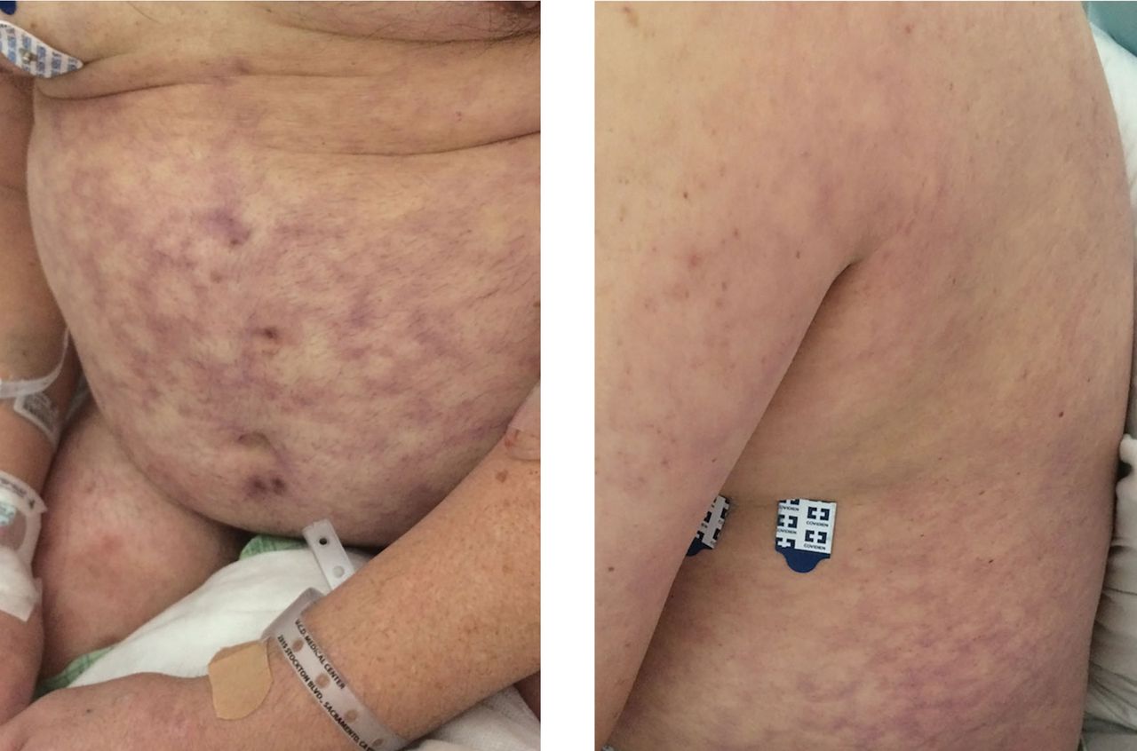
On hospital day 2, the patient developed macular, violaceous lesions with a reticular pattern on the abdomen and back. The extensive distribution of the lesions prompted an evaluation for an underlying systemic condition.

Livedo reticularis can be the manifestation of a wide range of conditions: hematologic and hypercoagulable states, embolic events, connective tissue disease, infection, vasculitis, malignancy, neurologic and endocrine conditions, and medication effects.1 Our patient had no recent history of vascular procedures or peripheral eosinophilia to suggest cholesterol embolization, and he had not recently started taking any new medications. His current medications included aspirin 81 mg, atorvastatin 40 mg, amlodipine 10 mg, and insulin glargine 20 units. Tests for cryoglobulin and antiphos-pholipid antibodies were negative. There was no evidence of malignancy, and evaluations for infectious and autoimmune diseases were negative.

Biopsy study of a skin lesion showed features consistent with livedo reticularis, with no evidence of vasculitis. The lesions resolved without definitive therapy by hospital day 3. This, in addition to other features of the lesions (eg, uniformity, unbroken reticular segments) and the extensive negative workup for systemic disease, suggested primary livedo re-ticularis.

CAUSES, TYPES, SUBTYPES

Livedo reticularis results from changes in the cutaneous microvasculature, composed of central arterioles that drain into an interconnecting, netlike venous plexus.1,2 Conditions such as arteriolar deoxygenation and venous plexus venodilation that result in a prominent venous plexus can give rise to clinical livedo reticularis.3

Livedo reticularis is divided into 2 broad categories depending on whether it is associated with systemic disease or not (Figure 2).3,4 In the absence of systemic symptoms, it can be further divided into 3 subcategories: idiopathic, physiologic, and primary.1 It is considered idiopathic if it is persistent, whereas physiologic and primary forms have a fluctuating course.4 Physiologic livedo reticularis often occurs in the periphery due to exposure to cold temperatures that cause vasospasm that resolves in warm temperatures.

Figure 2
  • Download figure
  • Open in new tab
  • Download powerpoint
Figure 2

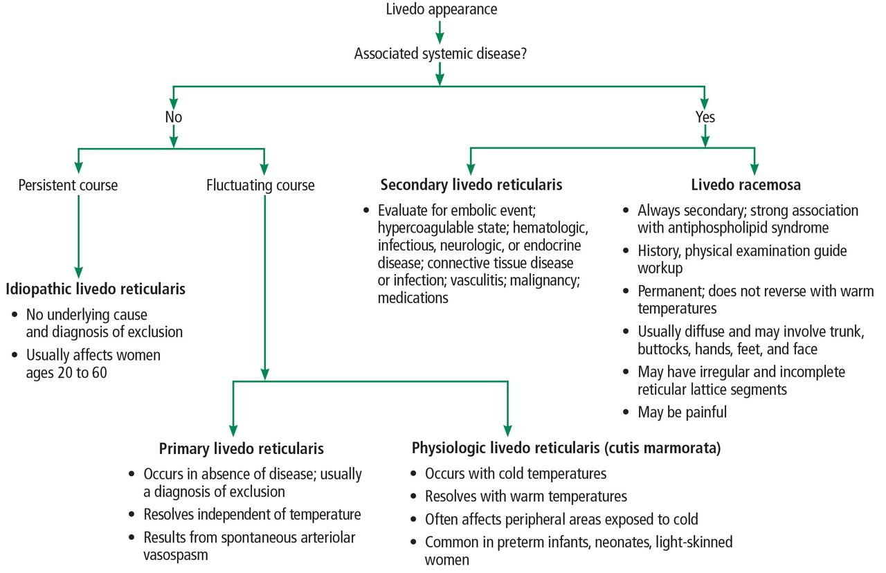
Algorithmic approach to the workup of livedo reticularis.

Primary livedo reticularis is thought to occur from spontaneous arteriolar vasospasm. It is a diagnosis of exclusion. An evaluation for underlying disease is important, as livedo re-ticularis can be associated with the range of conditions listed above.

In our patient, methamphetamine was considered a possible cause, but the findings of livedo reticularis were delayed and persisted longer than expected if they were drug-related.

Livedo racemosa

Distinguishing livedo reticularis from livedo racemosa is important. Livedo racemosa is always secondary and is often associated with an-tiphospholipid syndrome. It is present in 25% of cases of primary antiphospholipid syndrome and in up to 70% of cases of antiphospholipid syndrome associated with systemic lupus ery-thematosus.5 The reticular pattern of livedo racemosa is permanent and often has irregular and incomplete segments of reticular lattice, with a distribution that is more generalized, involving the trunk or buttocks (or both) in addition to the extremities.3,4 Consequently, a thorough history and physical examination are needed to guide additional workup.

  • Copyright © 2018 The Cleveland Clinic Foundation. All Rights Reserved.

REFERENCE

  1. ↵
    1. Gibbs MB,
    2. English JC 3rd.,
    3. Zirwas MJ
    . Livedo reticularis: an update. J Am Acad Dermatol 2005; 52:1009–1019.
    OpenUrlCrossRefPubMed
  2. ↵
    1. Kraemer M,
    2. Linden D,
    3. Berlit P
    . The spectrum of differential diagnosis in neurological patients with livedo reticularis and livedo racemosa. A literature review. J Neurol 2005; 252:1155–1166.
    OpenUrlCrossRefPubMed
  3. ↵
    1. Uthman IW,
    2. Khamashta MA
    . Livedo racemosa: a striking dermatological sign for the antiphospholipid syndrome. J Rheumatol 2006; 33:2379–2382.
    OpenUrlFREE Full Text
  4. ↵
    1. Dean SM
    . Livedo reticularis and related disorders. Curr Treat Options Cardiovasc Med 2011; 13:179–191.
    OpenUrlPubMed
  5. ↵
    1. Goldsmith LA,
    2. Katz SI,
    3. Gilchrest BA,
    4. Paller AS,
    5. Leffell DJ,
    6. Wolff K
    1. Chadachan V,
    2. Dean SM,
    3. Eberhardt RT
    . Cutaneous changes in peripheral arterial vascular disease. In: Goldsmith LA, , Katz SI, , Gilchrest BA, , Paller AS, , Leffell DJ, , Wolff K, eds. Fitzpatrick’s Dermatology in General Medicine.8th ed. New York, NY: McGraw-Hill Education; 2012.
PreviousNext
Back to top

In this issue

Cleveland Clinic Journal of Medicine: 85 (3)
Cleveland Clinic Journal of Medicine
Vol. 85, Issue 3
1 Mar 2018
  • Table of Contents
  • Table of Contents (PDF)
  • Index by author
Print
Download PDF
Article Alerts
Sign In to Email Alerts with your Email Address
Email Article

Thank you for your interest in spreading the word on Cleveland Clinic Journal of Medicine.

NOTE: We only request your email address so that the person you are recommending the page to knows that you wanted them to see it, and that it is not junk mail. We do not capture any email address.

Enter multiple addresses on separate lines or separate them with commas.
Primary livedo reticularis of the abdomen
(Your Name) has sent you a message from Cleveland Clinic Journal of Medicine
(Your Name) thought you would like to see the Cleveland Clinic Journal of Medicine web site.
CAPTCHA
This question is for testing whether or not you are a human visitor and to prevent automated spam submissions.
Citation Tools
Primary livedo reticularis of the abdomen
Michael Chew, Paul Aronowitz
Cleveland Clinic Journal of Medicine Mar 2018, 85 (3) 181-183; DOI: 10.3949/ccjm.85a.17047

Citation Manager Formats

  • BibTeX
  • Bookends
  • EasyBib
  • EndNote (tagged)
  • EndNote 8 (xml)
  • Medlars
  • Mendeley
  • Papers
  • RefWorks Tagged
  • Ref Manager
  • RIS
  • Zotero
Share
Primary livedo reticularis of the abdomen
Michael Chew, Paul Aronowitz
Cleveland Clinic Journal of Medicine Mar 2018, 85 (3) 181-183; DOI: 10.3949/ccjm.85a.17047
Twitter logo Facebook logo Mendeley logo
  • Tweet Widget
  • Facebook Like
  • Linkedin Share Button

Jump to section

  • Article
    • CAUSES, TYPES, SUBTYPES
    • REFERENCE
  • Figures & Data
  • Info & Metrics
  • PDF

Related Articles

  • No related articles found.
  • PubMed
  • Google Scholar

Cited By...

  • A reticular eruption on the thighs
  • Google Scholar

More in this TOC Section

  • Tinea incognito
  • Prolonged venous filling time and dependent rubor in a patient with peripheral artery disease
  • Sarcoidosis with diffuse purplish erythematous plaques on the hands
Show more The Clinical Picture

Similar Articles

Subjects

  • Dermatology
  • Hematology
  • Oncology
  • Rheumatology
  • Vascular Medicine

Navigate

  • Current Issue
  • Past Issues
  • Supplements
  • Article Type
  • Specialty
  • CME/MOC Articles
  • CME/MOC Calendar
  • Media Kit

Authors & Reviewers

  • Manuscript Submission
  • Authors & Reviewers
  • Subscriptions
  • About CCJM
  • Contact Us
  • Cleveland Clinic Center for Continuing Education
  • Consult QD

Share your suggestions!

Copyright © 2025 The Cleveland Clinic Foundation. All rights reserved. The information provided is for educational purposes only. Use of this website is subject to the website terms of use and privacy policy. 

Powered by HighWire