


Article Figures & Data
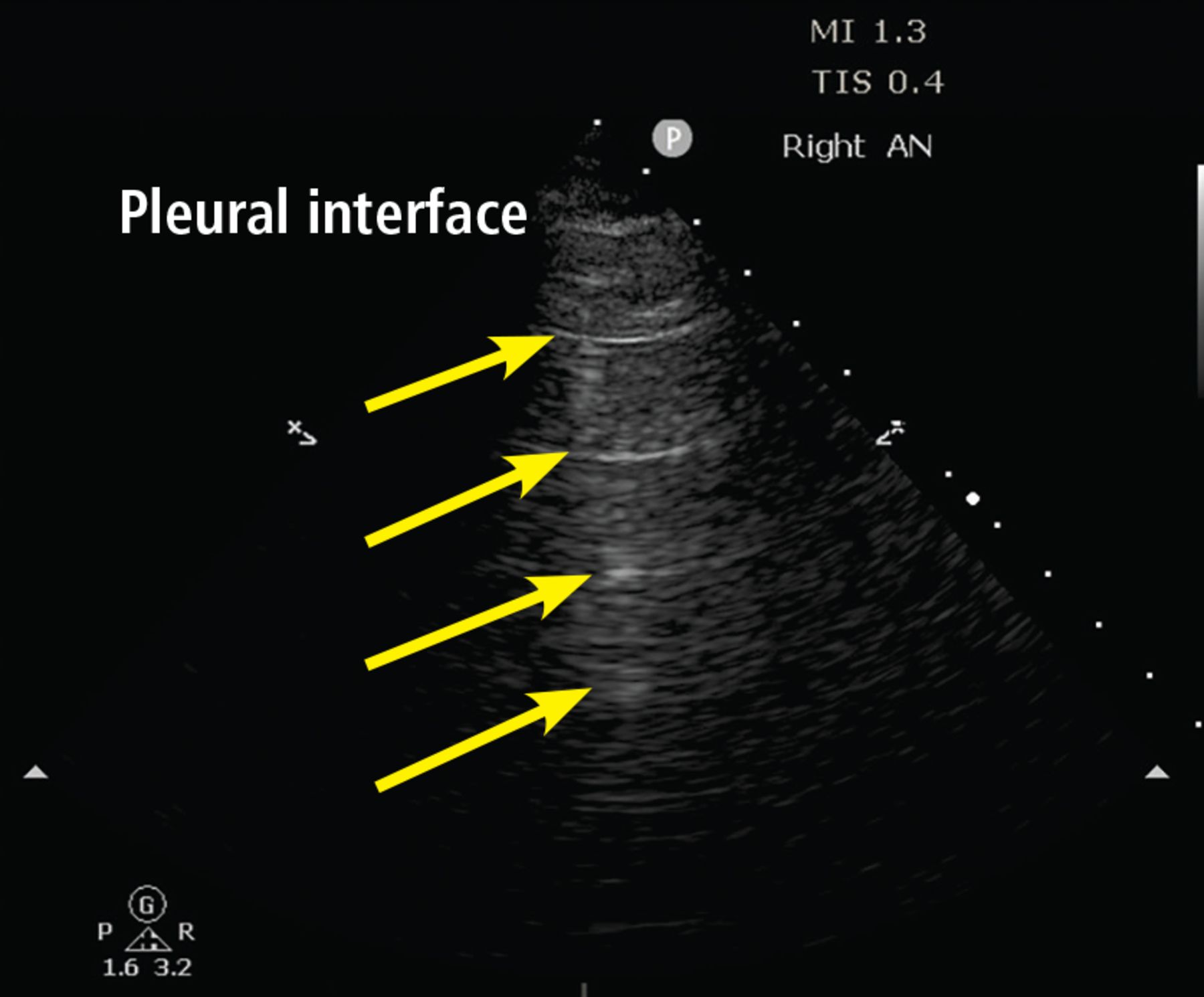
- Figure 1
A lines. The A-line pattern occurs in normal lung and in pneumothorax. Ultrasound waves (arrows) reflect off the pleural interface repeatedly, producing repeated horizontal lines throughout the field.
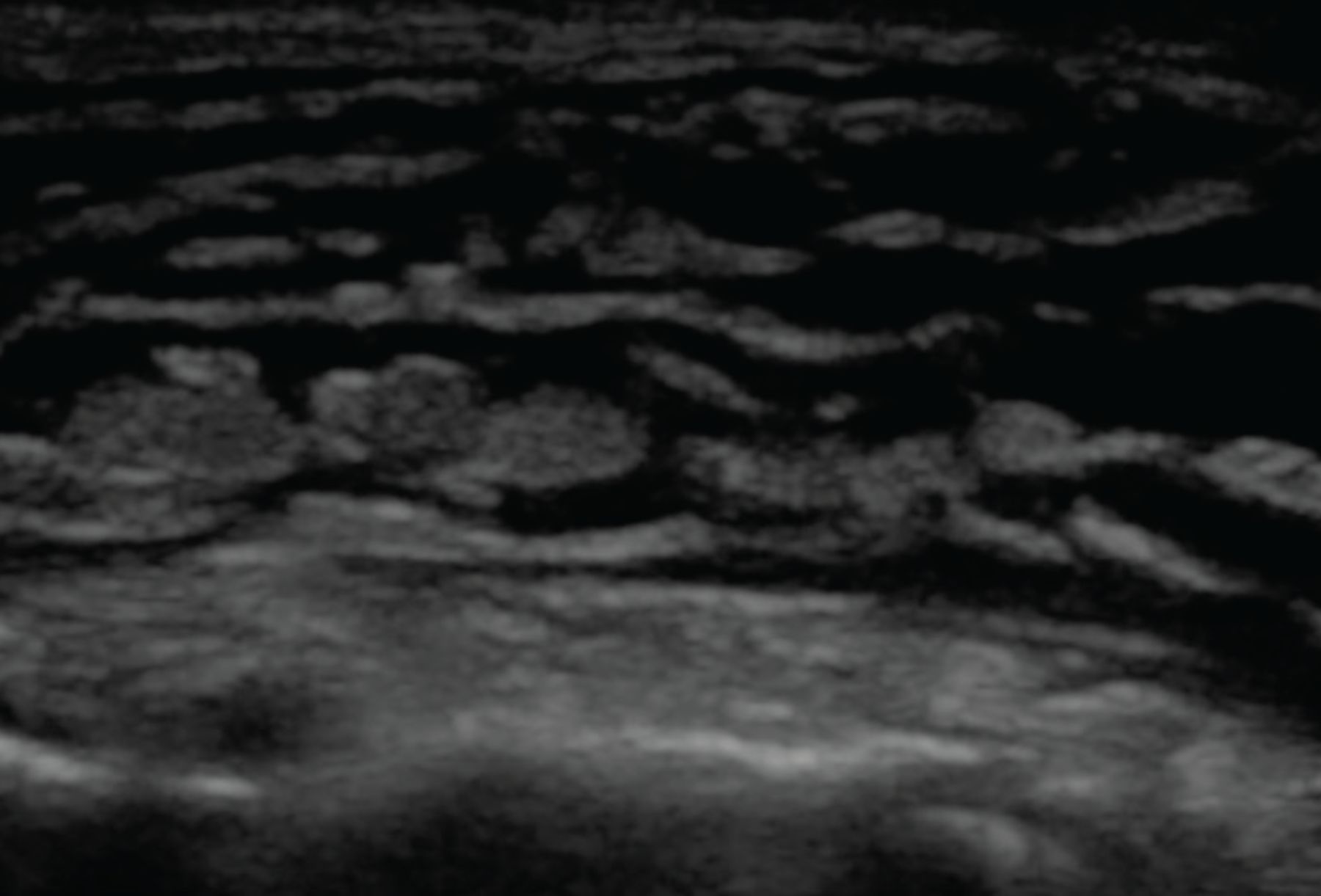
- Figure 2
B lines. The B-line pattern occurs in the setting of interstitial thickening by any cause, including cardiogenic pulmonary edema, noncardiogenic pulmonary edema, interstitial fibrosis, and interstitial pneumonia/pneumonitis. It is analogous to ground-glass opacity on computed tomography. It is demonstrated by vertical lines resembling the tail of a comet and extending to the bottom of the screen. In this image, confluent B lines (arrow) indicate significant interstitial involvement.
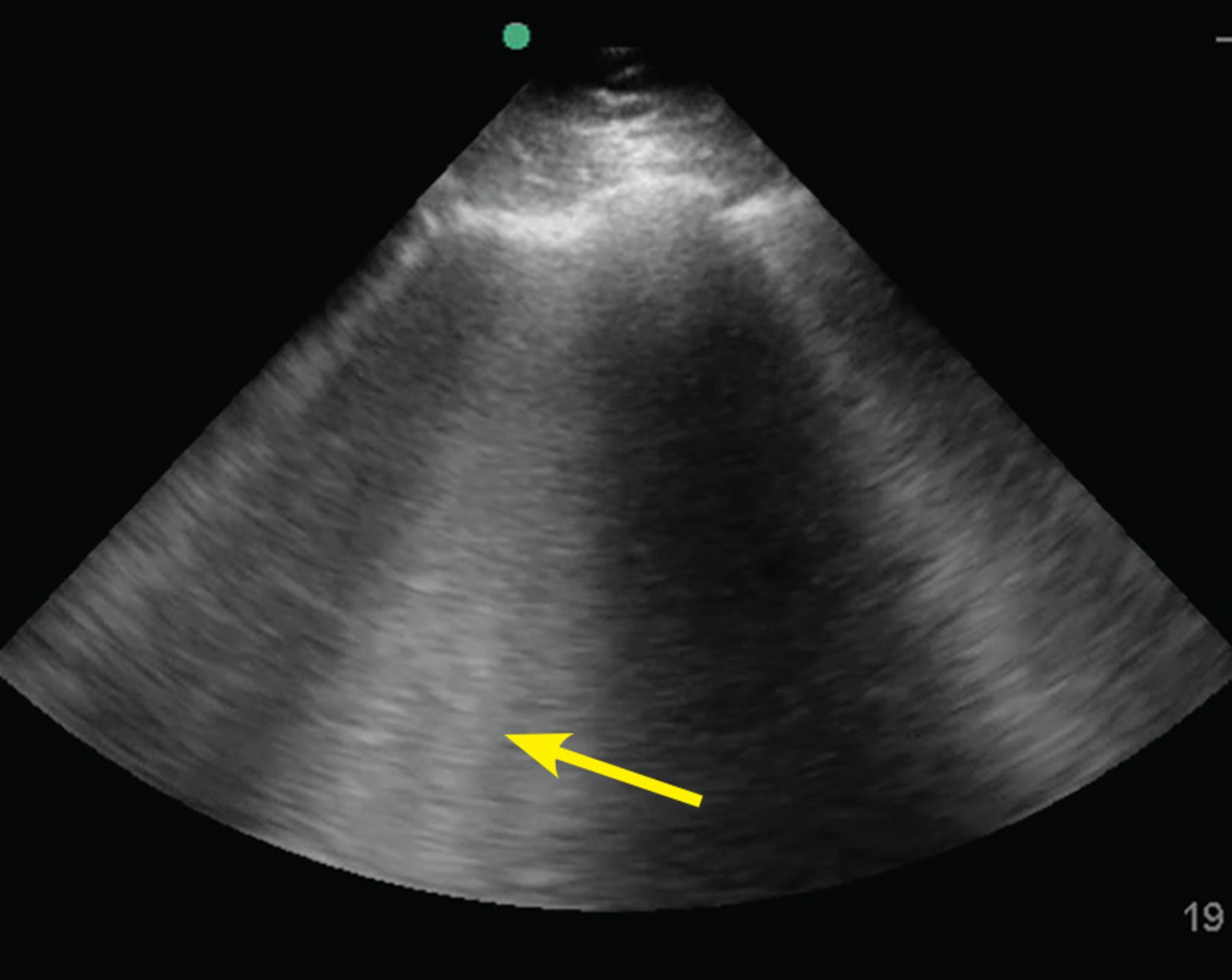
- Figure 3
Small peripheral (subpleural) consolidation. This is demonstrated by a small area of lung parenchyma visualized directly beneath the pleura (arrow). This pattern is common in bacterial or viral pneumonia, including COVID-19 pneumonia.
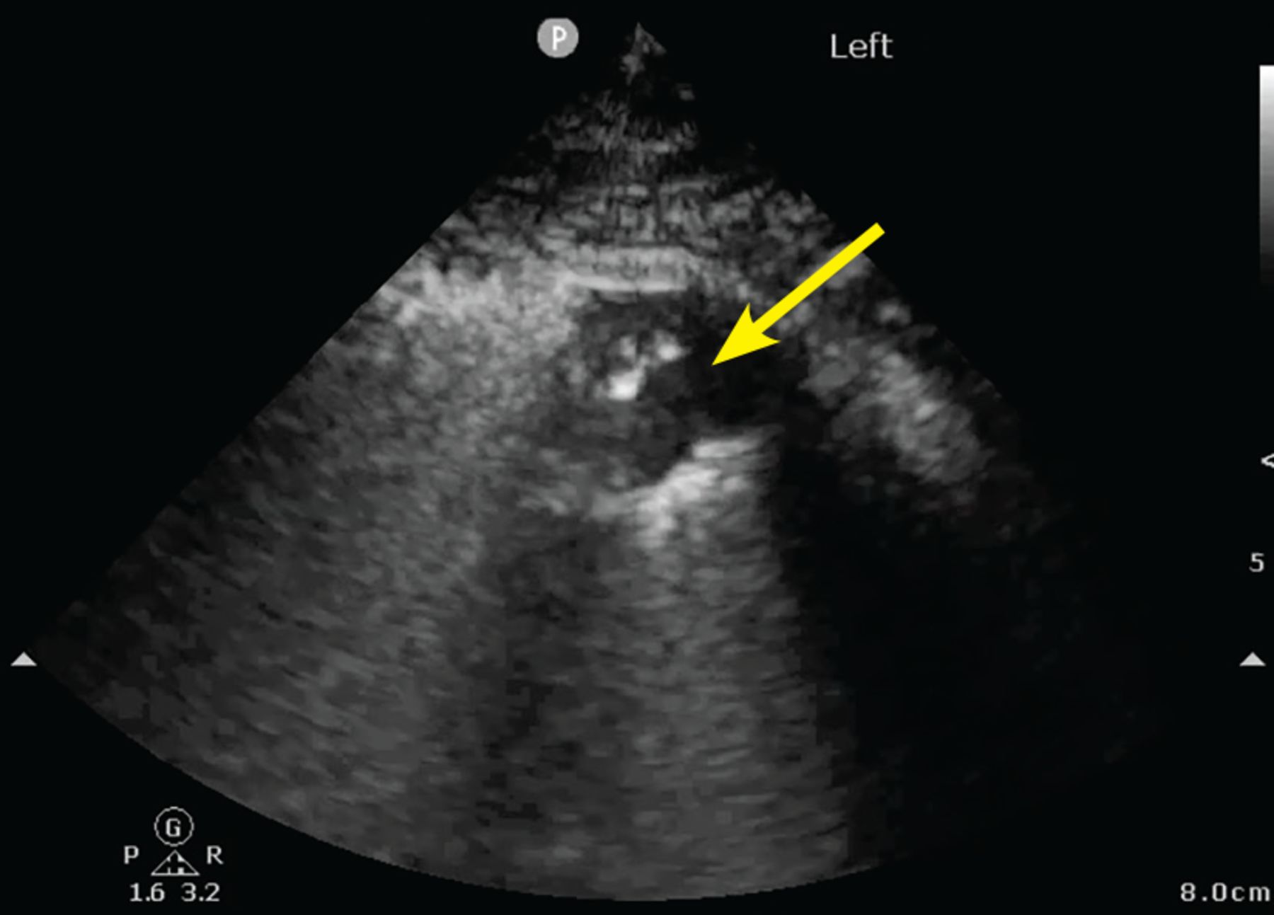
- Figure 4
Pleural effusion and consolidation.
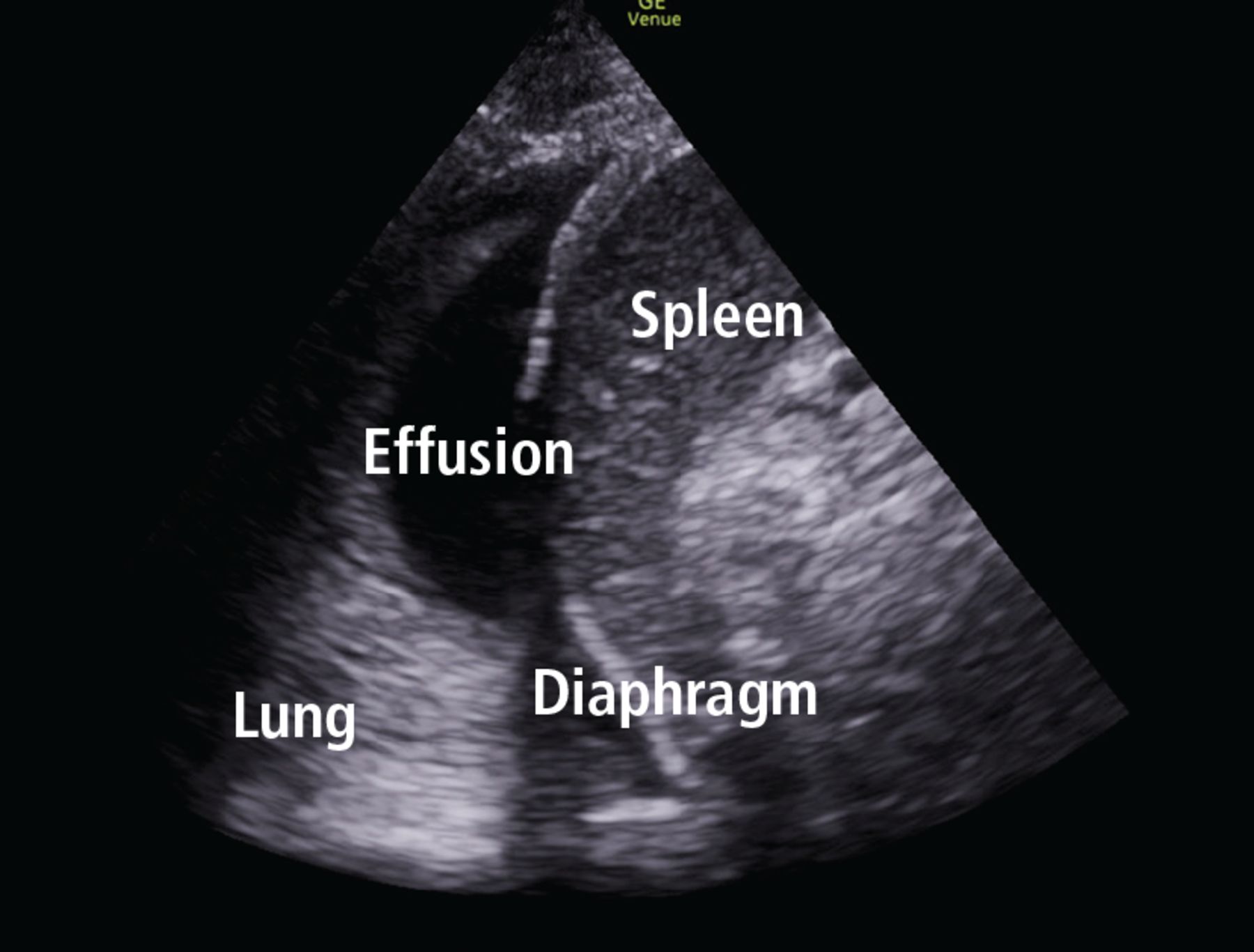
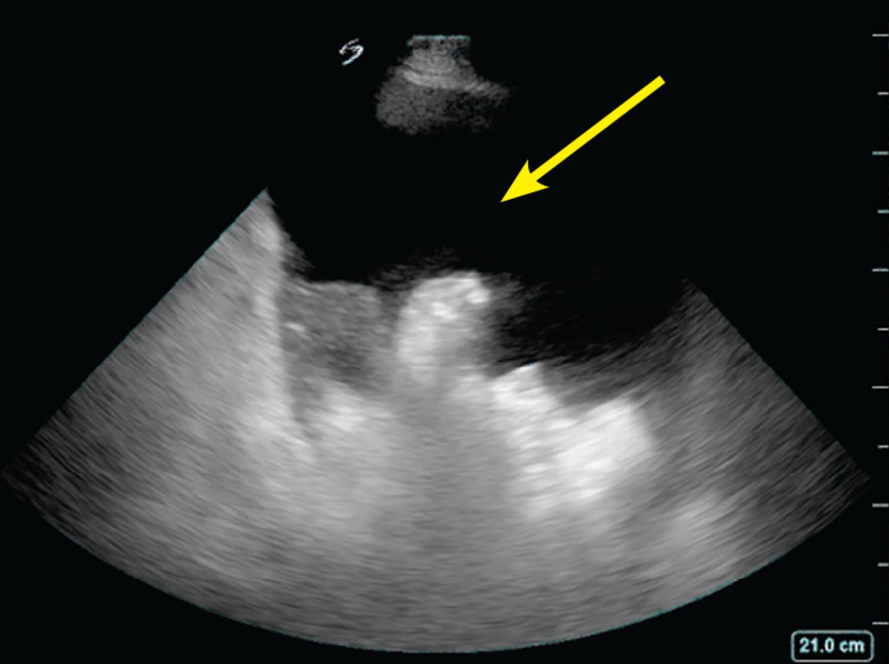
- Figure 5
Right lower quadrant with large ascites fluid pocket; Foley catheter in bladder.
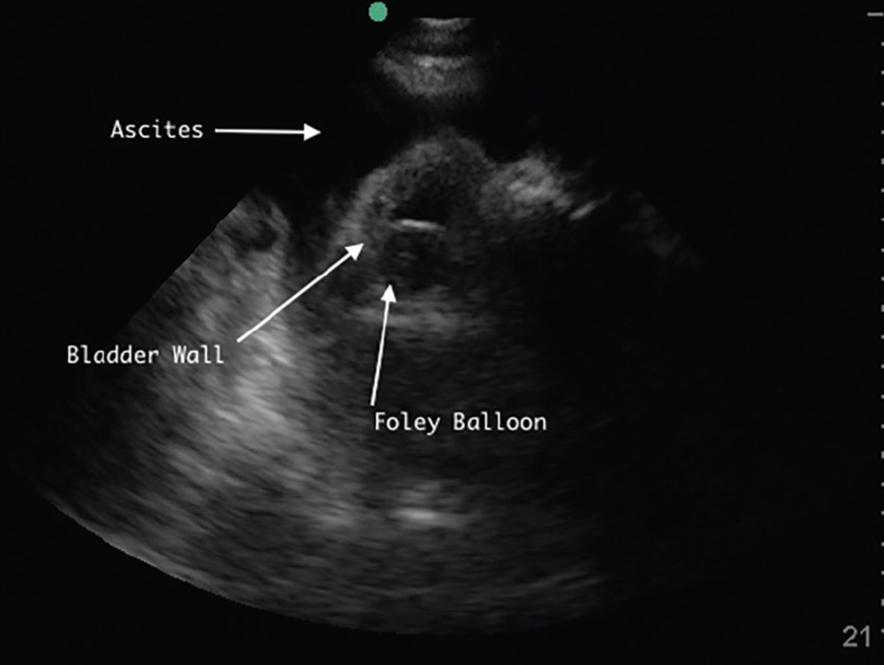
- Figure 6
Ascites pocket.
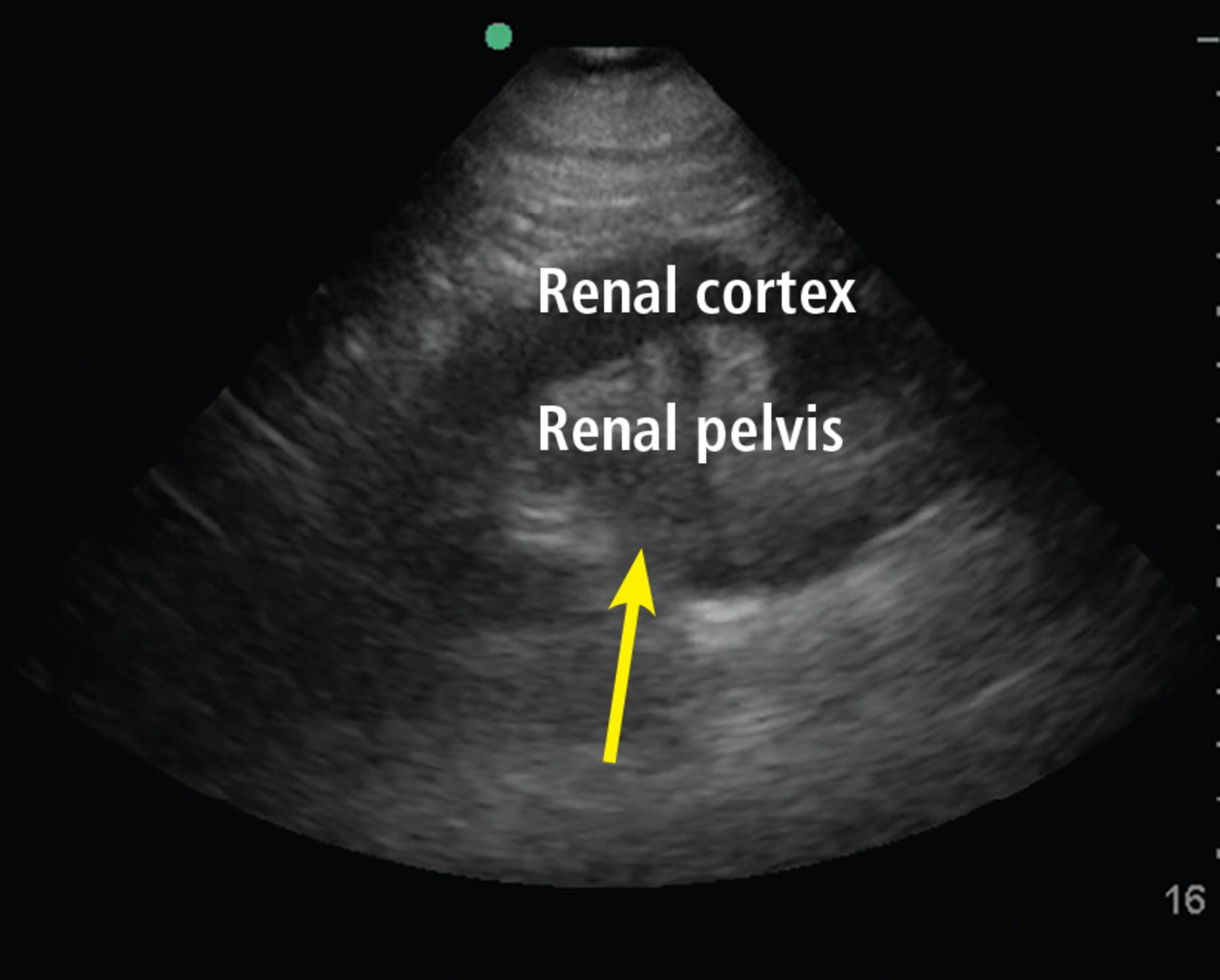
- Figure 7
Hydronephrosis. Hypoechoic (dark) fluid (arrow) is shown extending into the renal pelvis.
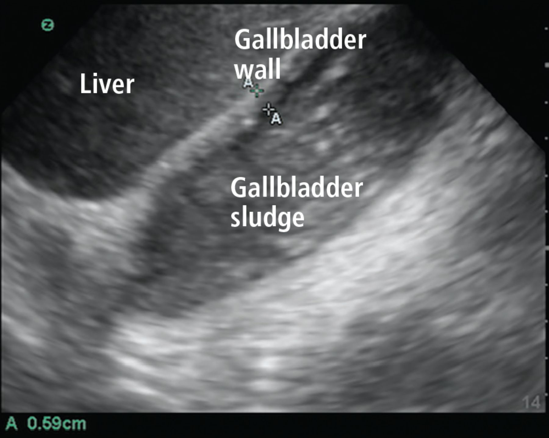
- Figure 8
Gallbladder containing sludge, with a thickened anterior wall, in a patient with acute cholecystitis.
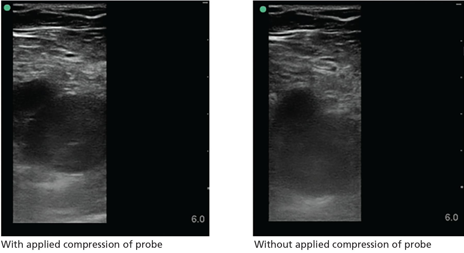
- Figure 9
Right common femoral vein deep vein thrombosis. The left image shows lack of compression of the vein with applied compression of the probe. The right image shows vein without compression.
- Figure 10
On ultrasonography, subcutaneous fluid is demonstrated as hypoechoic or anechoic (dark) layering within islands of subcutaneous tissue (gray). This occurs in any process leading to fluid within the subcutaneous tissue, including cellulitis and hydrostatic edema.


- TABLE 1
Point-of-care ultrasonography workflow compared with traditional consultative ultrasonography
Consultative ultrasonography POCUS Decision to perform ultrasonography Primary clinician Primary clinician Image acquisition Sonographer Image interpretation Sonographer
RadiologistClinical integration Radiologist
Primary clinician Diagnosis Meta-analysis No. of studies No. of patients Pooled sensitivity 95% confidence interval Pooled specificity 95% confidence interval Positive likelihood ratio Negative likelihood ratio Pleural effusion Yousefifard et al,11 2016 12 1,554 94% 88%–97% 98% 92%–100% 53.96 0.06 Acute cardiogenic pulmonary edema Maw et al,9 2019 7 1,075 94.1% 81.3%– 98.3% 92.4% 84.2%– 96.4% 12.38 0.06 Pneumonia Alzahrani et al,10 2017 20 2,513 85% 84%–87% 93% 92%–95% 12.14 0.16 Pneumothorax Alrajab et al,8 2013a 13 1,514 78.6% 68.1%–98.1% 98.4% 97.3%–99.5% 49.13 0.22 ↵a Included 1 study that used lung sliding sign alone, 12 studies that used lung sliding and comet tail signs, and 6 studies that included lung point in addition to the other 2 signs.
Cardiogenic pulmonary edema Noncardiogenic diffuse pulmonary interstitial edema Interstitial pneumonia or pneumonitis (bacterial, viral, or inflammatory) Interstitial fibrosis Distribution Diffuse
Usually bilateral and symmetric
Predominant in dependent regionsDiffuse or patchy
Often asymmetricFocal or patchy
Usually asymmetricDiffuse or patchy
Variable symmetrySpared areas Absent Often present Present Often present Number of B lines Variable Variable Variable Variable Pleura Smooth Irregular Irregular Irregular Subpleural consolidations Absent Present Present Typically absent Reduced lung sliding Absent May be present May be present May be present Pleural effusion Often present Typically absent May be present Typically absent ↵a Defining the terminology: diffuse = present throughout; patchy = present in many areas throughout, absent in other areas throughout; focal = present in one region but not in others; spared areas = regions of lung with A-line pattern (amid a background of B-line pattern).

- TABLE 5
Estimates of central venous pressure based on inferior vena cava size and collapsibility
Inferior vena cava size Percent collapse Estimated central venous pressure ≤ 2.1 cm > 50% 3 mm Hg ≤ 2.1 cm < 50% 8 mm Hg > 2.1 cm > 50% 8 mm Hg > 2.1 cm < 50% 15 mm Hg Based on reference 35.
- TABLE 6
Meta-analyses evaluating point-of-care ultrasonography for diagnosing deep vein thrombosis
Meta-analysis No. of studies No. of patients Pooled sensitivity 95% confidence interval Pooled specificity 95% confidence interval Positive likelihood ratio Negative likelihood ratio Burnside et al,50 2008 6 936 95% 87%–99% 96% 87%– 99% 23.75 0.05 Pomero et al,51 2013 16 2,379 96.1% 90.6%–98.5% 96.8% 94.6%–98.1% 30.03 0.04 West et al,52 2015 13 1,806 96.5% 90.1%–98.8% 96.8% 94.7% –98.0% 30.16 0.04

In this issue


Jump to section
- Article
- ABSTRACT
- DIRECT CLINICIAN INVOLVEMENT
- IMPROPER USE AND INTERPRETATION CAN CAUSE HARM
- LUNG AND PLEURAL ULTRASONOGRAPHY
- FOCUSED CARDIAC ULTRASONOGRAPHY
- ABDOMINAL ULTRASONOGRAPHY
- EVALUATION OF LOWER-EXTREMITY DEEP VEIN THROMBOSIS
- EVALUATING SKIN AND SOFT-TISSUE INFECTIONS
- ULTRASONOGRAPHY FOR PROCEDURAL GUIDANCE
- CONCLUSION
- DISCLOSURES
- REFERENCES
- Figures & Data
- Info & Metrics
Related Articles
Cited By...
- No citing articles found.