


A 40-year-old man with a history of diabetes mellitus and hypertension, but taking no medications, presented to the hospital with a 5-day history of fever, rash, and pruritus. The patient initially presented to an urgent care center, where he received intramuscular and oral antibiotics, but his symptoms continued to worsen.
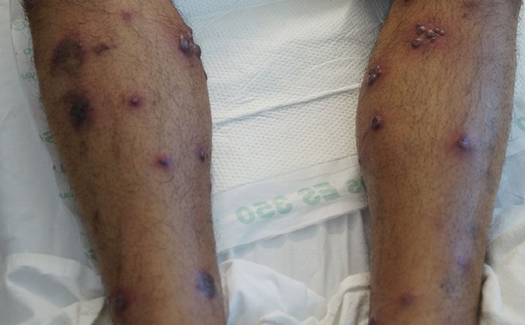
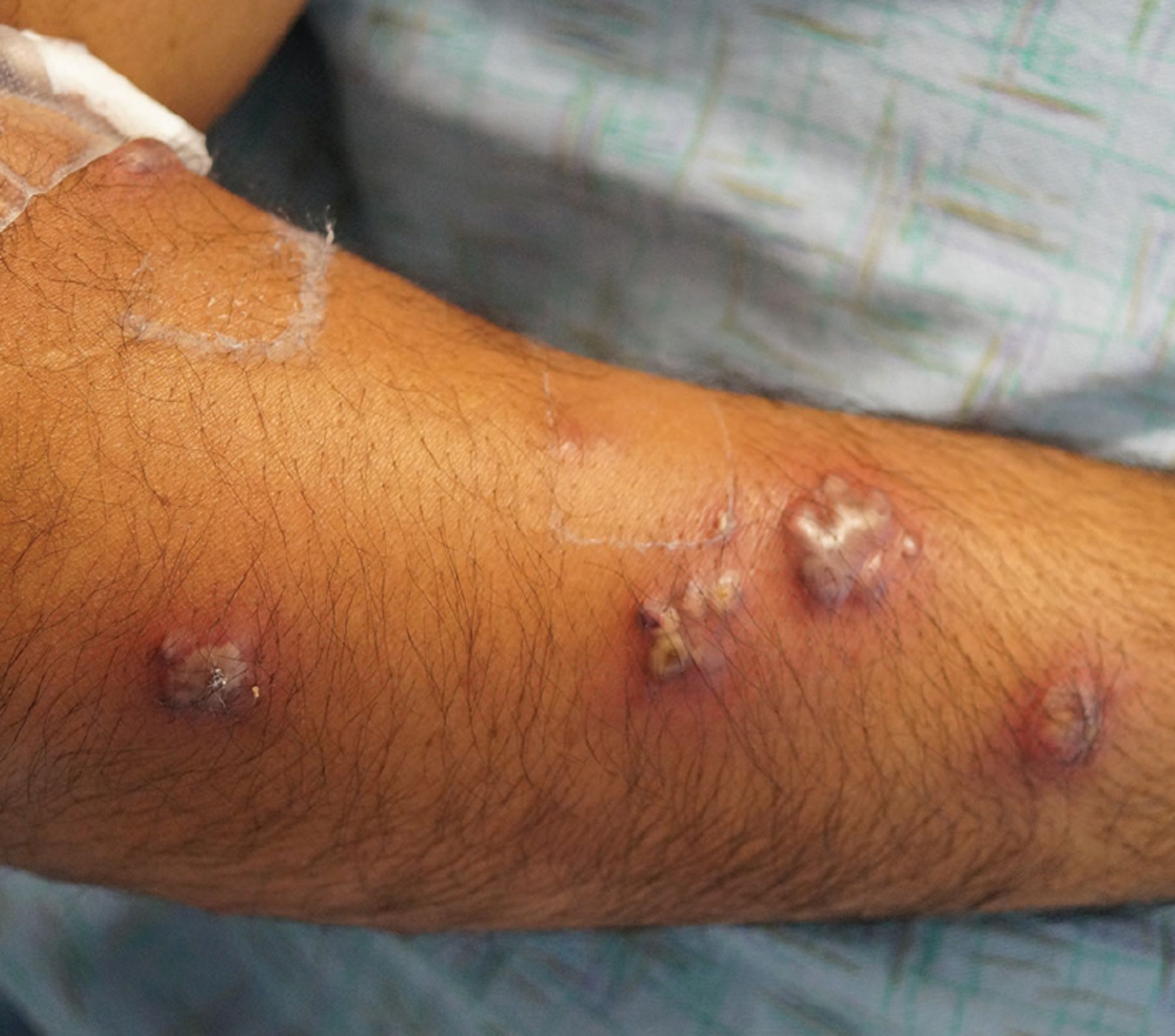
On physical examination, his temperature was 39.6°C, heart rate 138 beats per minute, blood pressure 93/70 mm Hg, and respiratory rate 18 breaths per minute. He had elevated, tender, inflammatory plaques, nodules, and pustules distributed throughout his upper and lower extremities, back, chest, abdomen, scalp, and dorsal aspects of both feet (Figures 1–3).
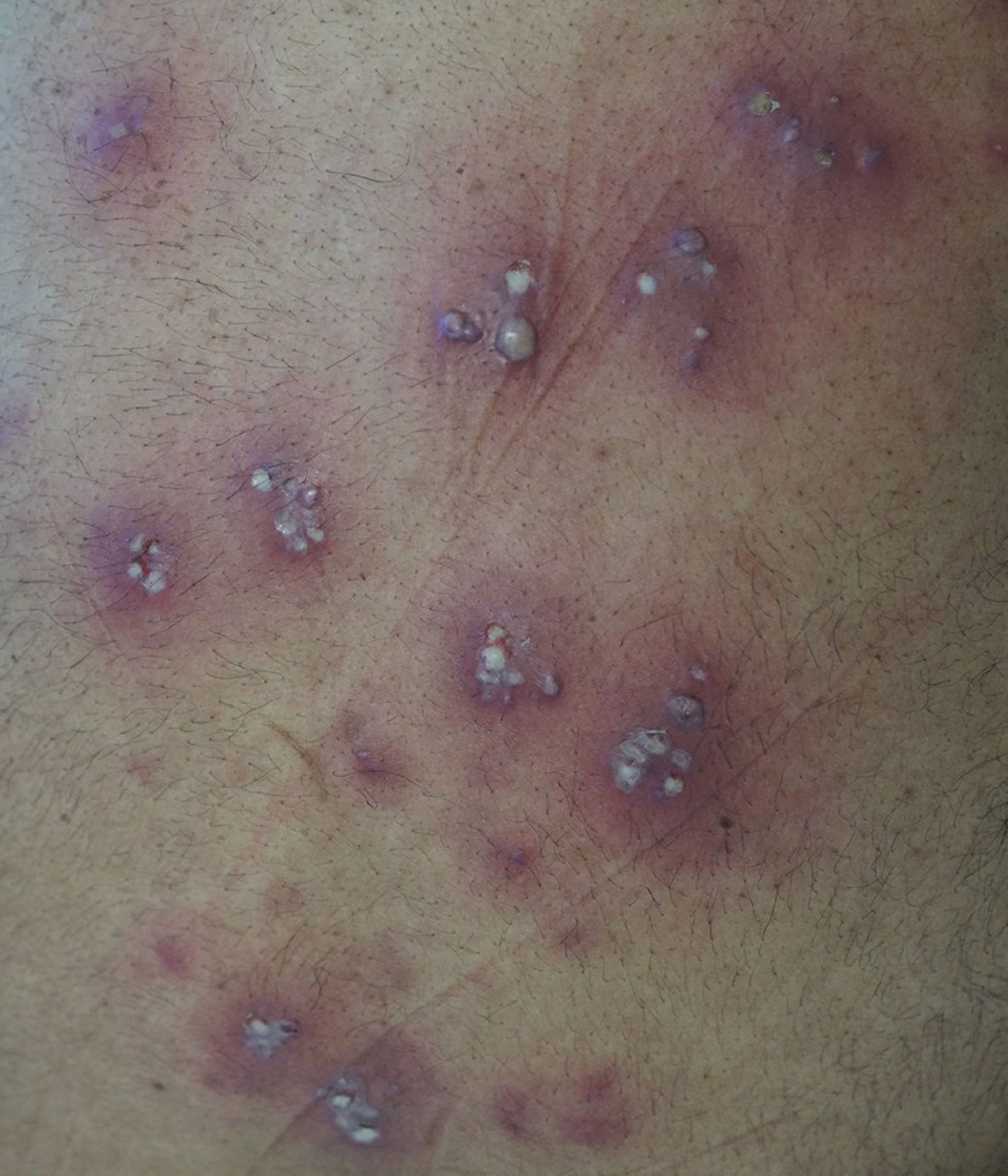
On the patient’s back were multiple firm erythematous nodules with pustular heads, as well as several erythematous plaques.

The patient’s lower extremities.

The patient’s right arm.
His white blood cell count was 15.2 × 10⁹/L (reference range 3.7–11.0 × 10⁹/L) with 60% neutrophils and 22% bands; the erythrocyte sedimentation rate was 48 mm/hour (0–15 mm/hour), and the C-reactive protein level was 10.4 mg/dL (< 0.9 mg/dL).
Because of concern for infectious dermatitis, his initial hospital treatment consisted of pain management, intravenous fluids, and intravenous vancomycin. However, a thorough infectious disease workup was unrevealing. Other considerations included a drug eruption, vasculitis, and various neutrophilic dermatoses (including pyoderma gangrenosum and Sweet syndrome). As a result, a skin biopsy was performed and revealed neutrophilic dermatitis without organisms, consistent with Sweet syndrome.
The patient began a 4-week course of prednisone in tapering doses (60 mg for 5 days, 40 mg for 5 days, 20 mg for 5 days, 10 mg for 5 days, then discontinued) and clobetasol 0.05% ointment twice daily for 7 days. He experienced rapid improvement of symptoms, which was noted during outpatient dermatology and primary care follow-up visits.
SWEET SYNDROME: KEY FEATURES
In 1964, R. D. Sweet, MD, first described acute febrile neutrophilic dermatosis, originally known as Gomm-Button disease, by its 4 cardinal features: fever; neutrophil polymer-phonuclear leucocytosis of the blood; raised painful plaques on the limbs, face, and neck; and histologically, a dense dermal infiltration with mature neutrophil polymorphs, with no evidence of infection and prompt response to corticosteroids.1
There are 3 subtypes of Sweet syndrome:
Classic or idiopathic
Malignancy-related, most commonly hematologic but also observed in solid tumor malignancies2–5
In this patient’s case, the abrupt appearance of fever, cutaneous lesions, and histopathologic findings, in the absence of malignancy or drug exposure and satisfaction of both major and 3 of 4 minor diagnostic criteria, were congruent with classic Sweet syndrome (Table 1).1,6-8 Notably, the patient did not have preceding symptoms suggestive of gastrointestinal or upper respiratory illness, was not on medications associated with Sweet syndrome, and never had signs or symptoms of hematologic or solid tumor malignancy.
Diagnostic criteria for classic Sweet syndrome
The pathogenesis of Sweet syndrome is multifactorial, but studies have suggested a hypersensitivity reaction that promotes neutrophil activation9 and higher serum levels of granulocyte colony-stimulating factor10 leading to increased circulating neutrophils, both of which may explain this patient’s leukocytosis with increased left shift.
Skin biopsy is the technical standard for diagnosis, but this patient’s response to systemic steroids and resolution of leukocytosis (his initial white blood cell count of 15.2 × 10⁹/L came down to 9.25 × 10⁹/L over 4 days) also supports the typical response to treatment.
STEROIDS ARE FIRST-LINE THERAPY
Systemic corticosteroid therapy is the first-line treatment, but some case reports11,12 have also noted accelerated improvement in cutaneous lesions and reduced dependence on systemic steroids with adjunct topical glucocorticoid use, although the efficacy has not been formally studied.
Given their low potential for harm, topical steroids were used as adjuvant therapy, and the patient had rapid overall clinical improvement and resolution of his rash. Colchicine and potassium iodide are also considered first-line treatment choices. Indomethacin, cyclosporine, dapsone, and clofazimine have all been used as second-line treatments.
DISCLOSURES
The authors report no relevant financial relationships which, in the context of their contributions, could be perceived as a potential conflict of interest.
- Copyright © 2021 The Cleveland Clinic Foundation. All Rights Reserved.
In this issue


Jump to section
Related Articles
Cited By...
- No citing articles found.